. 2013;8(2):e56387.
doi: 10.1371/journal.pone.0056387. Epub 2013 Feb 20.

Calreticulin induces dilated cardiomyopathy

Affiliations

Calreticulin induces dilated cardiomyopathy

Dukgyu Lee et al. PLoS One. 2013.

Erratum in

  • PLoS One. 2013;8(11). doi:10.1371/annotation/04c791d5-a71c-493d-b94a-a57b158c5538

Abstract

Background: Calreticulin, a Ca(2+)-buffering chaperone of the endoplasmic reticulum, is highly expressed in the embryonic heart and is essential for cardiac development. After birth, the calreticulin gene is sharply down regulated in the heart, and thus, adult hearts have negligible levels of calreticulin. In this study we tested the role of calreticulin in the adult heart.

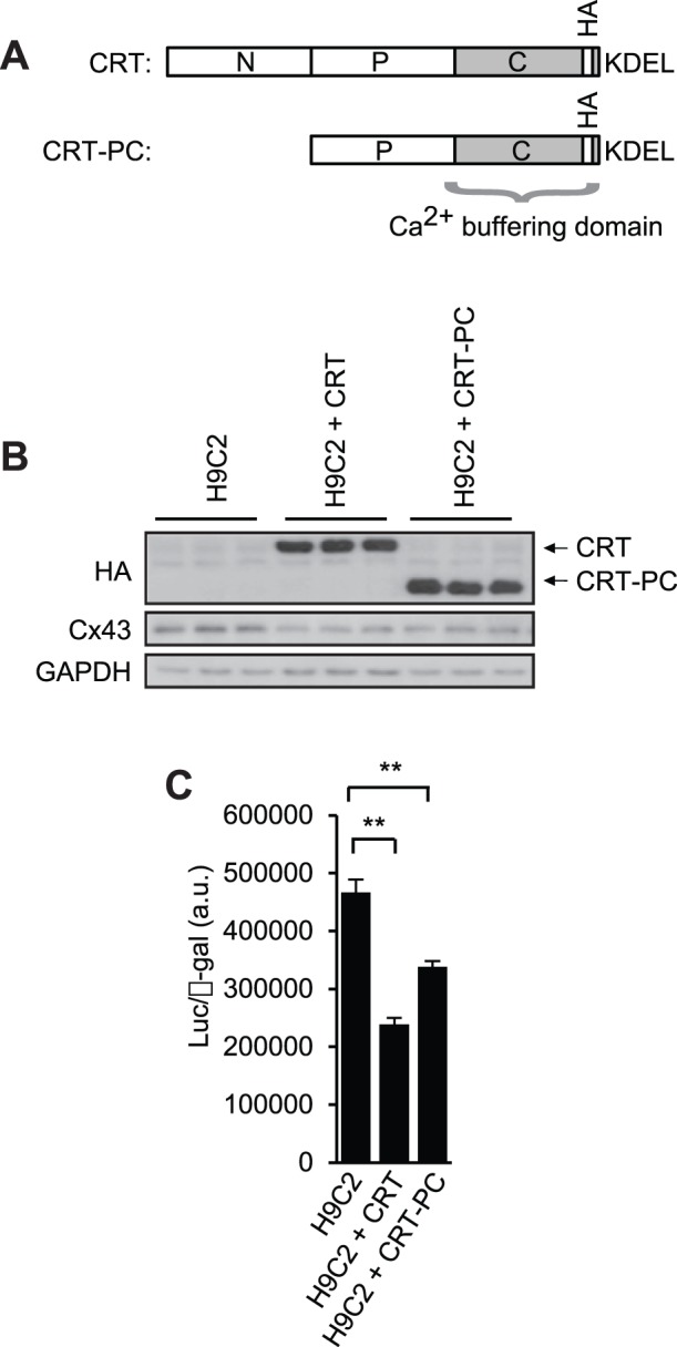
Methodology/principal findings: We generated an inducible transgenic mouse in which calreticulin is targeted to the cardiac tissue using a Cre/loxP system and can be up-regulated in adult hearts. Echocardiography analysis of hearts from transgenic mice expressing calreticulin revealed impaired left ventricular systolic and diastolic function and impaired mitral valve function. There was altered expression of Ca(2+) signaling molecules and the gap junction proteins, Connexin 43 and 45. Sarcoplasmic reticulum associated Ca(2+)-handling proteins (including the cardiac ryanodine receptor, sarco/endoplasmic reticulum Ca(2+)-ATPase, and cardiac calsequestrin) were down-regulated in the transgenic hearts with increased expression of calreticulin.

Conclusions/significance: We show that in adult heart, up-regulated expression of calreticulin induces cardiomyopathy in vivo leading to heart failure. This is due to an alternation in changes in a subset of Ca(2+) handling genes, gap junction components and left ventricle remodeling.


Conflict of interest statement

Competing Interests: The authors have declared that no competing interests exist.

Figures

Figure 1
Figure 1. Generation of cardiac-specific and inducible calreticulin transgenic mice.
(A) The CAT-loxP-CRT transgene consisted of a CAG promoter, the CAT gene flanked with loxP-sites followed by cDNA encoding HA-tagged calreticulin. Cardiac specific cre mice express cre recombinase (MerCreMer) under the control of αMHC promoter (αMHC-Cre). Double transgenic mice were generated by breeding CAT-loxP-CRT mice with αMHC-Cre mice to produce αMHC/CAT-loxP-CRT mice. Administration of tamoxifen triggers nuclear translocation of MerCreMer and cardiac-specific Cre mediated recombination and excision of the floxed CAT gene, resulting in the expression of HA-tagged calreticulin under the control of CAG promoter (αMHC/CRT mice). αMHC/CRT mice are referred to throughout the paper as calreticulin transgenic mice (CRT-TG). Arrows and arrow heads indicate the location of primers used for PCR-driven genomic DNA analysis of transgenic mice. The location of loxP sites is indicated. CAG, CMV early enhancer/chicken β actin; HA, hemagglutinin; CAT, chloramphenicol acetyltransferase, Cre, Cre recombinase (type I topoisomerase); αMHC, α-myosin heavy chain; Mer, modified estrogen receptor. CRT, calreticulin; KDEL, ER retention signal sequence. (B) Genotyping of the αMHC-Cre, CAT-loxP-CRT, and αMHC/CAT-loxP-CRT mice was carried out by PCR-driven amplification using primers shown in Figure 1A (arrows for CAT gene and arrow heads for MerCreMer gene). (C) Western blot analysis of expression of recombinant CRT (anti-HA) and endogenous CRT (anti-CRT) after feeding tamoxifen for 3 weeks. Glyceraldehyde 3-phosphate dehydrogenase (anti-GAPDH) was used as a loading control. (D) Tissue specific expression of recombinant calreticulin (anti-HA) in CRT-TG mice and expression of endogenous calreticulin (anti-CRT) from control mouse. Sk muscle, skeletal muscle.
Figure 2
Figure 2. Gross morphology, echocardiography and electrocardiographic presentations of hearts with increased expression of calreticulin.
(A) Hematoxylin and eosin staining of hearts from control (CAT-loxP-CRT), αMHC-Cre and CRT-TG mice fed tamoxifen for 3 weeks. Histological analysis of αMHC/CAT-loxP-CRT double transgenic not fed tamoxifen is also included. Scale bar, 1 mm. TAM, tamoxifen; LV, left ventricle; RV, right ventricle. (B) Representative M-mode echocardiography images of control (CAT-loxP-CRT) and CRT-TG hearts from mice fed tamoxifen for 3 weeks. ESD, end systolic diameter; EDD, end diastolic diameter. (C) Representative images of transmitral flow velocity pattern in the pulmonary venous flow in control and CRT-TG hearts. E, E-wave; A, A-wave. (D) Representative electrocardiography recording images of hearts from control and CRT-TG mice fed tamoxifen for 3 weeks (n = 10).
Figure 3
Figure 3. Attenuation of sarcoplasmic reticulum associated proteins in hearts of CRT-TG mice.
(A) Cardiac tissues from control and CRT-TG mice fed tamoxifen for 3 weeks were harvested, lysed followed by SDS-PAGE, transferred to nitrocellulose membrane, and probed with specific antibodies. Anti-GAPDH antibodies were used as a loading control. CNX, calnexin; PDI, protein disulfide isomerase. (B) Quantitative analysis of Western blots of sarcoplasmic reticulum associated proteins. **p<0.01 (n = 4). (C) Western blot analysis of SR-associated proteins from control and CRT-TG hearts. SR membrane vesicles were isolated from hearts of control and CRT-TG mice fed tamoxifen for 3 weeks followed by SDS-PAGE and Western blot analysis with specific antibodies. RYR2, cardiac ryanodine receptor; SERCA2a, cardiac sarcoplasmic/endoplasmic reticulum Ca2+ ATPase; PLN, phospholamban, p-PLN(S16/T17), phospho-phospholamban at Ser16/Thr17 amino acid residues, NCX1, sodium/calcium exchanger-1. GAPDH was used as a loading control. (D) Quantitative analysis of SR-associated proteins from control and CRT-TG hearts. **p<0.01 (n = 4). For junctin quantitative analysis in D: p>0.25.
Figure 4
Figure 4. Down-regulation of calsequestrin transcript and protein in hearts from CRT-TG mice.
(A) Semi-quantitative RT-PCR analysis of calsequestrin (CASQ2) in hearts isolated from control and CRT-TG mice fed tamoxifen. Gapdh, glyceraldehyde 3-phosphate dehydrogenase. (B) Western blot analysis with anti-HA (CRT-HA, for recombinant calreticulin) and anti-CASQ2 antibodies. Control and CRT-TG mice were fed tamoxifen for 1, 2 and 3 weeks, following which hearts were harvested and processed for Western blot analysis. GAPDH was used as a loading control. HA, hemagglutinin; CASQ2, cardiac calsequestrin. (C) Quantitative analysis of expression of CASQ2 in mice with induced expression of calreticulin in adult heart. (mean±SEM; n = 3). CASQ2, cardiac calsequestrin; CRT-HA, calreticulin hemagglutinin. Quantitation of CRT-HA and CASQ2 expression depicted in (B) *p<0.05 (n = 3).
Figure 5
Figure 5. Ca2+ signaling molecules in hearts from CRT-TG mice.
(A) Semi-quantitative RT-PCR analysis of mRNA encoding Ca2+ signaling genes in control and CRT-TG animals. Calm, calmodulin; CaNA, calcineurin A; MEF2c, myocyte enhancing factor 2c; GADPH, glyceraldehyde 3-phosphate dehydrogenase. (B) Quantitative analysis of RT-PCR of mRNA encoding Ca2+ signaling genes. **p<0.01 (n = 4).
Figure 6
Figure 6. Reduced expression of connexins in hearts from CRT-TG mice.
(A) Real-time Q-PCR analysis of Cx43 transcript level in control and CRT-TG hearts. *p<0.05 (n = 5). (B) Western blot analysis of hearts from control and CRT-TG mice probed with anit-CRT, anti-Cx43, anti-phospho-Cx43(S262), anti-phospho-Cx43(S368), anti-PKCε, and anti-Cx45 antibodies. GAPDH was used as a loading control. HA, hemagglutinin; PKCε, protein kinase C-epsilon. (C) Quantitative analysis of the level of Cx43, p-Cx43(S262), phospho-Cx43(S368), Cx45, and Cx45/Cx43 ratio. **p<0.01 (n = 3). (D) Distribution pattern of Cx43 gap junctions in ventricular myocardium in control (i, ii) and CRT-TG mice (iii, iv, v). i, ii, longitudinal sections from control hearts, at low (i) and high magnifications, (ii). The arrows in ii indicate a regular pattern of Cx43 intercalated disk staining. Scale bars, 200 µm (i) and 100 µm (ii). iii, iv, v, longitudinal sections from CRT-TG hearts, at low (iii) and high (iv, v) magnifications. In iii, Cx43 patchy staining with areas of near complete lack of Cx43. In iv, enlarged view of an area with a weak anti-Cx43 staining. In v, areas of disorganized Cx43 punctate staining (open pink arrows). The arrows indicate a regular pattern of Cx43 intercalated disk staining. Scale bars: 200 µm for iii, 100 µm for iv and 50 µm for v. (E) Western blot analysis of hearts from control and CRT-TG mice probed with anti-ZO-1 antibodies. GAPDH was used as a loading control. ZO-1; zona occludens-1. (F) Quantitative analysis of ZO-1 expression. *p<0.05 (n = 3).
Figure 7
Figure 7. Suppression of Cx43 promoter by calreticulin.
(A) A schematic representation of calreticulin and calreticulin domains. CRT, calreticulin full length tagged with HA epitope; CRT-PC, P+C domain of calreticulin tagged with HA epitope. HA, hemagglutinin; KDEL, ER retrieval amino acid sequence. (B) Western blot analysis with anti-HA antibodies (reporting recombinant calreticulin and calreticulin PC domain) and anti-Cx43 antibodies of cell lysates from H9C2 cells (H9C2), H9C2 cells expression full length calreticulin (H9C2+CRT), and H9C2 cells expression Ca2+ buffering PC domain of calreticulin (H9C2+CRT-PC). HA, hemagglutinin; CRT, calreticulin; CRT-PC, calreticulin P+C domain. GAPDH was used as a loading control. (C) Control H9C2 cells (H9C2), H9C2 cell lines expression full length calreticulin (H9C2+CRT) or H9C2 cells expression Ca2+ buffering PC domain of calreticulin (H9C2+CRT-PC) were transfected with luciferase reporter vector under control of the Cx43 promoter and β-galactosidase expression vector. Cell lysates were harvested and assayed for luciferase activity. **p<0.01 (n = 9).

References

    1. Michalak M, Opas M (2009) Endoplasmic and sarcoplasmic reticulum in the heart. Trends Cell Biol 19: 253–259. - PubMed
    1. Groenendyk J, Sreenivasaiah PK, Kim do H, Agellon LB, Michalak M (2010) Biology of endoplasmic reticulum stress in the heart. Circ Res 107: 1185–1197. - PubMed
    1. Mesaeli N, Nakamura K, Zvaritch E, Dickie P, Dziak E, et al. (1999) Calreticulin is essential for cardiac development. J Cell Biol 144: 857–868. - PMC - PubMed
    1. Nakamura K, Robertson M, Liu G, Dickie P, Guo JQ, et al. (2001) Complete heart block and sudden death in mouse over-expressing calreticulin. J Clin Invest 107: 1245–1253. - PMC - PubMed
    1. Mery A, Aimond F, Menard C, Mikoshiba K, Michalak M, et al. (2005) Initiation of embryonic cardiac pacemaker activity by inositol 1,4,5-trisphosphate-dependent calcium signaling. Mol Biol Cell 16: 2414–2423. - PMC - PubMed

Publication types

MeSH terms