. 2013;8(2):e56595.
doi: 10.1371/journal.pone.0056595. Epub 2013 Feb 21.

Nuclear receptor NR4A2 orchestrates Th17 cell-mediated autoimmune inflammation via IL-21 signalling

Affiliations

Nuclear receptor NR4A2 orchestrates Th17 cell-mediated autoimmune inflammation via IL-21 signalling

Ben J E Raveney et al. PLoS One. 2013.

Abstract

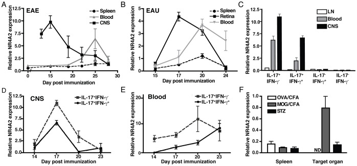
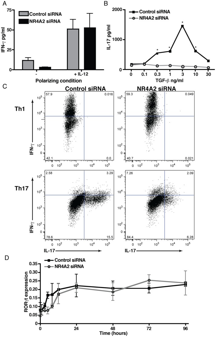
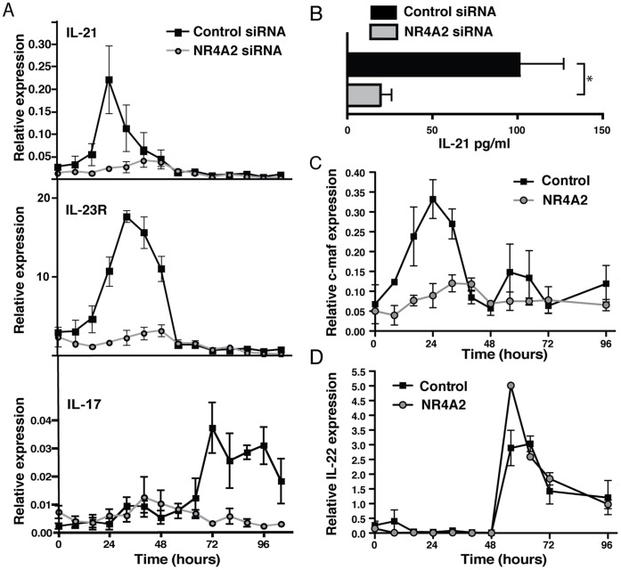
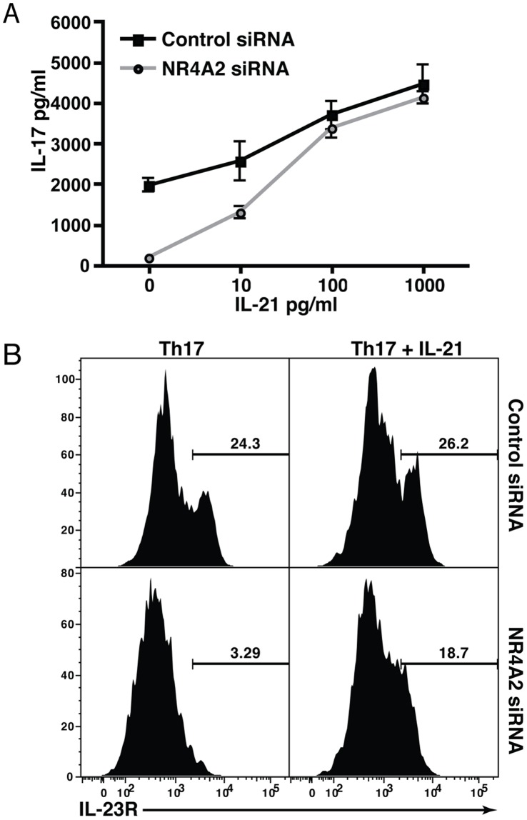
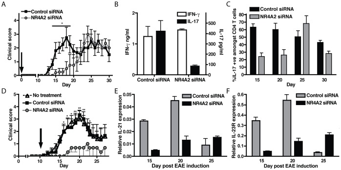
IL-17-producing CD4(+) T helper 17 (Th17) cells are pathogenic in a range of human autoimmune diseases and corresponding animal models. We now demonstrate that such T cells infiltrating the target organ during the induction of experimental autoimmune encephalomyelitis (EAE) and experimental autoimmune uveoretinitis (EAU) specifically express NR4A2. Further, we reveal a critical involvement of NR4A2 in Th17 cell functions and Th17 cell-driven autoimmune diseases. When NR4A2 expression was blocked with siRNA, full Th17 differentiation was prevented in vitro: although cells expressed the master Th17 regulator, RORγt, they expressed reduced levels of IL-23R and were unable to produce IL-17 and IL-21. Notably, Th17 differentiation in the absence of NR4A2 was restored by exogenous IL-21, indicating that NR4A2 controls full maturation of Th17 cells via autocrine IL-21 signalling. Preventing NR4A2 expression in vivo by systemic treatment with NR4A2-specific siRNA also reduced Th17 effector responses and furthermore protected mice from EAE induction. In addition, the lack of disease was associated with a reduction in autocrine IL-21 production and IL-23R expression. Similar modulation of NR4A2 expression was also effective as an intervention, reversing established autoimmune responses and ameliorating clinical disease symptoms. Thus, NR4A2 appears to control Th17 differentiation and so plays an essential role in the development of Th17-mediated autoimmune disease. As NR4A2 is also upregulated during human autoimmune disease, targeting NR4A2 may provide a new therapeutic approach in treating autoimmune disease.


Conflict of interest statement

Competing Interests: The authors have declared that no competing interests exist.

Figures

Figure 1
Figure 1. Autoimmune induction of NR4A2 in CD4+ T cells is associated with IL-17-secreting T cells.
EAE or EAU was induced in C57BL/6 mice by immunization with MOG35–55 or IRBP1–20 peptide in CFA, respectively. CD4+ T cells were purified from spleen, blood, or target organ (CNS or retina) on the indicated days and RNA was isolated. A and B: NR4A2 expression was quantified by real time PCR relative to GAPDH for T cells from EAE (A) or EAU (B). Timepoints correspond to a minimum of 5 animals and data are representative of 3 independent experiments. CD4+ T cells from mice with EAE were restimulated with PMA/ionomycin for 3 hours and 4 populations of cytokine secreting cells (IL-17+IFN-γ-, IL-17+IFN-γ+, IL-17-IFN-γ+, and IL-17-IFN-γ-) were sorted by flow cytometry using IFN-γ and IL-17 cytokine secretion assay kits. C: NR4A2 expression by populations of cytokine-secreting CD4+ T cells was quantified by real time PCR at day 15 post-EAE induction for lymph nodes (LN) and CNS-infiltrating cells (CNS), and day 25 for blood T cells. D and E: NR4A2 expression by IL-17+IFN-γ or IL-17+IFN-γ+ CNS-infiltrating T cells (D) or blood T cells (E) was measured by real time PCR at a range of timepoints. Data are representative of 2 independent experiments. F: Th1-mediated diabetes was induced in C57BL/6 mice by 5 daily low dose STZ treatments. Other groups of C57BL/6 mice were immunized with peptides in CFA plus PTX either OVA323–339 (OVA/CFA) or MOG35–55 (MOG/CFA). On day 22, NR4A2 expression was assessed by real time PCR amongst CD4+ T cells from spleen and leukocytes isolated from the relevant target organ (ND, OVA/CFA; CNS, EAE; pancreas, STZ). Timepoints correspond to a minimum of 5 animals and data are representative of 2 independent experiments.
Figure 2
Figure 2. NR4A2 knockdown prevents IL-17 secretion but not RORγt upregulation.
Naïve CD4+ T cells were transfected by electroporation with NR4A2-specific siRNA or scrambled control siRNA. Cells were then activated with 5 µg/ml plate-bound CD3-specific mAb and 0.5 µg/ml soluble CD28-specific mAb. A: IFN-γ production by cells activated in the presence or absence of 10 ng/ml IL-12 after 96 hours of culture. B: IL-17 production by cells activated in the presence of 20 ng/ml IL-6, 20 ng/ml IL-23, and TGF-β at a range of concentrations after 96 hours of culture. Significant differences between control and NR4A2 siRNA-treatments were tested with a student’s t-test, *p<0.05. C: IL-17 and IFN-γ intracellular cytokine staining for transfected T cells (control siRNA, left plots; NR4A2 siRNA, right plots) cultured for 96 hours in the presence of 10 ng/ml IL-12 (Th1 conditions, top row plots) or 20 ng/ml IL-6, 20 ng/ml IL-23, and 3 ng/ml TGF-β (Th17 conditions, bottom row plots). D: RORγt RNA expression as measured by real time PCR by activated T cells cultured under Th17 polarizing conditions at a range of timepoints. Data are representative of 5 independent experiments.
Figure 3
Figure 3. Absence of NR4A2 is associated with a lack of IL-21 production by Th17 cells.
Naïve CD4+ T cells were transfected by electroporation with NR4A2-specific siRNA or scrambled control siRNA and were activated with 5 µg/ml plate-bound CD3-specific mAb and 0.5 µg/ml soluble CD28-specific mAb in the presence of 20 ng/ml IL-6, 20 ng/ml IL-23, and 3 ng/ml TGF-β. A: RNA levels of IL-21, IL-23R, and IL-17 were quantified by real time PCR at the indicated timepoints following activation. Data are representative of 3 independent experiments. B: IL-21 supernatant concentration was measured by ELISA at 96 hours. Data are representative of 3 independent experiments. *p<0.05. C: RNA expression of c-maf quantified by real time PCR. D: RNA expression of IL-22 quantified by real time PCR. Data are representative of 2 independent experiments.
Figure 4
Figure 4. Exogenous IL-21 restores IL-17 production in the absence of NR4A2.
Naïve CD4+ T cells were transfected by electroporation with NR4A2-specific siRNA or scrambled control siRNA and were activated with 5 µg/ml plate-bound CD3-specific mAb and 0. 5 µg/ml soluble CD28-specific mAb in the presence of 20 ng/ml IL-6, 20 ng/ml IL-23, and 3 ng/ml TGF-β. To some wells, recombinant IL-21 was added as indicated. A: IL-17 was measured in the supernatants of control or NR4A2 siRNA-treated T cells by ELISA after 96 hours of culture under Th17 polarizing conditions in the presence or absence of IL-21 at the indicated concentrations. *p<0.05. Data are representative of 3 independent experiments. B: IL-23R expression was assessed by intracellular flow cytometric staining after 96 hours of culture under Th17 polarizing conditions in the presence (right plots) or absence (left plots) of 100 pg/ml recombinant IL-21 for control siRNA-treated T cells (top) and NR4A2 siRNA-treated T cells (bottom row). Data are representative of 2 independent experiments.
Figure 5
Figure 5. Systemic administration of NR4A2-specific siRNA reduces EAE severity.
siRNA, either NR4A2-specific or control, was stabilized in a collagen matrix and administered i.v. to groups of C57BL/6 mice at the time of EAE induction. EAE was scored clinically (A) and at day 15 post-EAE induction, production of IL-17 and IFN-γ by CNS-infiltrating leukocytes restimulated with 20 µg/ml MOG peptide for 96 hours were assessed by ELISA (B). CNS-infiltrating T cells were also assessed for IL-17 production at a range of timepoints by intracellular flow cytometry (C). Data are representative of 3 independent experiments. Control or NR4A2-specific siRNA was applied to MOG-immunized mice at day 10 post-disease induction and disease was scored clinically (D). Timepoints correspond to a minimum of 5 animals and data are representative of 2 independent experiments. IL-21 and IL-23R expressions amongst CNS-infiltrating T cells were measured by real time PCR (E&F). Data are representative of 2 independent experiments. Clinical scores in panels A) and D) were tested with a two-way ANOVA test. *p<0.01, **p<0.001.

References

    1. Sospedra M, Martin R (2005) Immunology of multiple sclerosis. Annu Rev Immunol 23: 683–747. - PubMed
    1. Bettelli E, Korn T, Kuchroo VK (2007) Th17: the third member of the effector T cell trilogy. Curr Opin Immunol 19: 652–657. - PMC - PubMed
    1. Damsker JM, Hansen AM, Caspi RR (2010) Th1 and Th17 cells: adversaries and collaborators. Ann N Y Acad Sci 1183: 211–221. - PMC - PubMed
    1. Korn T, Oukka M, Kuchroo V, Bettelli E (2007) Th17 cells: effector T cells with inflammatory properties. Semin Immunol 19: 362–371. - PMC - PubMed
    1. Goverman J (2009) Autoimmune T cell responses in the central nervous system. Nat Rev Immunol 9: 393–407. - PMC - PubMed

Publication types

MeSH terms

Substances