. 2013;8(2):e56841.
doi: 10.1371/journal.pone.0056841. Epub 2013 Feb 20.

Ultrasound-mediated stimulation of microbubbles after acute myocardial infarction and reperfusion ameliorates left-ventricular remodelling in mice via improvement of borderzone vascularization

Affiliations

Ultrasound-mediated stimulation of microbubbles after acute myocardial infarction and reperfusion ameliorates left-ventricular remodelling in mice via improvement of borderzone vascularization

Jonas Dörner et al. PLoS One. 2013.

Abstract

Aims: Post-infarction remodelling (PIR) determines left-ventricular (LV) function and prognosis after myocardial infarction. The aim of this study was to evaluate transthoracic ultrasound-mediated microbubble stimulation (UMS) as a novel gene- and cell-free therapeutic option after acute myocardial infarction and reperfusion (AMI/R) in mice.

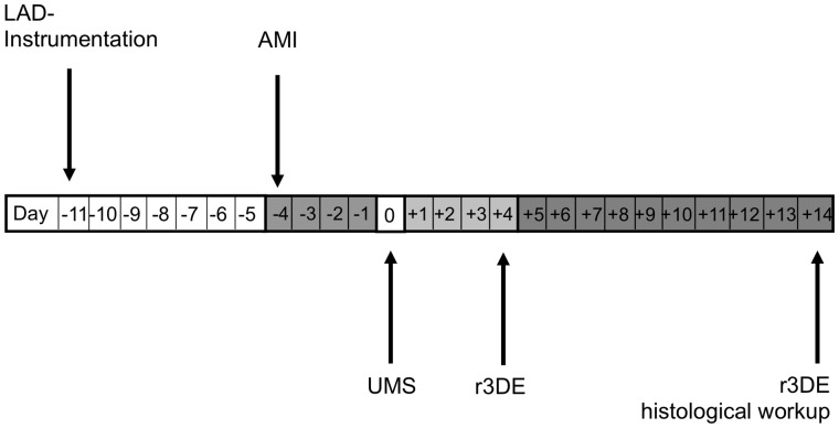
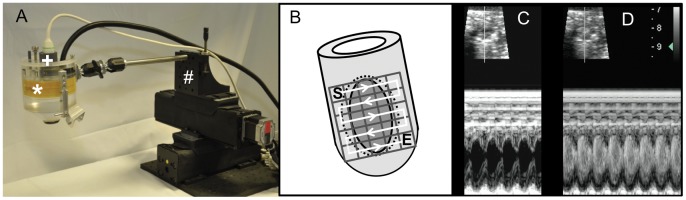
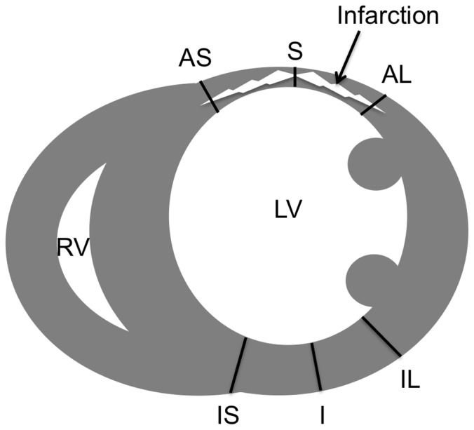
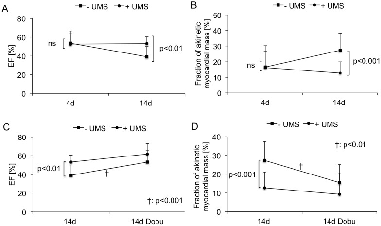
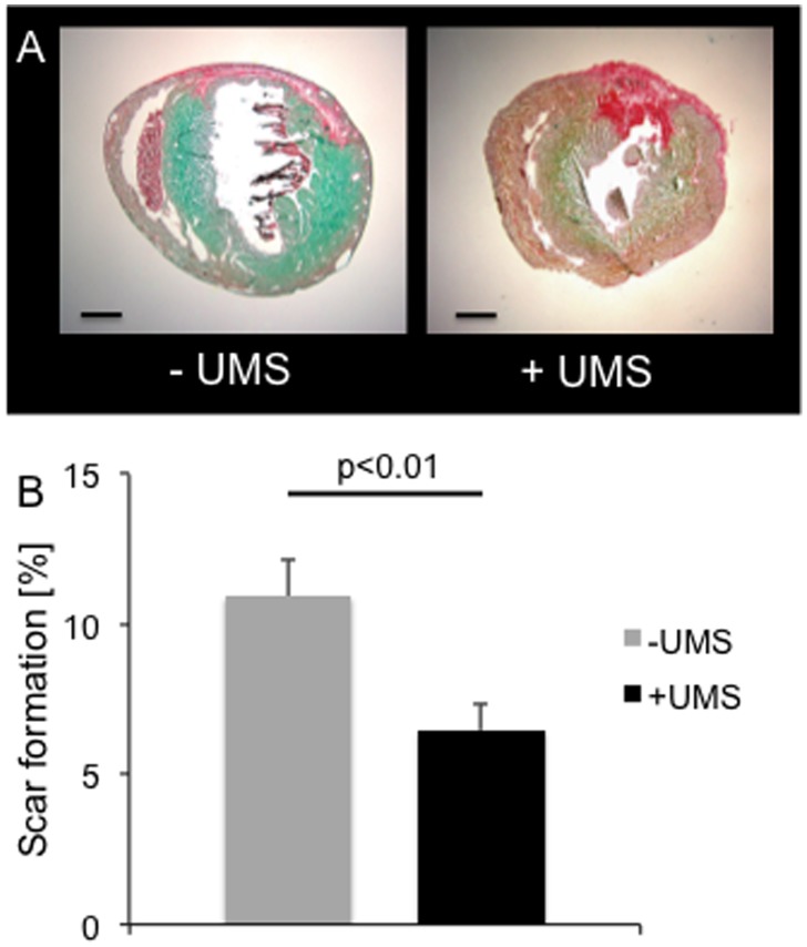
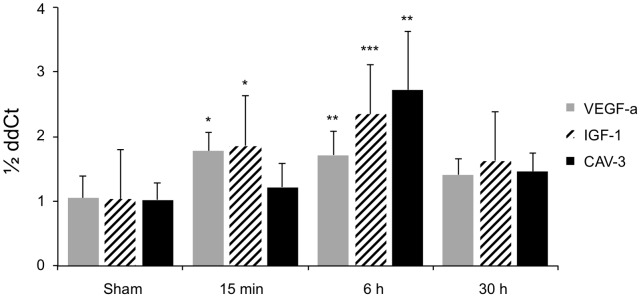
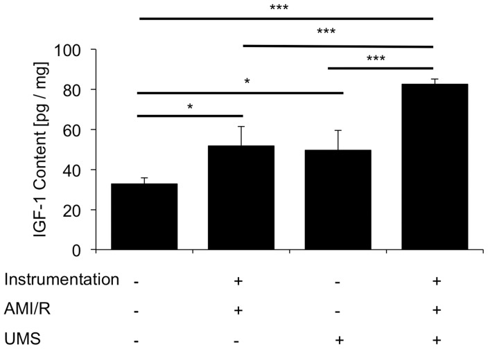
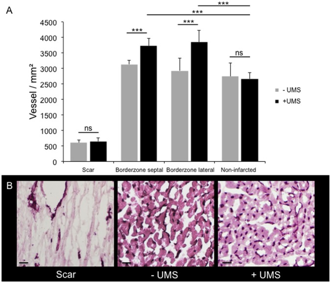
Methods and results: For myocardial delivery of UMS, a novel therapeutic ultrasound-system (TIPS, Philips Medical) and commercially available microbubbles (BR1, Bracco Suisse SA) were utilized in a closed-chest mouse model. UMS was performed as myocardial post-conditioning (PC) on day four after 30 minutes of coronary occlusion and reperfusion. LV-morphology, as well as global and regional function were measured repeatedly with reconstructive 3-dimensional echocardiography applying an additional low-dose dobutamine protocol after two weeks. Scar size was quantified by means of histomorphometry. A total of 41 mice were investigated; 17 received PC with UMS. Mean ejection fraction (EF) prior UMS was similar in both groups 53%±10 (w/o UMS) and 53%±14 (UMS, p = 0.5), reflecting comparable myocardial mass at risk 17%±8 (w/o UMS), 16%±13 (UMS, p = 0.5). Two weeks after AMI/R, mice undergoing UMS demonstrated significantly better global LV-function (EF = 53%±7) as compared to the group without PC (EF = 39%±11, p<0.01). The fraction of akinetic myocardial mass was significantly lower among mice undergoing UMS after AMI/R [27%±10 (w/o UMS), 13%±8 (UMS), p<0.001)]. Our experiments showed a fast onset of transient, UMS-induced upregulation of vascular-endothelial and insulin-like growth factor (VEGF-a, IGF-1), as well as caveolin-3 (Cav-3). The mice undergoing PC with UMS after AMI/R showed a significantly lower scar size. In addition, the microvascular density was significantly higher in the borderzone of UMS-treated animals.

Conclusion: UMS following AMI/R ameliorates PIR in mice via up-regulation of VEGF-a, IGF-1 and Cav-3, and consecutive improvement of myocardial borderzone vascularization.


Conflict of interest statement

Competing Interests: The authors have the following interest. Thierry Bettinger is employed by Bracco Suisse SA and provided the commercially available micro bubbles (BR1) for this study. There are no further patents, products in development or marketed products to declare. This does not alter the authors’ adherence to all the PLOS ONE policies on sharing data and materials, as detailed online in the guide for authors.

Figures

Figure 1
Figure 1. Experimental protocol.
The instrumentive surgery of the left anterior descending coronary artery (LAD-Instrumentation) was performed seven days prior to the acute myocardial infarction and reperfusion (AMI/R) to avoid a pro-inflammatory influence of trauma on post-infarction remodelling (PIR). The treatment group received UMS four days after AMI/R. On day +4 and +14, a reconstructive 3-dimensional echocardiography (r3DE) was performed to quantify global and regional left-ventricular function. Additionally, r3DE was carried out with low-dose dobutamine on day +14. Ultimately, hearts were harvested for histological workup.
Figure 2
Figure 2. Therapeutic imaging probe system (TIPS) for ultrasound mediated stimulation of microbubbles.
(A) Hybrid ultrasound probe system with electromechanically coupled diagnostic (+) and therapeutic probe (*) enabling simultaneous high-resolution imaging for targeted application of UMS. To allow a standardized application to a small, moving target organ, the system is coupled to a computer-programmed stepper motor (#). (B) “En face” heart model to visualize the computer-programmed grid on the anterolateral heart wall. The grid consists of 25 pulses total, administered every 1 mm and 5 pulses per row, respectively starting basal (S) and following the white line to the apex till E (end). UMS was targeted on the anterior left-ventricular wall and the anterior borderzone tissue (area within the dotted circle) after anterolateral ischemia. (C)+(D) Ultrasound B-Mode (upper image) and M-Mode (lower image) short axis view before (C) and after (D) microbubble application in a mouse without myocardial infarction. Hence, this hybrid scanhead allows standardized and targeted myocardial delivery of UMS in mice.
Figure 3
Figure 3. Schematic illustration for determination of hypertrophy.
The illustration shows the six locations of wall thickness measurements in a mid-ventricular histological short axis section with scarring of the anterolateral left-ventricular wall. S: scar; AS: anteroseptal borderzone; AL: anterolateral borderzone. Control regions: IS: inferoseptal wall; I: inferior wall; IL: inferolateral wall.
Figure 4
Figure 4. Global and regional left-ventricular function.
(A) Mean left-ventricular ejection fraction (LV-EF) was moderately reduced four days after acute myocardial infarction and reperfusion (AMI/R) prior to ultrasound-mediated stimulation of microbubbles (UMS) in both groups. (B) Regional LV-function was quantified by means of reconstructive 3-dimensional echocardiography (r3DE) and is expressed as fraction of akinetic myocardial mass. UMS-treated animals (circles) demonstrated functional improvement two weeks after AMI/R, as compared to controls (squares). (C, D) Global and regional LV-function were obtained prior and during pharmacological stimulation with low-dose dobutamine on day +14. Inotropic response was preserved in both groups and revealed a significant increase in LV-EF and decrease in fraction of akinetic myocardial mass. Both non-invasive measures are parameters indicating preserved myocardial viability after AMI and reperfusion. In all, UMS improved LV-function after AMIR/R without impact on myocardial viability.
Figure 5
Figure 5. Histological analyses.
Myocardial scar formation was determined by means of histomorphometry. (A) Representative images of short axis mouse heart sections stained with picrosirius-red and fast-green without (left) and with (right) UMS-treatment. The collagenous tissue is coloured red whereas myocardium is green. (B) UMS-treated mice demonstrated a significantly lower collagenous scar burden compared to control mice.
Figure 6
Figure 6. Delivery of myocardial UMS in mice.
UMS-mediated myocardial delivery of fluorescent nanospheres was measured and quantified by fluorescence microscopy. Myocardial delivery of fluorescent nanospheres with UMS was feasible in mice and demonstrated a dose-dependent effect.
Figure 7
Figure 7. RNA-levels after UMS.
UMS increases VEGF-a, IGF-1 and Cav-3 mRNA-levels within 15 min and reaches its peak expression after 6 hours. A prolonged upregulation could not be observed longer than 30 hours after UMS as compared to controls. The displayed p-values refer to the comparison with sham-treated animals.
Figure 8
Figure 8. Myocardial protein concentration of IGF-1 after UMS.
Insulin-like growth factor 1 (IGF-1) was measured with a quantitative ELISA. UMS application not only increased IGF-1 content in control hearts, but also demonstrated a significant upregulation of IGF-1 on top of acute myocardial infarction and reperfusion (AMI/R).
Figure 9
Figure 9. Microvascular density assessed by CD31 staining.
(A) Microvascular density was assessed in the myocardial scar, both adjacent borderzones, and non-infarcted regions (posterior left-ventricular wall). No differences between untreated and UMS-treated animals were found with respect to the scar tissue and the non-infarcted regions. However, the myocardial borderzone tissue of UMS-treated mice revealed a significantly increased microvascular density as compared to non-treated animals. (B) Representative CD31 stained histological images of scar, untreated borderzone (−UMS), and UMS-treated borderzone (+UMS) (from left to right). CD31 positive vessels can be identified by their dark colour. Note the higher microvascular density of the UMS-treated mice compared to the non-treated group. In contrast, scar displayed the lowest microvascular density.

References

    1. Pfeffer MA, Braunwald E (1990) Ventricular remodeling after myocardial infarction. Experimental observations and clinical implications. Circulation 81: 1161–1172. - PubMed
    1. Sutton MG, Sharpe N (2000) Left ventricular remodeling after myocardial infarction: pathophysiology and therapy. Circulation 101: 2981–2988. - PubMed
    1. Gaudron P, Eilles C, Kugler I, Ertl G (1993) Progressive left ventricular dysfunction and remodeling after myocardial infarction. Potential mechanisms and early predictors. Circulation 87: 755–763. - PubMed
    1. Cittadini A, Monti MG, Petrillo V, Esposito G, Imparato G, et al. (2011) Complementary therapeutic effects of dual delivery of insulin-like growth factor-1 and vascular endothelial growth factor by gelatin microspheres in experimental heart failure. Eur J Heart Fail 13: 1264–1274. - PubMed
    1. Tsutsumi YM, Horikawa YT, Jennings MM, Kidd MW, Niesman IR, et al. (2008) Cardiac-specific overexpression of caveolin-3 induces endogenous cardiac protection by mimicking ischemic preconditioning. Circulation 118: 1979–1988. - PMC - PubMed

Publication types

MeSH terms