. 2013;8(2):e57112.
doi: 10.1371/journal.pone.0057112. Epub 2013 Feb 20.

An extract of Artemisia dracunculus L. inhibits ubiquitin-proteasome activity and preserves skeletal muscle mass in a murine model of diabetes

Affiliations

An extract of Artemisia dracunculus L. inhibits ubiquitin-proteasome activity and preserves skeletal muscle mass in a murine model of diabetes

Heather Kirk-Ballard et al. PLoS One. 2013.

Abstract

Impaired insulin signaling is a key feature of type 2 diabetes and is associated with increased ubiquitin-proteasome-dependent protein degradation in skeletal muscle. An extract of Artemisia dracunculus L. (termed PMI5011) improves insulin action by increasing insulin signaling in skeletal muscle. We sought to determine if the effect of PMI5011 on insulin signaling extends to regulation of the ubiquitin-proteasome system. C2C12 myotubes and the KK-A(y) murine model of type 2 diabetes were used to evaluate the effect of PMI5011 on steady-state levels of ubiquitylation, proteasome activity and expression of Atrogin-1 and MuRF-1, muscle-specific ubiquitin ligases that are upregulated with impaired insulin signaling. Our results show that PMI5011 inhibits proteasome activity and steady-state ubiquitylation levels in vitro and in vivo. The effect of PMI5011 is mediated by PI3K/Akt signaling and correlates with decreased expression of Atrogin-1 and MuRF-1. Under in vitro conditions of hormonal or fatty acid-induced insulin resistance, PMI5011 improves insulin signaling and reduces Atrogin-1 and MuRF-1 protein levels. In the KK-A(y) murine model of type 2 diabetes, skeletal muscle ubiquitylation and proteasome activity is inhibited and Atrogin-1 and MuRF-1 expression is decreased by PMI5011. PMI5011-mediated changes in the ubiquitin-proteasome system in vivo correlate with increased phosphorylation of Akt and FoxO3a and increased myofiber size. The changes in Atrogin-1 and MuRF-1 expression, ubiquitin-proteasome activity and myofiber size modulated by PMI5011 in the presence of insulin resistance indicate the botanical extract PMI5011 may have therapeutic potential in the preservation of muscle mass in type 2 diabetes.


Conflict of interest statement

Competing Interests: The authors have declared that no competing interests exist.

Figures

Figure 1
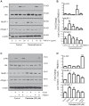
Figure 1. PMI5011 regulates expression of Atrogin-1 and MuRF-1 in skeletal muscle in a PI3K/Akt dependent manner.
(A) C2C12 myotubes were incubated with the indicated concentrations of PMI5011 and whole cell extracts were harvested 16 hours thereafter. The levels of IRS-1, pAkt, 19S proteasome subunit RPN2, Atrogin-1 and MuRF-1 were assayed by western blot analysis. The fold change in Atrogin-1 and MuRF-1 expression was analyzed from three separate experiments. The fold change in Atrogin-1 and MuRF-1 protein expression compared to expression in the absence of PMI5011 was analyzed from three independent experiments. (B) C2C12 myotubes were preincubated with PMI5011 (10 µg/ml) for 16 hours as indicated prior to the addition of wortmannin (200 nM). After a 1 hour preincubation with wortmannin, insulin (100 nM) was added as indicated. Whole cell extracts were harvested two hours thereafter and subjected to SDS-PAGE followed by western blot analysis. Inhibition of PI3K/Akt signaling by wortmannin was confirmed by loss of Akt phosphorylation. The fold change in Atrogin-1 and MuRF-1 protein expression in the presence of Wortmannin relative to the corresponding (−) wortmannin conditions was analyzed from three independent experiments. * p<0.05.
Figure 2
Figure 2. PMI5011 regulates expression of Atrogin-1 and MuRF-1 in two models of insulin resistance in vitro.
(A) C2C12 myotubes were incubated in the absence (DMSO) or presence of PMI5011 at the indicated concentrations for 16 hours prior to the addition of dexamethasone (1 µM). Twenty-four hour after adding dexamethasone, whole cell extracts were harvested and subjected to SDS-PAGE followed by western blot analysis to determine phospho-Akt, total Akt, Atrogin-1 and MuRF-1 protein levels. β-actin is included as a loading control. (B) The fold change over control for phospho-Akt, MuRF-1 and Atrogin-1 protein levels was analyzed from three independent experiments. * p<0.05 compared to control. (C) The C2C12 myotubes were incubated in the absence (DMSO) or PMI5011 (10 µg/ml) for 16 hours prior to adding palmitic acid (200 µM) as indicated. Twenty-four hours later, the cells were serum-deprived for 4 hours prior to insulin-stimulation (100 nM insulin) for 2 hours. Whole cell extracts were harvested and subjected to SDS-PAGE followed by western blot analysis of phospho-Akt, total Akt, Atrogin-1 and MuRF-1 protein levels. (D) The fold change over control for phospho-Akt, MuRF-1 and Atrogin-1 protein levels was analyzed from three independent experiments. * p<0.05,*** p<0.001 compared to control.
Figure 3
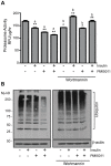
Figure 3. PMI5011 enhances the effect of insulin on proteasome activity and inhibits ubiquitylation in skeletal muscle.
C2C12 myotubes were incubated with 10 µg/ml PMI5011 for 16 hours. The cells were subsequently incubated with wortmannin (200 nM) for 1 hour prior to the addition of insulin (100 nM) for 2 hours as indicated. (A) The cells were harvested and assayed for the chymotrysin-like protease activity of the proteasome. Proteasome activity is reported as Relative Fluorescence Units (RFU) RFU/µg protein/hr. The data are reported as the mean −/+ standard deviation from triplicate measurements and are representative of three independent experiments. a = compared to control; b = compared to related treatment (−) wortmannin; *p<0.05, **p<0.01 (B) Whole cell extracts were also subjected to SDS-PAGE followed by western blot analysis using an anti-ubiquitin antibody to assay steady-state ubiquitylation levels. The data are representative of three independent experiments.
Figure 4
Figure 4. PMI5011 supplementation improves insulin sensitivity in vivo.
KK-Ay mice were singly housed and maintained on a low fat diet (control, N = 8) or a low fat diet containing 1% PMI5011 (w/w) (PMI5011, N = 8) for two months. (A) Food intake was measured daily and (B) body weight has measured each week. (C) Plasma insulin and (D) glucose levels were determined at baseline, 4 and 8 weeks. (E) The index of homeostasis model assessment of insulin resistance [HOMA-IR; insulin (mU/L) x glucose (mM)/22.5] was calculated from fasting glucose and insulin levels.
Figure 5
Figure 5. PMI5011 regulates proteasome and non-proteasome protease activity in skeletal muscle.
At the end of the study, the KK-Ay mice were fasted for 4 hours and insulin (1.5 U/kg) or an equal volume of sterile PBS was administered by intraperitoneal injection to a subgroup (N = 4) of the control or PMI5011 supplemented (N = 4) mice. Skeletal muscle tissue (gastrocnemious) was harvested 90 minutes thereafter. (A) Gene expression of two proteasome subunits, PSMA5 and PSMB3, was analyzed by realtime RT-PCR. (B) The chymotrypin-like, trypsin-like and caspase-like 26S proteasome activities were assayed in a buffer containing MgATP to maintain the 26S proteasome structure. (C) Non-proteasomal protease activity was assayed as the chymotrypsin-like, trypsin-like or caspase-like activity measured in the presence of epoxomicin (20 µM), a highly specific proteasome inhibitor. Proteasome and nonproteasome activities are reported as RFU/µg protein/hr. The data are reported as the mean −/+ standard deviation (4 animals/group). Statistical significance is compared to control. *p<0.05, ** p<0.01, ***p<0.001.
Figure 6
Figure 6. PMI5011 alters ubiquitin conjugation patterns in skeletal muscle.
Steady-state ubiquitylation were measured in (A) control (N = 4) and PMI5011 supplemented (N = 4) KK-Ay mice or (B) control (N = 4) and PMI5011 supplemented (N = 4) KK-Ay mice administered insulin (1.5 U/kg IP) at the end of the study with tissue harvested 90 minutes thereafter. Whole cell extracts were subjected to SDS-PAGE followed by western blot analysis using an anti-ubiquitin antibody. β-actin is included as a loading control. Statistical significance is compared to insulin-treated animals in (B). *p<0.05.
Figure 7
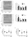
Figure 7. PMI5011 regulates Atrogin-1 and MuRF-1 gene and protein expression in skeletal muscle.
(A, B) Skeletal muscle from KK-Ay mice was processed for whole cells extracts and analyzed using SDS-PAGE followed by western blot analysis of phospho-Akt, total Akt, MuRF-1, Atrogin-1, phospho-FoxO3a and total FoxO3a. β-actin and quantitation of the total protein loaded via MemCode staining are included as loading controls. Fold change for phospho-Akt/total Akt, phospho-FoxO3a/total FoxO3a, MuRF-1/total protein and Atrogin-1/total protein is reported for PMI5011 relative to control (A) or PMI5011 combined with insulin relative to insulin alone (B). (C, D) Atrogin-1 and MuRF-1 gene expression was determined using realtime RT-PCR. Results are reported as the mean −/+ standard deviation (N = 4/group). * p<0.05, *** p<0.001. Significance is reported relative to control in (C, D).
Figure 8
Figure 8. Skeletal muscle myofiber size is larger with dietary intake of PMI5011.
(A) H&E staining of cross-section and longitudinal section of gastrocnemious skeletal muscle from control and PMI5011 supplemented KK-Ay mice. (B) The cross-sectional area of fifty myofibers/animal in each group was determined using ImageJ software. The statistical significance is reported as the mean −/+ standard deviation, p = 0.02.

References

    1. Bassel-Duby R, Olson EN (2006) Signaling pathways in skeletal muscle remodeling. Annu Rev Biochem 75: 19–37. - PubMed
    1. Wang X, Hu Z, Hu J, Du J, Mitch WE (2006) Insulin resistance accelerates muscle protein degradation: Activation of the ubiquitin-proteasome pathway by defects in muscle cell signaling. Endocrinology 147: 4160–4168. - PubMed
    1. Mitch WE, Goldberg AL (1996) Mechanisms of muscle wasting. The role of the ubiquitin-proteasome pathway. N Engl J Med 335: 1897–1905. - PubMed
    1. Park SW, Goodpaster BH, Lee JS, Kuller LH, Boudreau R, et al. (2009) Excessive loss of skeletal muscle mass in older adults with type 2 diabetes. Diabetes Care 32: 1993–1997. - PMC - PubMed
    1. Srikanthan P, Hevener AL, Karlamangla AS (2010) Sarcopenia Exacerbates Obesity-Associated Insulin Resistance and Dysglycemia: Findings from the National Health and Nutrition Examination Survey III. PLoS One 5: e10805. - PMC - PubMed

Publication types

MeSH terms