. 2013 Feb 21;20(2):285-95.
doi: 10.1016/j.chembiol.2012.12.007.

"Function-first" lead discovery: mode of action profiling of natural product libraries using image-based screening

Affiliations

"Function-first" lead discovery: mode of action profiling of natural product libraries using image-based screening

Christopher J Schulze et al. Chem Biol. .

Abstract

Cytological profiling is a high-content image-based screening technology that provides insight into the mode of action (MOA) for test compounds by directly measuring hundreds of phenotypic cellular features. We have extended this recently reported technology to the mechanistic characterization of unknown natural products libraries for the direct prediction of compound MOAs at the primary screening stage. By analyzing a training set of commercial compounds of known mechanism and comparing these profiles to those obtained from natural product library members, we have successfully annotated extracts based on MOA, dereplicated known compounds based on biological similarity to the training set, and identified and predicted the MOA of a unique family of iron siderophores. Coupled with traditional analytical techniques, cytological profiling provides an avenue for the creation of "function-first" approaches to natural products discovery.


Figures

Figure 1
Figure 1
Heat map depicting cytological profiling dendrogram with pertinent clusters highlighted. (A) Complete data set of 624 marine natural product prefractions and 1,920 discrete samples from the ICCB collection (480 compounds at 4 dilutions). Individual compounds are presented on the y-axis with individual features on the x-axis. (B) Detailed view of clusters A – G. In most instances, clusters contained both natural product prefractions and reference compounds from the ICCB library. Cluster A has been truncated in order to highlight the specific prefractions that were analyzed. See also Figure S1.
Figure 2
Figure 2
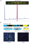
Identification and MOA characterization of prefraction 1516D from Cluster A. (A) HPLC trace (280 nm) and CP profiles for each timeslice for the peak library of prefraction 1516D. CP dendrogram presented with individual features on the y-axis, and timeslices on the x-axis. The active wells (minutes 42 – 44) showed a decrease in both EdU and pHH3 staining (highlighted with red box), that matched the CP fingerprint for the original prefraction. The blue features at minute 70 correspond to an unidentified media component contaminant that elutes at the end of the LCMS trace. (B) Full CP fingerprint and expansions of key individual features for prefraction 1516D. These expansions reveal strong negative deviations from control wells for features corresponding to DNA synthesis (EdU) and mitotic cells (pHH3). In addition, positive deviations from control wells are observed for features corresponding to nuclear size and shape, which is characteristic of cell cycle exit at G1/S. Analysis of the fluorescent images confirmed the phenotype predicted by the CP profile (blue = Hoechst, cyan = EdU, pink = pHH3). See also Figure S2.
Figure 3
Figure 3
Identification and characterization of microferrioxamines A – D (1-4) from prefraction 1522E. (A) HPLC trace (210 nm) and CP profiles for each timeslice for the peak library of prefraction 1522E. CP dendrogram presented with individual features on the y-axis, and timeslices on the x-axis. The original 1522E prefraction CP fingerprint is presented on the left for reference. (B) Structures of microferrioxamines A – D (1 – 4) and key COSY and HMBC correlations for the connection of the alternating cadaverine and succinyl units and the identification of the iso-branched alkyl terminus. Structures were determined by one- and two-dimensional NMR, and MSn experiments. See also Figure S3 and Table S2.
Figure 4
Figure 4
Cytological profiles of dilution series for bafilomycin A1 from both prefraction 1507E and ICCB library, and comparison to cytological profile for original 1507E prefraction. Bafilomyin A1 was isolated and characterized from prefraction 1507E. However, this prefraction did not cluster with the references standards of bafilomycin A1 (Figure 1B). Analysis of the images revealed few viable cells in the well, which precluded the generation of an accurate CP profile. Pure bafilomycin A1, isolated from 1507E, was screened in a dilution series and compared to the reference standard concentrations of bafilomycin A1 in cluster D and prefraction 1507E. Purified bafilomycin A1 showed higher correlation to the reference standard (Pearson correlation=0.67) than to the original prefraction (Pearson Correlation=0.46). The clustered heat map can be viewed in Figure S4.
Figure 5
Figure 5
Delineation of individual biological fingerprints from extracts containing multiple bioactive constituents. (A) HPLC trace (230 nm) and CP profiles for each timeslice for the peak library of prefraction 1526E. CP dendrogram presented with individual features on the y-axis, and timeslices on the x-axis. Combined CP and LCMS analyses reveal the presence of both staurosporine (minutes 2 – 4) and echinomycin (minutes 18 – 21), as two active constituents with differing modes of action in the same prefraction. Examination of the individual cytological profiles for these two compounds predicts that these compounds do not share the same MOA, and justifies the unique CP fingerprint observed for prefraction 1526E. (B) Fluorescence microscopy screening images from primary screening and peak library plates. Images for staurosporine and echinomycin show marked differences in cell morphology consistent with different MOAs. Both pure compound images differ from the phenotypes seen for both the original prefraction (1526E) and the corresponding control well. See also Figure S5.

References

    1. Ando T, Hirayama K, Takahashi R, Horino I, Etoh Y, Morioka H, Shibai H, Murai A. Cosmomycin D, a New Anthracycline Antibiotic. Agr. Biol. Chem. 1985;49:259–262.
    1. Arora SK. Molecular structure of heliomycin, an inhibitor of RNA synthesis. J. Antibiot. 1985;38:113–115. - PubMed
    1. Bowman E, Siebers A. Bafilomycins: a class of inhibitors of membrane ATPases from microorganisms, animal cells, and plant cells. P. Natl. Acad. Sci. USA. 1988;85:7972–7976. - PMC - PubMed
    1. Brockmann H, Schmidt-Kastner G. Valinomycin I, XXVII. Mitteil. über Antibiotica aus Actinomyceten. Chem. Ber. 1955;88:57–61.
    1. Cipollone a, Berettoni M, Bigioni M, Binaschi M, Cermele C, Monteagudo E, Olivieri L, Palomba D, Animati F, Goso C, et al. Novel anthracycline oligosaccharides: influence of chemical modifications of the carbohydrate moiety on biological activity. Bioorgan. Med. Chem. 2002;10:1459–1470. - PubMed

Publication types

MeSH terms