. 2013 Apr 12;112(8):1112-1120.
doi: 10.1161/CIRCRESAHA.111.300445. Epub 2013 Feb 25.

Super-resolution scanning patch clamp reveals clustering of functional ion channels in adult ventricular myocyte

Affiliations

Super-resolution scanning patch clamp reveals clustering of functional ion channels in adult ventricular myocyte

Anamika Bhargava et al. Circ Res. .

Abstract

Rationale: Compartmentation of ion channels on the cardiomyocyte surface is important for electric propagation and electromechanical coupling. The specialized T-tubule and costameric structures facilitate spatial coupling of various ion channels and receptors. Existing methods such as immunofluorescence and patch clamp techniques are limited in their ability to localize functional ion channels. As such, a correlation between channel protein location and channel function remains incomplete.

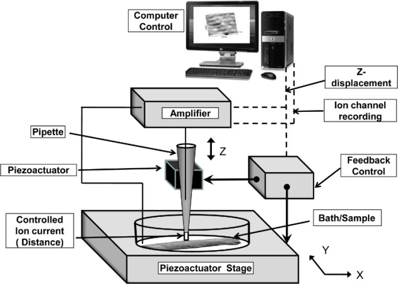
Objective: To validate a method that permits routine imaging of the topography of a live cardiomyocyte and study clustering of functional ion channels from a specific microdomain.

Methods and results: We used scanning ion conductance microscopy and conventional cell-attached patch clamp with a software modification that allows controlled increase of pipette tip diameter. The sharp nanopipette used for topography scan was modified into a larger patch pipette that could be positioned with nanoscale precision to a specific site of interest (crest, groove, or T-tubules of cardiomyocytes) and sealed to the membrane for cell-attached recording of ion channels. Using this method, we significantly increased the probability of detecting activity of L-type calcium channels in the T-tubules of ventricular cardiomyocytes. We also demonstrated that active sodium channels do not distribute homogenously on the sarcolemma instead, they segregate into clusters of various densities, most crowded in the crest region, that are surrounded by areas virtually free of functional sodium channels.

Conclusions: Our new method substantially increases the throughput of recording location-specific functional ion channels on the cardiomyocyte sarcolemma, thereby allowing characterization of ion channels in relation to the microdomain where they reside.


Figures

Figure 1
Figure 1. Setup for super-resolution scanning patch-clamp
The scanning/patch pipette is mounted on a piezo translation platform and connected to a patch-clamp amplifier. The amplifier's output drives a feedback control amplifier to control the pipette's piezo, which provides a Z only directional movement (up and down). It also provides a drive to the nanopositioning –piezoactuator- stage (bottom) raster scanning in two directions (X Y). Computer controlled software provides a user interface to control the setup. Sample is placed on the stage without any fixation or additional preparation.
Figure 2
Figure 2. Pipette clipping procedure
A) A sharp high resistance pipette is used to resolve the topographical structure of the cardiomyocyte. B) The pipette is moved to a cell free area on the dish and the fall rate is increased (as described in the text) to clip the pipette tip. C) The pipette is then returned to the structure of choice and patch-clamp can be performed with a pipette of wider tip than (A).
Figure 3
Figure 3. Schematic of super-resolution scanning patch-clamp
A) A single rat cardiomyocyte and pipette as seen optically. B) A 10 μm × 10 μm scan of a cardiomyocyte revealing topographic structures such as the Z-groove, T-tubule and crest. A depth profile along the x-y plane marked by the black dotted line is shown on the right hand side. Super-resolution scanning patch-clamp of T-tubule (C) and crest (D) allowed for detection (or lack thereof) of functional ion channels. The traces below represent LTCC activity which is usually seen in T-tubules of cardiomyocytes as opposed to crest.
Figure 4
Figure 4. Single LTCC activity in rat cardiomyocytes
A) A 10 μm × 10 μm scan of cardiomyocyte revealing topographic structures. The position of the pipette denotes a T-tubule where the pipette was lowered and a gigaseal was obtained. B) Representative traces of single LTCC activity recorded from a T-tubule at the given voltages using a pipette of 30 MΩ resistance. C) Several sweeps were recorded at −6.7 mV and average of 11 sweeps is shown below. D) Current-voltage relationship of single LTCC activity. A voltage dependence was observed in our experiments as expected (n= 5-13 for each data point). E) Percent of occurrence of functional LTCCs on the cardiomyocyte sarcolemma. p<0.02 when comparing the occurrence of channel activity in patches formed at the T-tubule versus the crest (Chi-square test).
Figure 5
Figure 5. Relationship between pipette resistance and probability of observing single LTCC activity
A) The graph depicts that clipping the pipette tip to 20 MΩ or lower greatly increases the chance of recording single LTCC activity in the T-tubules of cardiomyocytes. Lower X-axis is pipette resistance (Rp) and upper X axis denotes estimated pipette ID. B) Representative traces of LTCC activity recorded from a T-tubule at the given voltages using a pipette of 16 MΩ resistance. Note that 2 channels could be recorded at the same time. Level 1 and Level 2 denote the amplitude of two single channels.
Figure 6
Figure 6. Single Sodium channel activity in mouse cardiomyocytes
A) Current traces obtained from a T-tubule of an adult mouse cardiomyocyte, using super-resolution scanning patch-clamp. Traces have been inverted for consistency with other publications. Voltage steps to −70 mV from a holding potential of −120 mV (polarity referred to the intracellular space). Horizontal dotted lines reflect current steps indicative of unitary events. B) Histogram of unitary current amplitude and best-fit Gaussian (red line) corresponding to mean unitary current for that particular family of events. C) Cumulative data for unitary current amplitude at various command voltages. Dotted line represents the extrapolation used to calculate the reversal potential and slope unitary conductance of the channels. Each data point corresponds to average of 114, 124, 97 and 130 events captured during voltage steps to −80, −70, −60 and −50 mV, respectively.
Figure 7
Figure 7. Histograms indicating sodium channel clustering as a function of recording location
Ordinates: Percent of recordings (relative to total number of attempts). Abscissae, number of open channels detected. Number of open channels in each recorded patch was estimated from the average peak current amplitude (ten sweeps) during a voltage step to −30 mV from a holding potential of −120 mV, and an estimated unitary conductance of 10 pS (see data in Figure 6). Total number of attempts: 12, 10 and 13 for T-tubule (A), z-groove (B) or crest (C) respectively. p<0.02 when comparing the occurrence of more than 20 channels in crest versus z-groove and T-tubules (Chi square test).

Comment in

References

    1. Balijepalli RC, Foell JD, Hall DD, Hell JW, Kamp TJ. Localization of cardiac L-type Ca2+ channels to a caveolar macromolecular signaling complex is required for beta(2)-adrenergic regulation. Proc Natl Acad Sci U S A. 2006;103:7500–7505. - PMC - PubMed
    1. Scriven DR, Dan P, Moore ED. Distribution of proteins implicated in excitation-contraction coupling in rat ventricular myocytes. Biophys J. 2000;79:2682–2691. - PMC - PubMed
    1. Rybin VO, Xu X, Lisanti MP, Steinberg SF. Differential targeting of beta-adrenergic receptor subtypes and adenylyl cyclase to cardiomyocyte caveolae. A mechanism to functionally regulate the camp signaling pathway. J Biol Chem. 2000;275:41447–41457. - PubMed
    1. Bers DM. Cardiac excitation-contraction coupling. Nature. 2002;415:198–205. - PubMed
    1. Bito V, Heinzel FR, Biesmans L, Antoons G, Sipido KR. Crosstalk between L-type Ca2+ channels and the sarcoplasmic reticulum: Alterations during cardiac remodelling. Cardiovasc Res. 2008;77:315–324. - PubMed

Publication types