. 2013 May;21(5):973-85.
doi: 10.1038/mt.2013.31. Epub 2013 Feb 26.

Hepatocyte-targeted RNAi therapeutics for the treatment of chronic hepatitis B virus infection

Affiliations

Hepatocyte-targeted RNAi therapeutics for the treatment of chronic hepatitis B virus infection

Christine I Wooddell et al. Mol Ther. 2013 May.

Abstract

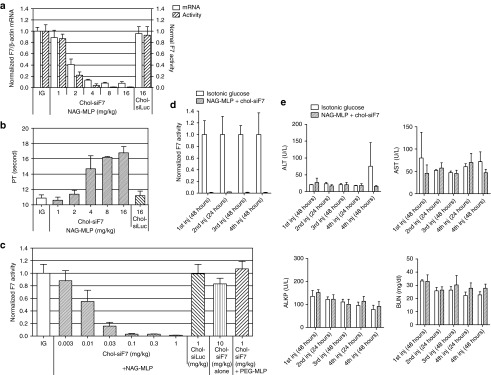
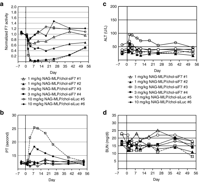
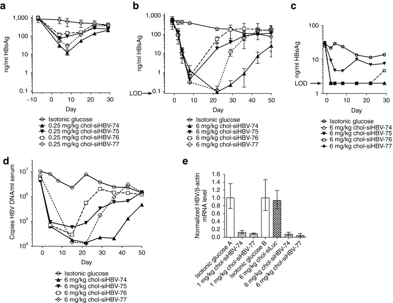
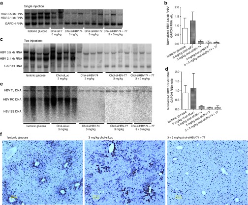
RNA interference (RNAi)-based therapeutics have the potential to treat chronic hepatitis B virus (HBV) infection in a fundamentally different manner than current therapies. Using RNAi, it is possible to knock down expression of viral RNAs including the pregenomic RNA from which the replicative intermediates are derived, thus reducing viral load, and the viral proteins that result in disease and impact the immune system's ability to eliminate the virus. We previously described the use of polymer-based Dynamic PolyConjugate (DPC) for the targeted delivery of siRNAs to hepatocytes. Here, we first show in proof-of-concept studies that simple coinjection of a hepatocyte-targeted, N-acetylgalactosamine-conjugated melittin-like peptide (NAG-MLP) with a liver-tropic cholesterol-conjugated siRNA (chol-siRNA) targeting coagulation factor VII (F7) results in efficient F7 knockdown in mice and nonhuman primates without changes in clinical chemistry or induction of cytokines. Using transient and transgenic mouse models of HBV infection, we show that a single coinjection of NAG-MLP with potent chol-siRNAs targeting conserved HBV sequences resulted in multilog repression of viral RNA, proteins, and viral DNA with long duration of effect. These results suggest that coinjection of NAG-MLP and chol-siHBVs holds great promise as a new therapeutic for patients chronically infected with HBV.


Figures

Figure 1
Figure 1
Efficacy and safety of NAG-MLP delivery of chol-siRNA in mice. (a,b) ICR mice were injected intravenously with NAG-MLP at 1–16 mg/kg and 2 mg/kg chol-siRNA targeting either mouse F7 or luciferase as a control (n = 4). After 48 hours, serum and plasma were collected and RNA was isolated from the liver. (a) F7 mRNA was measured relative to endogenous β-actin mRNA, open bars; and F7 activity was measured in serum, diagonally striped bars. F7 activity and mRNA were normalized to that in mice injected with isotonic glucose (IG). (b) Prothrombin time was measured in plasma. (c) Cholesterol-siRNA dose titration: ICR mice were injected intravenously with 6 mg/kg NAG-MLP or with 6 mg/kg of MLP with polyethylene glycol (PEG) attached instead of NAG (PEG-MLP); n = 4. Chol-siRNAs targeting either mouse F7 (0.003–1 mg/kg) or luciferase (1 mg/kg) were coinjected with NAG-MLP. One group received 10 mg/kg chol-siF7 alone. F7 activity in serum was measured 48 hours later and was normalized to that in mice injected with isotonic glucose. (d,e) SV129 mice were injected four times with 6 mg/kg NAG-MLP and 5 mg/kg chol-siF7 (n = 4). The first and second injections were 14 days apart and remaining injections were 7 days apart. (d) F7 activity and (e) clinical chemistries in serum were measured 24 and/or 48 hours after each injection. ALT, alanine transaminase; AST, aspartate transaminase; ALKP, alkaline phosphatase; BUN, blood urea nitrogen; Inj, Injection. Data bars indicate mean + SD.
Figure 2
Figure 2
Efficacy and safety of NAG-MLP delivery of chol-siRNA in nonhuman primates. (ad) Cynomolgus monkeys were injected with 1, 3, or 10 mg/kg NAG-MLP and 2 mg/kg chol-siRNA targeting either primate F7 or luciferase (n = 2). Serum and plasma were collected at the indicated times and assayed for (a) F7 activity and (b) prothrombin time, respectively. Serum collected at the indicated times was assayed for (c) alanine transaminase and (d) blood urea nitrogen. Each data set represents one animal (NHPs #1 to #6). ALT, alanine transaminase; BUN, blood urea nitrogen; PT, prothrombin time.
Figure 3
Figure 3
Selection of siRNA sequences targeting human hepatitis B virus (HBV). (a) Locations of the open reading frames within the human HBV genome are shown above the purple line that represents the genomic DNA and locations of the mRNAs are shown below this. Numbering of HBV bases begins with position 1 at the EcoR I site. The direct repeats (DR1 and DR2) and the epsilon stem-loop are features of the pregenomic RNA important for replication. The conserved target sites of all screened siRNAs (140) and the four most efficacious siRNAs are indicated by red bars. Blue bars indicate siRNA sequences that target the 3.5 kb RNA in the terminally redundant region. The graph indicates percentage conservation of 17-mers in 2,754 full-length HBV sequences (genotypes A–D). Schematic was adapted from Chen et al. (b) 140 siRNAs directed against conserved target sites were selected in silico, synthesized and screened at 10 nmol/l in Cos-7 green monkey kidney cells that had been transfected with firefly and Renilla luciferase-expressing psiCHECK-HBV. The Renilla/firefly luciferase expression ratio was normalized to that in cells transfected with a control siRNA.
Figure 4
Figure 4
Efficacy and the duration of knockdown after coinjection of chol-siHBVs and NAG-MLP in the pHBV mouse model of chronic hepatitis B virus (HBV) infection. NOD-SCID mice were given a hydrodynamic tail vein injection with (a,e) 13.5 µg pHBV1.3 or (bd) 10 µg pHBV1.3. Three or more weeks thereafter, mice were given one 200 µl IV coinjection of 6 mg/kg NAG-MLP and 0.25 mg/kg, 1 mg/kg or 6 mg/kg chol-siHBV-74, -75, -76, or 77 (n = 3–4). (a,b) HBsAg and (c) HBeAg in serum were measured by enzyme linked immunosorbent assay at the indicated times relative to injection on day 1; LOD, limit of detection. (d) DNA was isolated from serum and the concentration of HBV genomes was quantitated by qPCR. (e) RNA was isolated from the liver 14 days after chol-siRNA injection. The relative amount of HBV transcripts was determined by RT-qPCR using a probe that was within the S gene and normalizing to the endogenous β-actin mRNA. At each dose level the chol-siRNA injected mice were compared with isotonic glucose-injected mice with similar initial HBsAg levels (isotonic glucose groups A and B). Standard deviation bars are shown for HBsAg and HBV RNA quantitation. Serum HBV DNA and HBeAg levels for each group were determined by combining equal proportions of serum from each mouse within the group to obtain sufficient pooled sample at each time point (n = 3–4).
Figure 5
Figure 5
Efficacy of NAG-MLP and chol-siHBV coinjection in transgenic HBV mice. HBV1.3.32 transgenic mice were injected once (a,b, males) or twice (cf, females) with 6 mg/kg NAG-MLP and 3 mg/kg of the indicated chol-siRNA or a combination of 3 mg/kg chol-siHBV-74 and 3 mg/kg chol-siHBV-77 (n = 2–4). (a) RNA filter hybridization (northern blot) analysis of 3.5 kb and 2.1 kb HBV RNA from livers of mice injected once and killed 7 days later for evaluation. Glyceraldehyde-3-phosphate dehydrogenase (GAPDH) was used as an internal control for RNA loading per lane. (b) RT-qPCR analysis of the 3.5 kb HBV RNA is shown relative to the GAPDH mRNA in mice that received one NAG-MLP and chol-siRNA injection, and then normalized to the control groups, mean + SD. (cf) Mice were coinjected with NAG-MLP and chol-siRNA on days 1 and 8. They were killed 7 days after the second injection for evaluation of (c,d) HBV RNA and (e) DNA in liver. (c) Northern blot analysis of RNAs and (d) RT-qPCR analysis of the 3.5 kb HBV RNA in mice that received two NAG-MLP and chol-siRNA coinjections. RT-qPCR analysis is normalized to the control groups and shown as mean + SD. (e) HBV replicative intermediates were evaluated by DNA filter hybridization (Southern blot). (f) Immunohistochemical detection of HBcAg in hematoxylin-stained sections of liver. Scale bar = 100 µm. HBV RC DNA, HBV relaxed circular DNA; HBV SS DNA, HBV single-stranded DNA; Tg, transgene.

References

    1. Seeger C, Zoulim F., and, Mason WS.2007Hepadnaviruses Fields BN, Knipe DM., and, Howley PM.eds.). Fields Virology5th edn. Wolters Kluwer Health/Lippincott Williams & Wilkins: Philadelphia
    1. Chen JD, Yang HI, Iloeje UH, You SL, Lu SN, Wang LY, Risk Evaluation of Viral Load Elevation and Associated Liver Disease/Cancer in HBV (REVEAL-HBV) Study Group et al. Carriers of inactive hepatitis B virus are still at risk for hepatocellular carcinoma and liver-related death. Gastroenterology. 2010;138:1747–1754. - PubMed
    1. Yuen MF., and, Lai CL. Treatment of chronic hepatitis B: Evolution over two decades. J Gastroenterol Hepatol. 2011;26 Suppl 1:138–143. - PubMed
    1. Heathcote EJ, Marcellin P, Buti M, Gane E, De Man RA, Krastev Z.et al. (2011Three-year efficacy and safety of tenofovir disoproxil fumarate treatment for chronic hepatitis B Gastroenterology 140132–143. - PubMed
    1. Funk ML, Rosenberg DM., and, Lok AS. World-wide epidemiology of HBeAg-negative chronic hepatitis B and associated precore and core promoter variants. J Viral Hepat. 2002;9:52–61. - PubMed

MeSH terms