. 2013 Apr 26;288(17):11718-30.
doi: 10.1074/jbc.M113.452276. Epub 2013 Feb 25.

Biochemical and biophysical characterization of the sialyl-/hexosyltransferase synthesizing the meningococcal serogroup W135 heteropolysaccharide capsule

Affiliations

Biochemical and biophysical characterization of the sialyl-/hexosyltransferase synthesizing the meningococcal serogroup W135 heteropolysaccharide capsule

Angela Romanow et al. J Biol Chem. .

Abstract

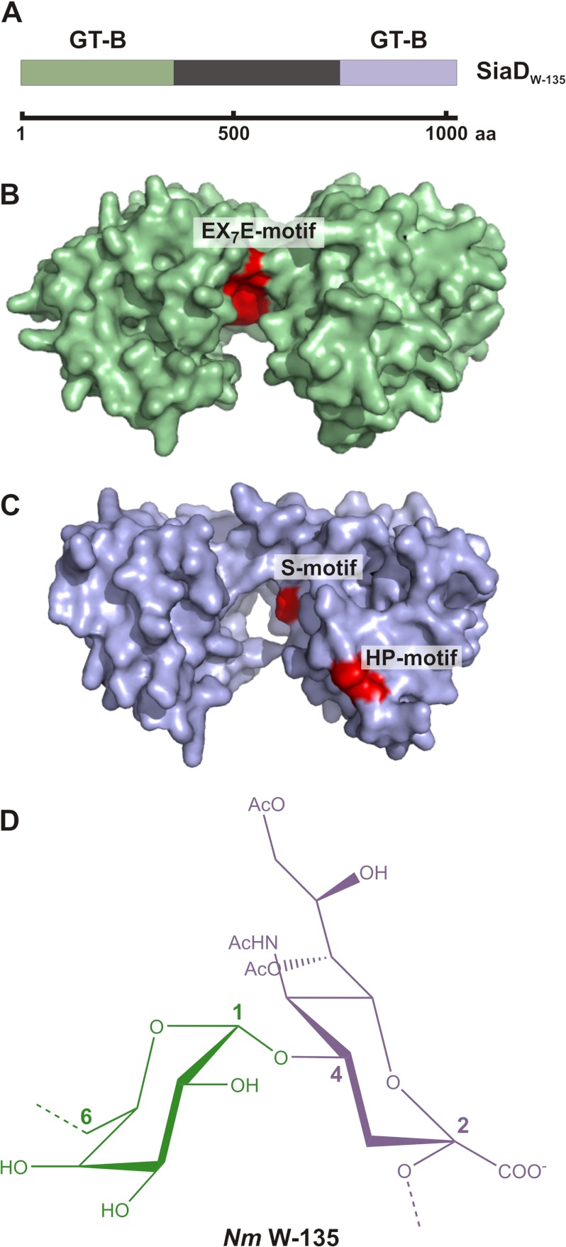
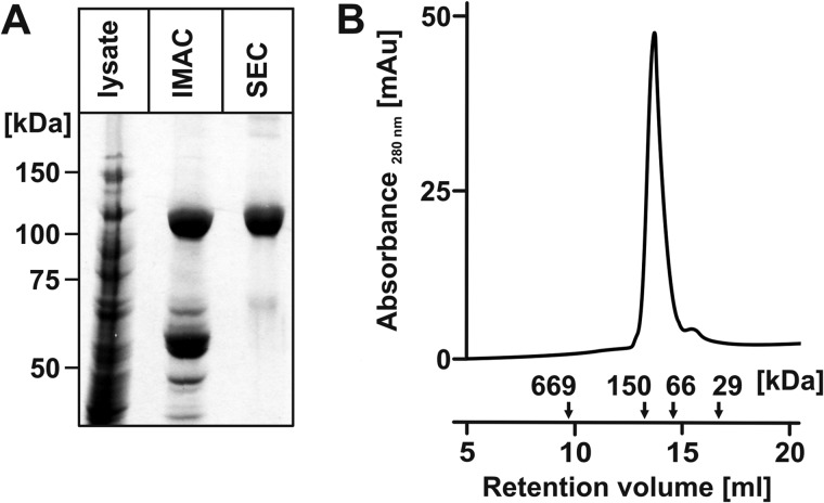
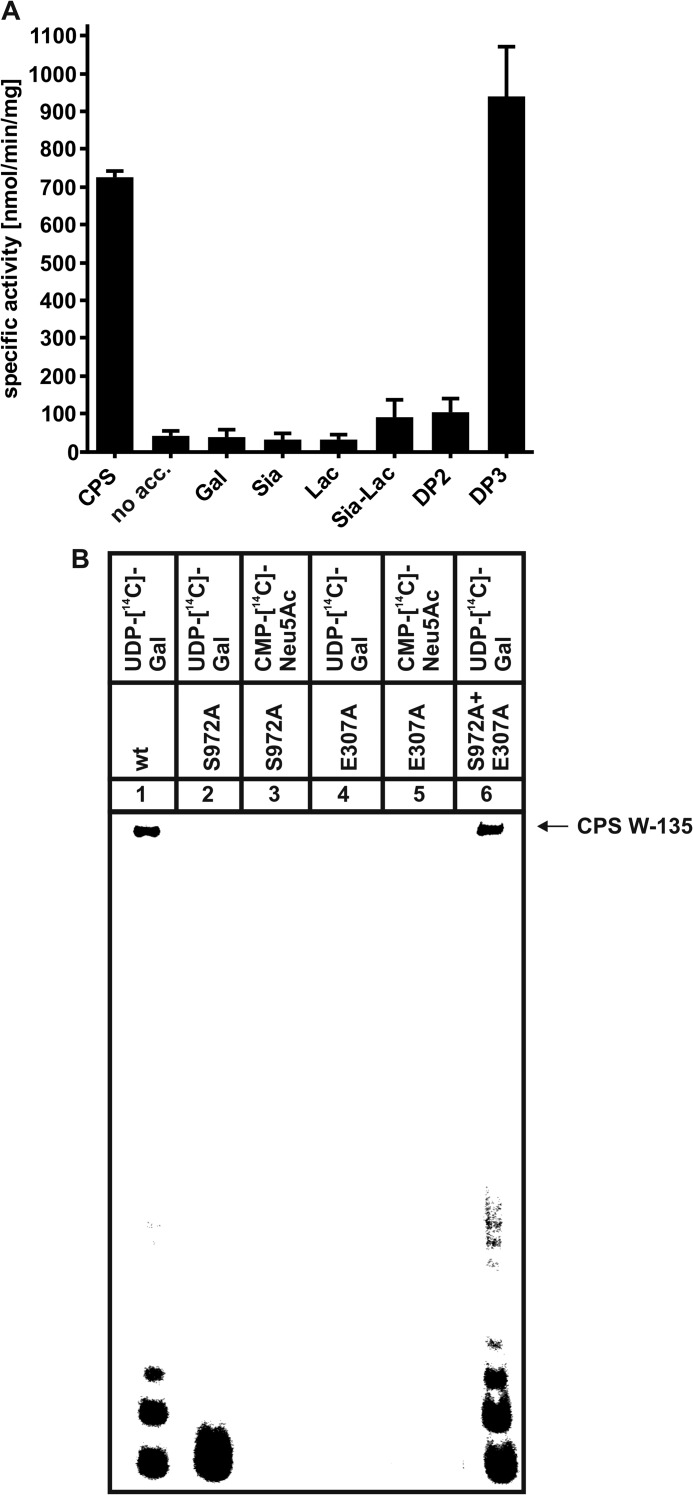
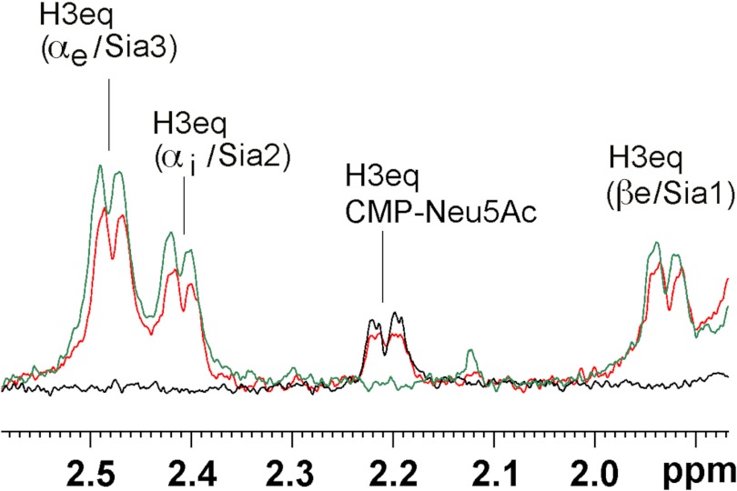
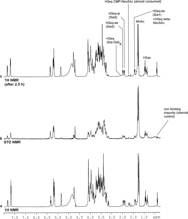
Neisseria meningitidis (Nm) is a leading cause of bacterial meningitis and sepsis. Crucial virulence determinants of pathogenic Nm strains are the polysaccharide capsules that support invasion by hindering complement attack. In NmW-135 and NmY the capsules are built from the repeating units (→ 6)-α-D-Gal-(1 → 4)-α-Neu5Ac-(2 →)n and (→ 6)-α-D-Glc-(1 → 4)-α-Neu5Ac-(2 →)n, respectively. These unusual heteropolymers represent unique examples of a conjugation between sialic acid and hexosyl-sugars in a polymer chain. Moreover, despite the various catalytic strategies needed for sialic acid and hexose transfer, single enzymes (SiaDW-135/Y) have been identified to form these heteropolymers. Here we used SiaDW-135 as a model system to delineate structure-function relationships. In size exclusion chromatography active SiaDW-135 migrated as a monomer. Fold recognition programs suggested two separate glycosyltransferase domains, both containing a GT-B-fold. Based on conserved motifs predicted folds could be classified as a hexosyl- and sialyltransferase. To analyze enzyme properties and interplay of the two identified glycosyltransferase domains, saturation transfer difference NMR and mutational studies were carried out. Simultaneous and independent binding of UDP-Gal and CMP-Sia was seen in the absence of an acceptor as well as when the catalytic cycle was allowed to proceed. Enzyme variants with only one functionality were generated by site-directed mutagenesis and shown to complement each other in trans when combined in an in vitro test system. Together the data strongly suggests that SiaDW-135 has evolved by fusion of two independent ancestral genes encoding sialyl- and galactosyltransferase activity.


Figures

FIGURE 1.
FIGURE 1.
GT-B folds predicted in SiaDW-135 and structure of the biosynthetic product. A, schematic illustration of full-length SiaDW-135. The protein comprises 1037 amino acids. The N- and C-terminal domains (shaded in green and blue, respectively) have been predicted to individually attain the GT-B fold. No structural information is available for the linker region (black) that connects the N- and C-terminal domains. B and C, GT-B folds as identified by Phyre. Based on the presence of the conserved motifs EX7E as well as S and HP, hexosyl- and sialyltransferase activity could be attributed to the GT-B folds. D, chemical structure of the repeating unit forming the W-135 capsular polymer.
FIGURE 2.
FIGURE 2.
Recombinant epitope-tagged SiaDW-135 is a monomer. A, SiaDW-135 was expressed with C-terminal His6 tag. The Coomassie-stained SDS-PAGE shows that a highly purified protein fraction was obtained in two steps including Ni2+ chelating (IMAC) and size exclusion chromatography (SEC). B, the oligomerization status of recombinant SiaDW-135 was determined after calibration of the Superdex 200 column used in SEC. In the presence of 300 mm NaCl, SiaDW-135 eluted with an apparent molecular mass of 120 kDa, corresponding to the monomeric protein. Standards were: thyroglobulin (669 kDa), alcohol dehydrogenase (150 kDa), albumin (66 kDa), and carbonic anhydrase (29 kDa).
FIGURE 3.
FIGURE 3.
Recombinant SiaDW-135 synthesizes high molecular weight W-135 polymer chains from short oligosaccharide primers. A, to test if recombinant SiaDW-135 can elongate short oligomer primers, CPS isolated from NmW-135 was hydrolyzed (CPS(h)) and used to prime the polymerase reaction. The production of long polymer chains demonstrated an active enzyme. High percentage PAGE and Alcian blue/silver staining was used to display starting, as well as synthesis products. B, CPS(h) produced from CPS was used in a time course experiment. Here products were analyzed on a 10% SDS-PAGE to additionally visualize the enzyme.
FIGURE 4.
FIGURE 4.
Site-directed mutagenesis identified amino acid residues crucial for enzymatic activity. A, in vivo activity of the mutant capsule polymerases (as indicated) was tested with Nm strain WUE171 as recipient. Mutant genes were introduced by homologous recombination and capsule production was quantified in a whole cell ELISA. Values are given as means of three independent experiments in relationship to the wild-type recipient (100%). The capsule-deficient strain WUE 2661-Δ CPS served as a negative control. B, display of recombinant purified wild-type and mutant forms of SiaDW-135 in a Coomassie Blue-stained 10% SDS-PAGE. C, activity of recombinant purified wild-type and mutant capsule polymerases (proteins as shown in B) was determined in a radioactive incorporation assay with CPS(h) as acceptor. Of note, combination of the inactive mutants E307A and S972A in one reaction mixture restored activity to 70% of wild-type.
FIGURE 5.
FIGURE 5.
A, the trimer of α(2,8)-linked sialic acid residues (DP3) is efficiently elongated by SiaDW-135. As defined W-135 CPS oligomers were not readily available, the compounds as listed were analyzed for their capacity to prime the SiaDW-135 reaction in the radioactive incorporation assay. In this experiment DP3 was found to be efficiently elongated by SiaDW-135, whereas DP2 and 2,3-sialyllactose (Sia-Lac) were only weak acceptors. The monosaccharides Gal and Sia as well as the disaccharide lactose (Lac) were not used by the enzyme. Specific activities were calculated from three independent experiments. B, asking if DP3 is an acceptor for both glycosyltransferase domains present in SiaDW-135, the transfer reactions catalyzed by the single mutants E307A and S972A were analyzed in comparison to the wild-type enzyme. Both nucleotide sugars were present in these reactions, with only one radioactively labeled as indicated. Long polymer chains were synthesized in reaction mixtures with wild-type SiaDW-135 (lane 1) and both single mutants (lane 6). In contrast, a single radioactive spot, representing the transfer of one [14C]Gal onto DP3 (lane 2) by S972A (comprises an active galactosyltransferase domain) was observed, whereas all other reactions (lanes 3-5) remained negative. These data clearly demonstrate that DP3 is selectively recognized by the galactosyltransferase domain and that formation of long CPS requires the iterative activity between galactosyl- and sialyltransferase domain.
FIGURE 6.
FIGURE 6.
DP1–DP3 are entirely bound by SiaDW-135. 1H and STD NMR were used to study the binding of Sia and oligoSia structures to SiaDW-135. All three compounds (DP1, DP2, and DP3) were entirely bond to the enzyme. However, in the case of DP1 ∼50% of the sugar attained β-configuration (at 1.95 ppm), indicating that the monomer binds to the donor sugar (CMP-Sia) binding site (a–f). In DP2, the dimer of α(2,8)-linked sialic acid residues (g–l), and DP3, the trimer of α(2,8)-linked sialic acid residues (m–p), all sugar units are in contact with the protein. The signals are highlighted for H3eq and N-acetamido protons (NHAc). Although in both compounds the nonreducing end sugar receives the highest energy transfer, only DP3 was found to be an efficient primer to start the enzyme reaction. q, schematic representation of DP3. Differences in saturation transfer are illustrated by the letter size of 3Heq protons. Spectra were recorded at 280 K, 600 MHz using deuterated Tris buffer (20 mm, pH 8.3, and 20 mm MgCl2). Protein resonances were saturated using 40 Gaussian pulses of 5 ms duration at −1.00 ppm. The off-resonance frequency was set to 33 ppm.
FIGURE 7.
FIGURE 7.
STD NMR spectra of SiaDW-135 in complex with the nucleotide sugar donor substrates. To determine epitopes in the donor sugars CMP-Neu5Ac (a and b) and UDP-Gal (c and d) that make contact to the protein, an STD NMR study was performed. In line with previous data this experiment demonstrated the importance of H1 Rib for nucleotide sugar binding. This position together with H5 in the nucleotide base received the highest saturation in both UDP-Gal and CMP-Neu5Ac. In accordance with the specificity of SiaDW-135 for galactose H4 Gal received 57% saturation and, remarkably, also the opposite site of the pyranose ring, the anomeric proton H1 Gal, received considerable saturation (62%). For experimental details, see Fig. 6.
FIGURE 8.
FIGURE 8.
CMP-Neu5Ac and UDP-Gal bind independently to SiaDW-135. To find out if binding of one nucleotide sugar impacts binding of the second, the STD NMR spectrum of CMP-Neu5Ac in complex with SiaDW-135 was recorded in the absence (a) and presence (b) of an equimolar concentration of UDP-Gal. No change in the position or intensity of any signal was seen, thus indicating independent binding of the nucleotide sugars. To highlight this fact, an overlay is shown for the intense signal produced by the tightly bound N-acetamido group (c). For experimental details, see Fig. 6.
FIGURE 9.
FIGURE 9.
STD NMR spectra of SiaDW-135 in complex with CMP-Neu5Ac and the acceptor DP3. To evaluate if binding of the acceptor DP3 influences the binding of CMP-Neu5Ac, three STD NMR spectra were overlaid. In black and green the spectra obtained for SiaDW-135 in complex with CMP-Neu5Ac and DP3, respectively, are shown. The spectrum obtained when both substrates were present in equimolar concentrations is shown in red. The absolute STD NMR intensities revealed only a minor (∼15%) reduction in the saturation transfer to the H3eq proton of CMP-Neu5Ac when DP3 was present. Similarly, a reduction of ∼20% was seen for the H3eq protons of DP3 under these conditions. The faintness of the differences observed in the presence of the second substrate argued for independent binding sites. For experimental details, see Fig. 6.
FIGURE 10.
FIGURE 10.
1H and STD NMR analysis of SiaDW-135 in complex with acceptor and donor substrates. The STD NMR analysis was carried out with SiaDW-135 in complex with CMP-Neu5Ac, UDP-Gal, and DP3 (a and b). The absolute STD NMR intensities revealed specific and independent binding of all three substrates (b). Consumption of CMP-Neu5Ac and parallel appearance of a product peak after the 2.5-h acquisition time (c) clearly showed that the enzyme reaction proceeded. For experimental details, see Fig. 6.
FIGURE 11.
FIGURE 11.
Bacterial genome regions comprising putative sialyltransferases with homology to SiaDW-135. With the sialyltransferase domain identified in SiaDW-135 as template, a BLAST search was carried out and returned a number of bacterial proteins with weak homology. Although no function has yet been attributed to these proteins, all are encoded by genes (designated homologue CT and marked in red) that in the genome occur in close company with two other genes encoding enzymes of the sialylation pathway: the Neu5Ac9P-synthase (gene marked in green) and the CMP-Neu5Ac synthetase (gene marked in yellow). Bacteria harboring these homologues are named and the respective gene regions are schematically shown.

References

    1. Rosenstein N. E., Perkins B. A., Stephens D. S., Popovic T., Hughes J. M. (2001) Meningococcal disease. N. Engl. J. Med. 344, 1378–1388 - PubMed
    1. Stephens D. S., Greenwood B., Brandtzaeg P. (2007) Epidemic meningitis, meningococcaemia, and Neisseria meningitidis. Lancet 369, 2196–2210 - PubMed
    1. Raymond N. J., Reeves M., Ajello G., Baughman W., Gheesling L. L., Carlone G. M., Wenger J. D., Stephens D. S. (1997) Molecular epidemiology of sporadic (endemic) serogroup C meningococcal disease. J. Infect. Dis. 176, 1277–1284 - PubMed
    1. Taha M. K., Achtman M., Alonso J. M., Greenwood B., Ramsay M., Fox A., Gray S., Kaczmarski E. (2000) Serogroup W135 meningococcal disease in Hajj pilgrims. Lancet 356, 2159. - PubMed
    1. Decosas J., Koama J. B. (2002) Chronicle of an outbreak foretold. Meningococcal meningitis W135 in Burkina Faso. Lancet Infect. Dis. 2, 763–765 - PubMed

Publication types

MeSH terms