. 2013 Jan 29:3:313.
doi: 10.3389/fpls.2012.00313. eCollection 2012.

Investigating the role of viral integral membrane proteins in promoting the assembly of nepovirus and comovirus replication factories

Affiliations

Investigating the role of viral integral membrane proteins in promoting the assembly of nepovirus and comovirus replication factories

Hélène Sanfaçon. Front Plant Sci. .

Abstract

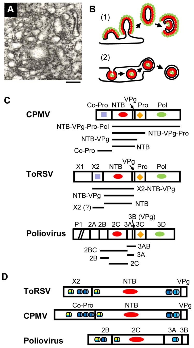
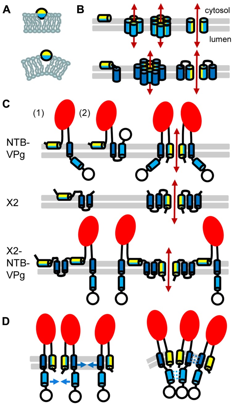
Formation of plant virus membrane-associated replication factories requires the association of viral replication proteins and viral RNA with intracellular membranes, the recruitment of host factors and the modification of membranes to form novel structures that house the replication complex. Many viruses encode integral membrane proteins that act as anchors for the replication complex. These hydrophobic proteins contain transmembrane domains and/or amphipathic helices that associate with the membrane and modify its structure. The comovirus Co-Pro and NTP-binding (NTB, putative helicase) proteins and the cognate nepovirus X2 and NTB proteins are among the best characterized plant virus integral membrane replication proteins and are functionally related to the picornavirus 2B, 2C, and 3A membrane proteins. The identification of membrane association domains and analysis of the membrane topology of these proteins is discussed. The evidence suggesting that these proteins have the ability to induce membrane proliferation, alter the structure and integrity of intracellular membranes, and modulate the induction of symptoms in infected plants is also reviewed. Finally, areas of research that need further investigation are highlighted.

Keywords: integral membrane proteins; intracellular membranes; membrane remodeling; picornavirales; plant–virus interactions; protein–membrane interactions; secoviridae; viral replication complexes.


Figures

FIGURE 1
FIGURE 1
Membrane replication proteins encoded by CPMV, ToRSV and poliovirus. (A) Electron micrograph showing the proliferation of single-membrane vesicles in ToRSV-infected Nicotiana clevelandii plants. The bar indicates 200 nm. (B) Models for the formation of viral replication complexes. (1) In cells infected with poliovirus or coxsackie B3 virus, viral integral membrane proteins (red ovals) induce positive curvature of the membrane allowing the budding of tubular structures. Other viral replication proteins (e.g., polymerase, green ovals) interact with the viral membrane proteins. Host factors and viral RNA (not shown) associate with the replication complex by protein–protein and protein–RNA interactions. Single-membrane vesicles may bud out and form double-membrane vesicles after the internal collapse of the single-membrane vesicle and subsequent membrane fusion to allow its circularization. Late in infection, double-membrane vesicles are predominant in picornavirus-infected cells. This model is based on electron tomography observations from Belov et al. (2012) and Limpens et al. (2011). (2) In cells infected with many plant and animal viruses, induction of negative membrane curvature results in membrane invagination and formation of spherules in the lumen of the membrane. In plant, spherules have been observed in association with membranes from the ER (brome mosaic virus), chloroplast (turnip yellow mosaic virus), peroxisome (tomato bushy stunt virus) and mitochondria (carnation Italian ringspot virus). The spherules are connected to the cytoplasm by a neck. Viral integral membrane proteins (red ovals) line the interior of the spherule. The viral polymerase (green ovals) as well as other viral proteins, host factors and the viral RNA (not shown) are enclosed in the spherule. Release of the vesicle in the lumen of the membrane may be followed by budding of a double-membrane vesicle into the cytoplasm. This model has been discussed in recent reviews (den Boon and Ahlquist, 2010; Laliberte and Sanfacon, 2010; Nagy and Pogany, 2012). (C) Organization of replication protein domains in the polyproteins of CPMV, ToRSV, and poliovirus. The RNA1-encoded polyproteins of CPMV and ToRSV are shown. For poliovirus, the polyprotein encoded by the single genomic RNA is shown, although the P1 region (containing the structural proteins) is truncated as indicated by the diagonal bars. Vertical lines represent the protease cleavage sites. Conserved motifs are: RNA-dependent RNA polymerase (Pol, green ovals), protease (Pro, orange diamond), nucleotide-binding protein (NTB, red oval), Co-Pro and X2 (purple square). Horizontal bars under each polyprotein represent integral membrane proteins that have been detected in virus-infected cells. The mature ToRSV X2 protein is shown with a question mark. Although likely, its presence in infected cells could not be confirmed due to the lack of antibodies. (D) The regions of the polyprotein containing the putative membrane anchors are shown for each virus. Predicted membrane-association domains are indicated with blue barrels (hydrophobic helices) or with yellow/blue barrels (amphipathic helices, with the yellow half representing the polar/charged hydrophilic side of the helix and the blue half representing the hydrophobic side of the helix).
FIGURE 2
FIGURE 2
Topology model for ToRSV membrane replication proteins. (A) Model for the parallel insertion of an amphipathic helix. The hydrophobic side of the helix (blue) inserts in one leaflet of the lipid bilayer while the polar/charged hydrophilic side of the helix (yellow) is exposed to the cytosolic face of the membrane. This insertion displaces the lipid headgroup, causing the acyl chain to reorient and inducing positive membrane curvature. (B) Model for the oligomerization of amphipathic helices and formation of an aqueous pore. In the top panel, an amphipathic helix is inserted parallel to the lipid bilayer (horizontal gray lines) of the membrane (left). Formation of an aqueous pore (double-ended red arrow) requires oligomerization of four or six amphipathic helices (middle). In the aqueous pore, the hydrophilic side of the helix (yellow) is exposed toward the pore, while its hydrophobic side (blue) is oriented toward the membrane lipid bilayer. A simplified representation of the pore shows only two molecules to better visualize each side of the amphipathic helix relative to the pore (right). In the bottom panel, a membrane protein consisting of an amphipathic helix and a hydrophobic helix (blue) is shown. After initial membrane insertion of the monomer with the amphipathic helix parallel to the membrane (left), an aqueous pore is formed by oligomerization of the amphipathic helix (middle). The hydrophobic helix of each molecule is located on the outside of the pore alongside the amphipathic helix (model shown for a hexamer). Hydrophobic interactions between the hydrophobic side of the amphipathic helix and the hydrophobic helix stabilize pore formation. A simplified representation of the pore shows only two molecules (right). (C) Predicted topologies for NTB–VPg, X2, and X2–NTB–VPg shown for monomers (left) or oligomers (right). Two possible topologies are shown for NTB–VPg monomers (1 and 2, see text). To simplify the figure, only two molecules are shown in the oligomer models. However, at least four molecules would be necessary to form an aqueous pore (as shown in B). The open circle represents the VPg domain and the red oval indicates the conserved NTB motif. (D) Model for the induction of positive membrane curvature by hydrophobic interactions of membrane proteins oligomers, shown for NTB–VPg. On the left, blue arrows represent possible hydrophobic interactions. These interactions (shown by broken blue lines on the right) would induce positive membrane curvature. Similar hydrophobic interactions are predicted to occur in X2 or X2–NTB–VPg oligomers (not shown).

References

    1. Agirre A., Barco A., Carrasco L., Nieva J. L. (2002). Viroporin-mediated membrane permeabilization. Pore formation by nonstructural poliovirus 2B protein. J. Biol. Chem. 277 40434–40441 - PubMed
    1. Belov G. A., Feng Q., Nikovics K., Jackson C. L., Ehrenfeld E. (2008). A critical role of a cellular membrane traffic protein in poliovirus RNA replication. PLoS Pathog. 4:e1000216. 10.1371/journal.ppat.1000216 - DOI - PMC - PubMed
    1. Belov G. A., Nair V., Hansen B. T., Hoyt F. H., Fischer E. R, Ehrenfeld E. (2012). Complex dynamic development of poliovirus membranous replication complexes. J. Virol. 86 302–312 - PMC - PubMed
    1. Bienz K., Egger D., Pfister T. (1994). Characteristics of the poliovirus replication complex. Arch. Virol. Suppl. 9 147–157 - PubMed
    1. Cameron C. E., Oh H. S., Moustafa I. M. (2010). Expanding knowledge of P3 proteins in the poliovirus lifecycle. Future Microbiol. 5 867–881 - PMC - PubMed

LinkOut - more resources