. 2013 Mar 12;110(11):4374-9.
doi: 10.1073/pnas.1217505110. Epub 2013 Feb 25.

Lifespan of neurons is uncoupled from organismal lifespan

Affiliations

Lifespan of neurons is uncoupled from organismal lifespan

Lorenzo Magrassi et al. Proc Natl Acad Sci U S A. .

Abstract

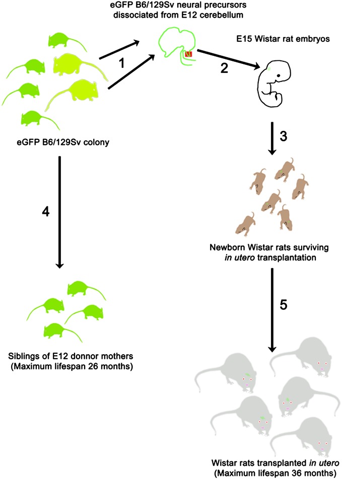
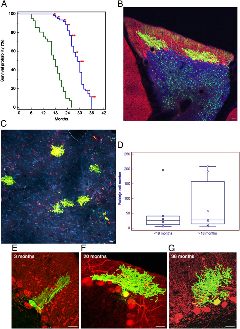
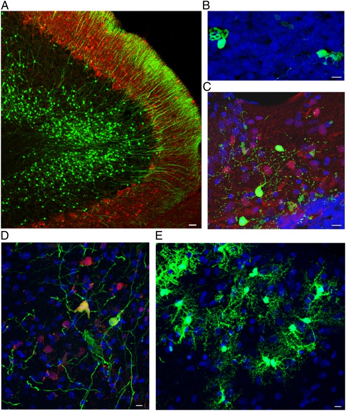
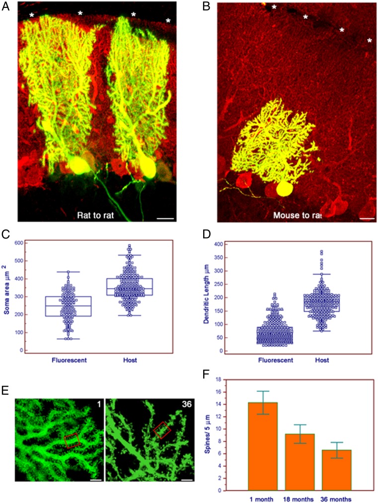
Neurons in mammals do not undergo replicative aging, and, in absence of pathologic conditions, their lifespan is limited only by the maximum lifespan of the organism. Whether neuronal lifespan is determined by the strain-specific lifetime or can be extended beyond this limit is unknown. Here, we transplanted embryonic mouse cerebellar precursors into the developing brain of the longer-living Wistar rats. The donor cells integrated into the rat cerebellum developing into mature neurons while retaining mouse-specific morphometric traits. In their new environment, the grafted mouse neurons did not die at or before the maximum lifespan of their strain of origin but survived as long as 36 mo, doubling the average lifespan of the donor mice. Thus, the lifespan of neurons is not limited by the maximum lifespan of the donor organism, but continues when transplanted in a longer-living host.


Conflict of interest statement

The authors declare no conflict of interest.

Figures

Fig. 1.
Fig. 1.
Schematic outline of the experiment. (1) E12 fetuses were obtained from pregnant mothers in our colony of EGFP B6/129Sv mice, and the cerebellar primordium was dissected and mechanically dissociated into a single cell suspension. (2) A total of 5 × 10⁴ E12 cerebellar precursor cells were injected through a glass microneedle into the developing cerebellum of E15 Wistar rats. All fetuses contained in the uterine horns were injected. (3) From our transplantation experiments, we obtained 69 live-born Wistar rats. (4) A total of 59 of these rats were allowed to survive until moribund and unlikely to survive for more than 2 d; at that time, they were perfused, and their cerebella and brainstem were collected for histological processing.
Fig. 2.
Fig. 2.
Developing mouse cerebellar neurons transplanted in utero into the developing rat CNS integrate and survive to the rat host maximum lifespan, doubling the average lifespan of the neurons in the donor mice strain. (A) Kaplan–Meier plot showing lifespan of siblings of donor mice and rat host. Rats perfused electively were excluded from the survival curve. Each red dot indicates a transplanted rat in which we found mouse cells. (B) Parasagittal section from confocal microscopy of a 20-mo-old rat cerebellum. Fluorescences are as follows: EGFP (green), anti-calbindin antibody (red), overlapping fluorescence (yellow), and DAPI nuclear staining (blue). Two grafted mouse PCs are integrated in the host PC and molecular layers, and granular cells of mouse origin are visible in the granular layer (asterisks). (Scale bar: 25 μm.) (C) Same as Fig. 1A, but coronal section of the brainstem and fourth ventricle of a 36-mo-old graft, in which six ectopic calbindin/EGFP-positive PCs are visible in the brainstem outside the dorsal cochlear nucleus. Interrupted line indicates the ventricle. (Scale bar: 50 μm.) (D) Plot of the number of PCs found in the transplanted animals at different ages (<19 mo, rats perfused before 19 mo of age; >18 mo, rats perfused after 18 mo to 36 mo of age). Each square represents a single animal: <19 mo, n = 6; >18 mo, n = 7. Differences in mean and median number of PCs in the two groups were not significant (P = 0.5968, unpaired Student t test; P = 0.5338, Mann–Whitney test). Horizontal line indicates the median; central box encloses values from the lower to upper quartile; vertical line extends from the minimum to the maximum value, excluding outside and far out values. (EG) Parasagittal sections as in Fig. 1A but without DAPI. Rat cerebella at 3, 20, and 36 mo of age showing mouse-derived PCs. The span of mouse PC dendrite did not reach the top of the host molecular layer and remained approximately the same at all survival times. (Scale bar: 25 μm.)
Fig. 3.
Fig. 3.
Together with PCs, all other neural and glial phenotypes derived from transplanted mouse cerebellar precursors survived, as long as the host rat did. All of the following examples are from rats surviving beyond 20 mo. Confocal images of sagittal sections of brainstem and cerebellum of transplant-bearing rats. (A) EGFP-positive granule cells engrafted in the recipient internal granular layer. The axons of these neurons (parallel fibers) can be seen in the overlying molecular layer. Green indicates EGFP; red indicates calbindin. (Scale bar: 25 μm.) (B) Unipolar brush cells in the host granular layer displaying the typical dendritic structure. Green indicates EGFP; blue indicates DAPI nuclear staining. (Scale bar: 10 μm.) (C) Molecular layer interneurons and basket and stellate cells located in the recipient molecular layer of the cerebellar cortex. Green indicates EGFP; red indicates parvalbumin; blue indicates DAPI nuclear staining. (Scale bar: 10 μm.) (D) Neurons in the deep nuclei show multipolar morphology and express NeuN. Green indicates EGFP; red indicates NeuN; blue indicates DAPI nuclear staining. (Scale bar: 10 μm.) (E) Astrocytes scattered through the cerebellar white matter. Green indicates EGFP; blue indicates DAPI nuclear staining. (Scale bar: 10 μm.)
Fig. 4.
Fig. 4.
Mouse PCs retain mouse morphometric characteristics when integrated in the rat cerebellum and undergo a progressive loss of dendritic spines with aging. (A) Parasagittal section stained as in Fig. 1E. Two rat PCs at 35 d after in utero transplantation into rat cerebellum. The grafted rat PC extends its dendrite to the top of the molecular layer (asterisks). (Scale bar: 25 μm.) (B) Same as Fig. 2A but mouse PC 35 d after in utero transplantation into rat cerebellum. Despite a fully mature PC phenotype, the mouse cell did not reach the top of the rat molecular layer (asterisks). (Scale bar: 25 μm.) (C) Plot of the areas of the PC soma of grafted mouse (marked as “M”) and host rat (“R”) cells. Cells were in the cerebellum. All survival times are pooled. Each square represents a single area (mouse, n = 160; rat, n = 308). The mean difference of 109.03 μm2 was highly significant (95% CI of difference, 94.56–123.50; P < 0.0001, unpaired Student t test and Mann–Whitney test). Lines and boxes are as in Fig. 1D. (D) Plot of the span of grafted mouse (“M”) and host rat (“R”) PC dendrites. Cells and times are as in Fig. 2C. Each square represents a single dendrite (mouse, n = 193; rat, n = 308). The mean difference of 90.94 μm was highly significant (95% CI, 82.85–99.03; P < 0.0001, unpaired Student t test and Mann–Whitney test). Lines and boxes are as in Fig. 1D. (E) Grafted mouse PC dendrites at 1 and 36 mo of age. We counted by optical sectioning all spines contained in randomly selected 5 μm of dendrite. (Scale bar and boxes: 5 μm.) (F) Histogram demonstrating progressive decrease in average number of spines at 1, 18, and 36 mo of age. Error bars indicate SDs.

Similar articles

Cited by

References

    1. Jeyapalan J-C, Sedivy J-M. Cellular senescence and organismal aging. Mech Ageing Dev. 2008;129(7-8):467–474. - PMC - PubMed
    1. Rodier F, Campisi J. Four faces of cellular senescence. J Cell Biol. 2011;192(4):547–556. - PMC - PubMed
    1. Rando T-A, Chang H-Y. Aging, rejuvenation, and epigenetic reprogramming: Resetting the aging clock. Cell. 2012;148(1-2):46–57. - PMC - PubMed
    1. Esiri M-M. Ageing and the brain. J Pathol. 2007;211(2):181–187. - PubMed
    1. Andersen B-B, Gundersen H-J-G, Pakkenberg B. Aging of the human cerebellum: A stereological study. J Comp Neurol. 2003;466(3):356–365. - PubMed

Publication types