. 2013 Mar 12;110(11):4297-302.
doi: 10.1073/pnas.1222248110. Epub 2013 Feb 25.

Quinone-dependent proton transfer pathways in the photosynthetic cytochrome b6f complex

Affiliations

Quinone-dependent proton transfer pathways in the photosynthetic cytochrome b6f complex

S Saif Hasan et al. Proc Natl Acad Sci U S A. .

Abstract

As much as two-thirds of the proton gradient used for transmembrane free energy storage in oxygenic photosynthesis is generated by the cytochrome b6f complex. The proton uptake pathway from the electrochemically negative (n) aqueous phase to the n-side quinone binding site of the complex, and a probable route for proton exit to the positive phase resulting from quinol oxidation, are defined in a 2.70-Å crystal structure and in structures with quinone analog inhibitors at 3.07 Å (tridecyl-stigmatellin) and 3.25-Å (2-nonyl-4-hydroxyquinoline N-oxide) resolution. The simplest n-side proton pathway extends from the aqueous phase via Asp20 and Arg207 (cytochrome b6 subunit) to quinone bound axially to heme c(n). On the positive side, the heme-proximal Glu78 (subunit IV), which accepts protons from plastosemiquinone, defines a route for H(+) transfer to the aqueous phase. These pathways provide a structure-based description of the quinone-mediated proton transfer responsible for generation of the transmembrane electrochemical potential gradient in oxygenic photosynthesis.


Conflict of interest statement

The authors declare no conflict of interest.

Figures
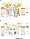
Fig. 1.
Fig. 1.
Dimeric cytochrome b6f complex of oxygenic photosynthesis from the cyanobacterium Nostoc PCC 7120. (A) Eight transmembrane polypeptides form the cytochrome b6f monomer. Cytochrome b6 (cyt b6, cyan) and subunit IV (SubIV, pink) with four and three transmembranes helices, respectively, are polytopic subunits. Cytochrome f (cyt f, yellow) and the Rieske ISP (orange) have one transmembrane helix and a large soluble domain. The Pet G (brown), L (wheat), M (green), and N (blue) are located on the periphery of the complex and consist of one transmembrane helix and no soluble domains. p- and n- side, electrochemically positive and negative side of the membrane. (B) Prosthetic groups in the b6f complex that function in electron transfer. The transmembrane pathway of electron transfer consists of 2 bis-histidine ligated hemes (bp and bn) and one covalently linked heme cn, all linked to the cytochrome b6 subunit. The p-side soluble domain of the ISP subunit has an iron-sulfur cluster ([2Fe-2S]), and the cytochrome f soluble domain has a covalently attached heme (f). n- and p-side proton transfer pathways are highlighted to show their position in the membrane. The n-side pathway consists of Asp20 (cyan and red sticks) and Arg207 (cyan and blue sticks) of cytochrome b6. The p-side water channel is shown in green extending from the Glu78 residue (pink and red sticks) of subunit IV. The lipid bilayer has been modeled in cartoon mode (orange).
Fig. 2.
Fig. 2.
N-side proton uptake pathways of the cytochrome b6f complex. (A) The D/R proton uptake pathway in the 2.70-Å Nostoc PCC 7120 cytochrome b6f structure. The Asp20→Arg207 route of proton transfer is demonstrated. The Arg207/heme cn interaction is also shown. Orange mesh, 2Fo-Fc map, 1.3 σ. (B and C) Arg207 as a ligand to the quinone analog inhibitors TDS (B) and NQNO (C) bound at the Qn-site. From the cocrystal structures of b6f with quinone analog inhibitors, it is inferred that the Arg207 side chain interacts with the natural quinone bound at the Qn-site. Polypeptide color code, same as in Fig. 1, Heme bp, brown lines; heme cn, blue lines; TDS and NQNO, green red and blue sticks; 2Fo-Fc map, 1.2 σ, orange mesh. The peripheral Pet G, L, M, and N subunits and the hydrocarbon tails of TDS and NQNO are not shown. (D) Potential proton uptake pathway mediated by water in the b6f complex. Water416 (red sphere) is coordinated by the Arg207 backbone carbonyl oxygen (a = 2.8 Å) and side chain (b = 3.3 Å), along with the propionate-A (c = 2.7 Å) carboxylic acid group of heme cn (blue) (Fig. S2 shows heme nomenclature). Wat416 is separated from the basic side chain of Lys24 (cytochrome b6, cyan) by a distance of 4.6 Å. The ISP transmembrane helix is omitted. (E and F) The E/D pathway of proton entry on the n-side of the b6f complex. (E) In the 2.70-Å structure, Glu29 (subunit IV) located on the n-side surface of cytochrome b6f interacts with a water molecule (“Wat,” red sphere), which forms a hydrogen bond with the acidic carboxylate side chain of Asp35 (subunit IV). (F) The Asp35 residue interacts with the quinone analog TDS bound at the Qn-site. 2Fo-Fc map, 1.2 σ, orange mesh. For simplicity, the TDS hydrocarbon tail is not shown. Color code, same as in Fig. 1. Transmembrane helices of cyt b6 (a–d) and subunit IV (e–g) are labeled.
Fig. 3.
Fig. 3.
Proposed proton exit pathways on the p-side of the cyt b6f complex. A hydrophilic portal (green surface) leads from Glu78 of subunit IV to the p-side surface, providing a route for diffusion of water. The portal is lined by Arg87 (cytochrome b6), Ser91 (cytochrome b6), Glu3 (PetG), and Asp58 (subunit IV). Glu78 is linked to water 316 (wat316 in the figure) through a hydrogen bond that lies at the end of this portal. The proton exit pathway is highlighted through the hydrophilic portal (green) by a broken arrow (black). The TDS quinone analog inhibitor has been introduced from PDB ID 2E76 to mark the position of the Qp-site. Orange mesh, 2Fo-Fc map, 1.3 σ. Transmembrane helices of cytochrome b6 (a–d) and subunit IV (e–g) are labeled.

References

    1. Deisenhofer J, Epp O, Miki K, Huber R, Michel H. Structure of the protein subunits in the photosynthetic reaction centre of Rhodopseudomonas viridis at 3Å resolution. Nature. 1985;318(6047):618–624. - PubMed
    1. Iwata S, Ostermeier C, Ludwig B, Michel H. Structure at 2.8 A resolution of cytochrome c oxidase from Paracoccus denitrificans. Nature. 1995;376(6542):660–669. - PubMed
    1. Ermler U, Fritzsch G, Buchanan SK, Michel H. Structure of the photosynthetic reaction centre from Rhodobacter sphaeroides at 2.65 A resolution: Cofactors and protein-cofactor interactions. Structure. 1994;2(10):925–936. - PubMed
    1. Hosler JP, Ferguson-Miller S, Mills DA. Energy transduction: Proton transfer through the respiratory complexes. Annu Rev Biochem. 2006;75:165–187. - PMC - PubMed
    1. Lyons JA, et al. Structural insights into electron transfer in caa3-type cytochrome oxidase. Nature. 2012;487(7408):514–518. - PMC - PubMed

Publication types

MeSH terms

LinkOut - more resources