. 2013 Mar 12;110(11):4386-91.
doi: 10.1073/pnas.1216867110. Epub 2013 Feb 25.

Genotype-phenotype correlations in neonatal epilepsies caused by mutations in the voltage sensor of K(v)7.2 potassium channel subunits

Affiliations

Genotype-phenotype correlations in neonatal epilepsies caused by mutations in the voltage sensor of K(v)7.2 potassium channel subunits

Francesco Miceli et al. Proc Natl Acad Sci U S A. .

Abstract

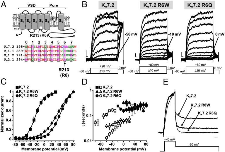
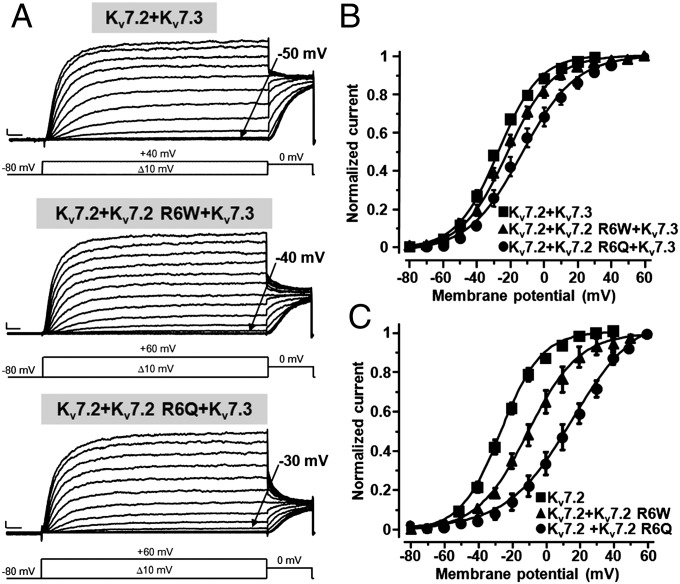
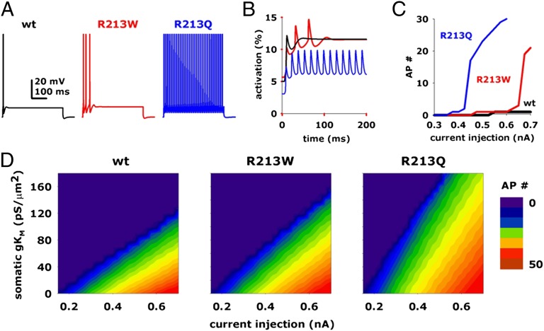
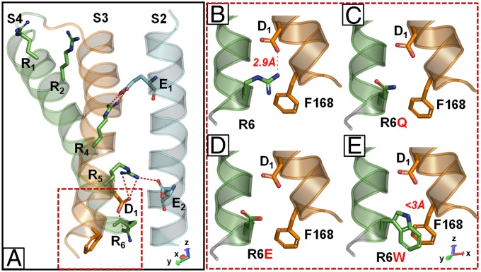
Mutations in the K(V)7.2 gene encoding for voltage-dependent K(+) channel subunits cause neonatal epilepsies with wide phenotypic heterogeneity. Two mutations affecting the same positively charged residue in the S4 domain of K(V)7.2 have been found in children affected with benign familial neonatal seizures (R213W mutation) or with neonatal epileptic encephalopathy with severe pharmacoresistant seizures and neurocognitive delay, suppression-burst pattern at EEG, and distinct neuroradiological features (R213Q mutation). To examine the molecular basis for this strikingly different phenotype, we studied the functional characteristics of mutant channels by using electrophysiological techniques, computational modeling, and homology modeling. Functional studies revealed that, in homomeric or heteromeric configuration with K(V)7.2 and/or K(V)7.3 subunits, both mutations markedly destabilized the open state, causing a dramatic decrease in channel voltage sensitivity. These functional changes were (i) more pronounced for channels incorporating R213Q- than R213W-carrying K(V)7.2 subunits; (ii) proportional to the number of mutant subunits incorporated; and (iii) fully restored by the neuronal K(v)7 activator retigabine. Homology modeling confirmed a critical role for the R213 residue in stabilizing the activated voltage sensor configuration. Modeling experiments in CA1 hippocampal pyramidal cells revealed that both mutations increased cell firing frequency, with the R213Q mutation prompting more dramatic functional changes compared with the R213W mutation. These results suggest that the clinical disease severity may be related to the extent of the mutation-induced functional K(+) channel impairment, and set the preclinical basis for the potential use of K(v)7 openers as a targeted anticonvulsant therapy to improve developmental outcome in neonates with K(V)7.2 encephalopathy.


Conflict of interest statement

The authors declare no conflict of interest.

Figures

Fig. 1.
Fig. 1.
Functional properties of homomeric KV7.2, KV7.2 R6W, and KV7.2 R6Q channels. (A) (Upper) Topological representation of a Kv7.2 subunit. The R213 (R6) residue is highlighted. (Lower) Sequence alignment of the S4 segments of the indicated Kv subunits (www.ebi.ac.uk/Tools/psa/). Residues are colored according to the following scheme: magenta, basic; blue, acid; red, nonpolar; and green, polar. (B) Macroscopic currents from KV7.2, KV7.2 R6W, and KV7.2 R6Q channels, in response to the indicated voltage protocols. Current scale, 100 pA; time scale, 0.1 s. The arrows indicate the threshold voltage for current activation. (C) Conductance/voltage curves. Continuous lines are Boltzmann fits to the experimental data. (D) Time constants for ionic current activation (weighed average of τf and τs; filled symbols) and deactivation (empty symbols) for the indicated channels (n = 4–9). (E) Normalized and superimposed current traces from KV7.2, KV7.2 R6W, and KV7.2 R6Q channels. Current scale, 100 pA; time scale, 100 ms.
Fig. 2.
Fig. 2.
Functional properties of heteromeric channels incorporating KV7.2 R6W or KV7.2 R6Q mutant subunits. (A) Macroscopic current traces from the indicated heteromeric channels in response to the indicated voltage protocols. Current scale, 200 pA; time scale, 0.1 s The arrows indicate the threshold voltage for current activation. (B and C) Conductance/voltage curves for the heteromeric channels formed by mutant subunits with KV7.2 and KV7.3 (B) or with KV7.2 (C). Continuous lines are Boltzmann fits to the experimental data. The cDNA ratios were: 1:1 for KV7.2+KV7.3, KV7.2+KV7.2 R6W, and KV7.2+KV7.2 R6Q, and 0.5:0.5:1 for KV7.2+KV7.2 R6W+KV7.3 and KV7.2+KV7.2 R6Q+KV7.3. Current scale, 200 pA; time scale, 100 ms.
Fig. 3.
Fig. 3.
Effect of retigabine on heteromeric KV7.2+KV7.3, KV7.2+KV7.2 R6W+KV7.3, or KV7.2+KV7.2 R6Q+KV7.3 channels. (A) Current responses from the indicated heteromeric channels to voltage ramps from −80 to 0 mV. C, control currents; RET, retigabine; W, washout. Current scale, 200 pA; time scale, 0.2 s. (B) Quantification of the effects of RET on the indicated heteromeric channels. Data are expressed as membrane potentials at which currents reached 20% of their peak value for controls (black bars) and after RET exposure (white bars). *P < 0.05 vs. corresponding controls; ns, not significantly different). Current scale, 200 pA; time scale, 200 ms.
Fig. 4.
Fig. 4.
Modeling of the effects of the KV7.2 R213W (R6W) and R213Q (R6Q) mutations on neuronal excitability. Time courses of the somatic membrane potential (A) and of the activated conductances (B; only the first 200 ms are shown) from CA1 neurons expressing KV7.2+KV7.3 (black traces), KV7.2+KV7.2 R6W+KV7.3 (red traces), or KV7.2+KV7.2 R6Q+KV7.3 (blue traces) heteromeric channels, during a 500-ms somatic current injection of 0.65 nA. (C) Input/output (I/O) curves from cells expressing KV7.2+KV7.3 (black trace), KV7.2+KV7.2 R6W+KV7.3 (red trace), or KV7.2+KV7.2 R6Q+KV7.3 (blue trace) heteromeric channels. Number of APs (AP#) is expressed as a function of the somatic current injected, using a 120 mS/μm2 peak IKM conductance. (D) Contour plots for number of APs elicited by a 500-ms somatic current injection from cells expressing KV7.2+KV7.3, KV7.2+KV7.2 R6W+KV7.3, or KV7.2+KV7.2 R6Q+KV7.3 heteromeric channels as a function of current injection and peak IKM conductance.
Fig. 5.
Fig. 5.
Homology models of KV7.2, KV7.2 R6W, KV7.2 R6Q, and KV7.2 R6E subunits. (A) Homology model of activated configuration of VSD of a single KV7.2 subunit; only S2, S3, and S4 segments are shown for clarity. Charged amino acids shown correspond to E130 (E1) and E140 (E2) in S2, D172 (D1) in S3, and R198 (R1), R201 (R2), R207 (R4), R210 (R5), and R213 (R6) in S4. (BE) Higher magnification of the boxed region in A in a single KV7.2 (B), KV7.2 R6Q (C), KV7.2 R6E (D), and KV7.2 R6W (E) subunit. To improve clarity, images in BE are shown after leftward rotation by 90° of the model shown in A.

References

    1. Wang HS, et al. KCNQ2 and KCNQ3 potassium channel subunits: Molecular correlates of the M-channel. Science. 1998;282(5395):1890–1893. - PubMed
    1. Brown DA, Adams PR. Muscarinic suppression of a novel voltage-sensitive K+ current in a vertebrate neurone. Nature. 1980;283(5748):673–676. - PubMed
    1. Soldovieri MV, Miceli F, Taglialatela M. Driving with no brakes: Molecular pathophysiology of Kv7 potassium channels. Physiology (Bethesda) 2011;26(5):365–376. - PubMed
    1. Cooper EC, Harrington E, Jan YN, Jan LY. M channel KCNQ2 subunits are localized to key sites for control of neuronal network oscillations and synchronization in mouse brain. J Neurosci. 2001;21(24):9529–9540. - PMC - PubMed
    1. Biervert C, et al. A potassium channel mutation in neonatal human epilepsy. Science. 1998;279(5349):403–406. - PubMed

Publication types

MeSH terms

LinkOut - more resources