. 2013 Mar;141(3):389-95.
doi: 10.1085/jgp.201210940.

The voltage-sensing domain of a phosphatase gates the pore of a potassium channel

Affiliations

The voltage-sensing domain of a phosphatase gates the pore of a potassium channel

Cristina Arrigoni et al. J Gen Physiol. 2013 Mar.

Abstract

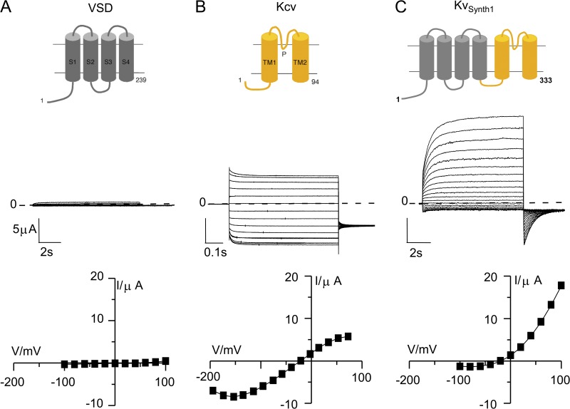
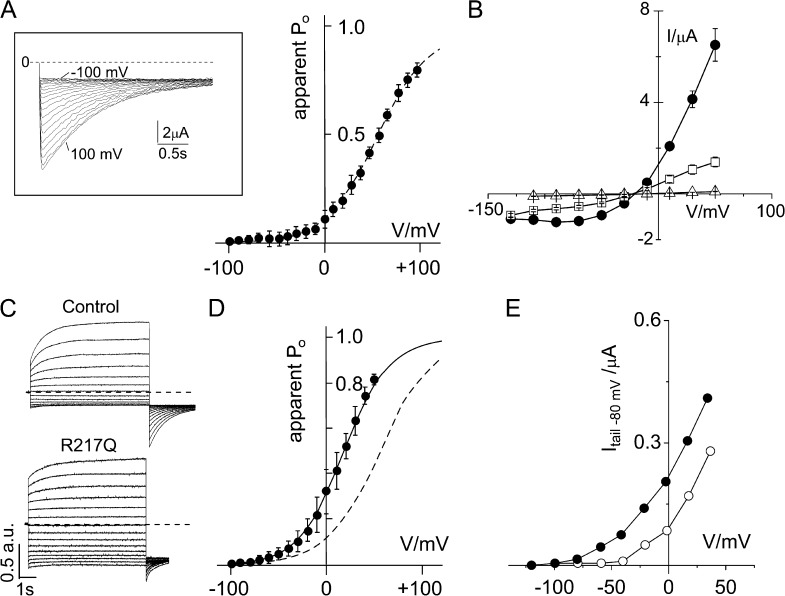
The modular architecture of voltage-gated K(+) (Kv) channels suggests that they resulted from the fusion of a voltage-sensing domain (VSD) to a pore module. Here, we show that the VSD of Ciona intestinalis phosphatase (Ci-VSP) fused to the viral channel Kcv creates Kv(Synth1), a functional voltage-gated, outwardly rectifying K(+) channel. Kv(Synth1) displays the summed features of its individual components: pore properties of Kcv (selectivity and filter gating) and voltage dependence of Ci-VSP (V(1/2) = +56 mV; z of ~1), including the depolarization-induced mode shift. The degree of outward rectification of the channel is critically dependent on the length of the linker more than on its amino acid composition. This highlights a mechanistic role of the linker in transmitting the movement of the sensor to the pore and shows that electromechanical coupling can occur without coevolution of the two domains.


Figures

Figure 1.
Figure 1.
The synthetic channel KvSynth1 is an outward rectifier. (Top) Cartoons of the individual components and of the fusion protein KvSynth1. (A) VSD is the voltage-sensing domain (amino acids 1–239) of Ci-VSP; S1–S4 indicate the four transmembrane domains of the VSD. (B) Kcv is the full-length (amino acids 1–94) viral K+ channel; TM1, TM2, and P indicate, respectively, the two transmembrane domains and the pore loop of Kcv. (C) KvSynth1 is the 333–amino acid fusion protein of VSD and Kcv. (Middle) Currents recorded by TEVC in oocytes injected with the respective constructs. Dotted line indicates zero current level. Voltage protocol: Vh, −20 mV; test voltages, +100 to −100 mV (−200 mV for Kcv); tail, −80 mV. Test pulse length: A and C, 8.5 s; B, 0.6 s. (Bottom) Corresponding steady-state I-V relationships. All experiments were performed in 50 mM [K+]out.
Figure 2.
Figure 2.
Biophysical and biochemical properties of KvSynth1. (A) KvSynth1 selects K+ over Na+. Current reversal potential recorded from the same oocyte was −20 mV in 50 mM [K+]out (•) and −98 mV in 50 mM [Na+]out (○). Voltage protocol: prepulse voltage step at +60 mV, followed by testing voltages from +60 to −180 mV. Arrowhead marks the point of data collection. (B) Single-channel properties of KvSynth1. (Left) Representative opening burst at different membrane voltages, with arrows denoting the baseline. The open probability in the presented sections is not representative. The low-pass filter was set to 20 kHz, the sampling rate was 100 kHz, and traces were digitally filtered at 2 kHz for clarity. (Right) Comparison of the I-V curves of KvSynth1 in 150 mM K+ (•) and Kcv in 100 mM K+ (▪). Symbols represent mean and standard deviation of three experiments. (C) Nernst plot: current reversal potential (Erev) of macroscopic KvSynth1 current plotted as a function of external K+ concentration (Lg[K+]out). Black line is the linear regression to Erev mean values (n = 3). (D) Effect of barium on KvSynth1 current. Steady-state currents recorded in (•) control solution (50 mM K+) and (○) plus 1 mM BaCl2. Currents recorded from a holding potential (Vh) of −20 mV to the indicated test voltages. Test pulse length was 8.5 s. The addition of barium blocks the inward current and moderately affects the outward current, as reported previously for Kcv channel (Plugge et al., 2000). (E) Western blot analysis performed on total protein extracts with a custom-made antibody (8D6) recognizing the tetramer, but not the monomer, of Kcv. The expected molecular mass of Kcv and KvSynth1 tetramers are 42.5 and 149 kD, respectively.
Figure 3.
Figure 3.
Voltage dependency of KvSynth1. (A) Activation curve of KvSynth1 constructed from tail currents (inset) after subtracting the instantaneous current offset. Voltage protocol as in Fig. 1 C. Datasets (n = 6) were jointly fitted to a two-state Boltzmann equation (dotted line) of the form y = (1 + ezF(V V1/2)/RT)−1, where z is the effective charge; V1/2 is the half-activation voltage; and F, R, and T have their usual thermodynamic meaning. Data were normalized to the extrapolated maximum current. Points represent mean ± SEM. V1/2 = 56 mV and z = 0.92. (B) Steady-state I-V relationships of the single mutant F305A (Δ), the double mutant R229Q/R232Q (□), and the WT KvSynth1 (•). Points represent mean ± SEM (n = 6). Currents were recorded from a holding potential (Vh) of −20 mV to the indicated test voltages (pulse length, 600 ms). (C) Exemplary currents recorded from WT KvSynth1 (control) and its R217Q mutant. To compare currents from different constructs, the traces have been normalized to the current value recorded at +60 mV and expressed in arbitrary units (a.u.). Voltage protocol as in Fig. 1 C. (D) Activation curve of the R217Q mutant (•) constructed as in A from four datasets: V1/2 = 18 mV and z = 1.1. The curve of the WT (dotted line) is replotted from A for comparison. (E) The activation of the R217Q mutant depends on the preconditioning voltage. The same R217Q-expressing oocyte was subjected to the following protocol: preconditioning (5 s) at either −100 or +40 mV and test pulses from −120 to +30 mV. Tail currents (at −80 mV) are plotted for preconditioning at −100 mV (○) or at +40 mV (•). The test pulses were kept short (1.2 s) to maintain the effect of the preconditioning voltages; hence, data in E do not reflect full activation of the channel.
Figure 4.
Figure 4.
The length of the linker connecting the VSD to the pore affects the degree of rectification in KvSynth1 constructs. (A) Expanded view of the sequences of the two sequences that where variably combined to form the KvSynth1 linker. (B) List of linkers that have been tested in KvSynth1, their amino acid (aa) length, and amino acid sequences (color coded as in A). (C) Exemplary current traces recorded from KvSynth1 constructs with the 4–, 6–, and 20–amino acid linkers and their corresponding I-V relationships (color coded). To compare currents from different constructs, the traces have been normalized to the current value recorded at +60 mV and expressed in arbitrary units (a.u.). Voltage protocol as in Fig. 1 C. (D) The degree of current rectification (I+60mV/I−100mV) is plotted as a function of linker length for all functional constructs. Data are mean ± SEM (n ≥ 3). Experimental data (with the exclusion of the value corresponding to the construct with a 4–amino acid linker that loses the rectification) have been interpolated with a logistic function (black line) in which the lower asymptote was set to 1, corresponding to the value measured in Kcv that lacks rectification (dotted line).

References

    1. Abenavoli A., DiFrancesco M.L., Schroeder I., Epimashko S., Gazzarrini S., Hansen U.P., Thiel G., Moroni A. 2009. Fast and slow gating are inherent properties of the pore module of the K+ channel Kcv. J. Gen. Physiol. 134:219–229 10.1085/jgp.200910266 - DOI - PMC - PubMed
    1. Alabi A.A., Bahamonde M.I., Jung H.J., Kim J.I., Swartz K.J. 2007. Portability of paddle motif function and pharmacology in voltage sensors. Nature. 450:370–375 10.1038/nature06266 - DOI - PMC - PubMed
    1. Ben-Abu Y., Zhou Y., Zilberberg N., Yifrach O. 2009. Inverse coupling in leak and voltage-activated K+ channel gates underlies distinct roles in electrical signaling. Nat. Struct. Mol. Biol. 16:71–79 10.1038/nsmb.1525 - DOI - PubMed
    1. Bosmans F., Martin-Eauclaire M.F., Swartz K.J. 2008. Deconstructing voltage sensor function and pharmacology in sodium channels. Nature. 456:202–208 10.1038/nature07473 - DOI - PMC - PubMed
    1. Caprini M., Ferroni S., Planells-Cases R., Rueda J., Rapisarda C., Ferrer-Montiel A., Montal M. 2001. Structural compatibility between the putative voltage sensor of voltage-gated K+ channels and the prokaryotic KcsA channel. J. Biol. Chem. 276:21070–21076 10.1074/jbc.M100487200 - DOI - PubMed

Publication types

MeSH terms

Substances