. 2013 Apr 1;190(7):3289-98.
doi: 10.4049/jimmunol.1203086. Epub 2013 Feb 25.

IRF7-dependent IFN-β production in response to RANKL promotes medullary thymic epithelial cell development

Affiliations

IRF7-dependent IFN-β production in response to RANKL promotes medullary thymic epithelial cell development

Dennis C Otero et al. J Immunol. .

Abstract

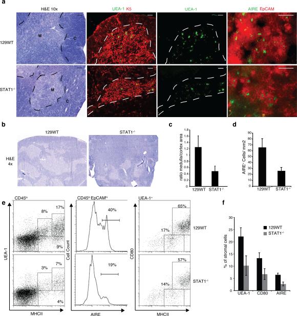
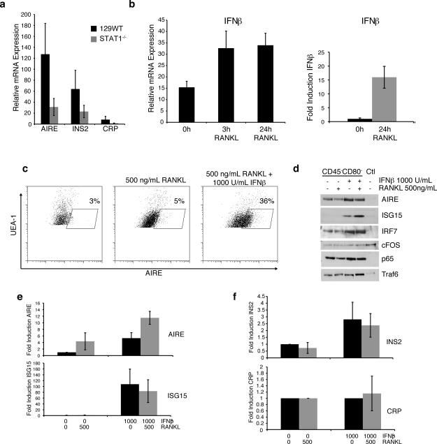
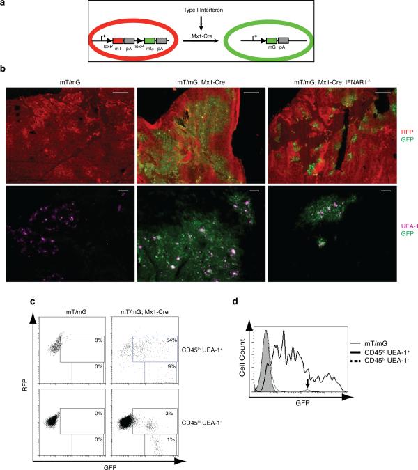
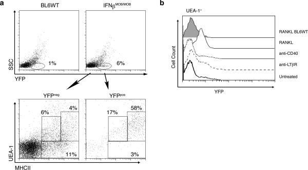
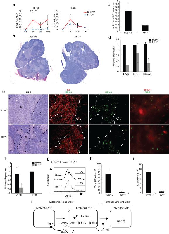
The contributions of IFN regulatory factor (IRF) 3/7 and the type I IFNs IFN-α/β to the innate host defense have been extensively investigated; however, their role in thymic development is less clear. In this study, we show that mice lacking the type I IFN receptor IFN-α/β receptor (IFNAR) or the downstream transcription factor STAT1 harbor a significant reduction in self-Ag-presenting, autoimmune regulator (AIRE)(+) medullary thymic epithelial cells (mTECs). Constitutive IFNAR signaling occurs in the thymic medulla in the absence of infection or inflammation. Receptor activator for NF-κB (RANK) ligand stimulation results in IFN-β upregulation, which in turn inhibits RANK signaling and facilitates AIRE expression in mTECs. Finally, we find that IRF7 is required for thymic IFN-β induction, maintenance of thymic architecture, and mTEC differentiation. We conclude that spatially and temporally coordinated cross talks between the RANK ligand/RANK and IRF7/IFN-β/IFNAR/STAT1 pathways are essential for differentiation of AIRE(+) mTECs.


Figures

Figure 1
Figure 1. Impaired medullary thymic architecture in IFNAR-/- and STAT1-/- mice
a) Thymic architecture in WT129 and STAT1-/- mice as revealed by H&E staining of thymic sections (left panels); Keratin 5 (red) and UEA-1 (green) staining (middle panels), bar = 100μm; EpCAM (red) and AIRE (green) staining (right panels), bar = 50μm; M, medulla; C, cortex. b) Low magnification (4x) of H&E stains. c) Ratio of medullary to cortical cellularity in thymic sections from WT and STAT1-/- mice (data collected using ImageJ (NIH) and represent avg +/-SD of 3 sections each from 3 mice (p<0.05). d) AIRE+ cells per unit area (quantified from thymus sections shown in Fig.1a, lower panels) e) mTEC populations in WT and STAT1-/- thymi as determined by flow cytometric expression analysis of MHCII and UEA-1 (left), as well as AIRE and EpCAM (middle) on CD45lo stromal cells; MHCII and CD80 expression on UEA-1hi gated stromal cells. f) mTEC populations in WT and STAT1-/- thymi (n=4) using the indicated markers.
Figure 2
Figure 2. IFNβ promotes AIRE expression following RANKL stimulation
a) AIRE, INS2, and CRP mRNA levels in thymic stromal cells from WT and STAT1-/- mice (avg+/-sd; n=4) b) Thymic epithelial cells from WT mice (left) or TE-71 cells (right) were stimulated with 500 ng/mL RANKL for the indicated times and IFNβ mRNA levels were determined by qPCR (n = 4). c) Thymic epithelial cells were left untreated or pretreated with 1000 U/mL IFNβ prior to stimulation with 500 ng/mL RANKL for 24 hrs. UEA-1+ cells were analyzed for intracellular AIRE expression by flow cytometry. d) Thymic epithelial cells were depleted of CD80+/hi cells and then stimulated with 500 ng/mL RANKL, 1000 U/mL IFNβ or both for 24hrs. Lysates were probed for AIRE, ISG15, IRF7, cFOS, p65, and Traf6 by Western blotting (representative of three experiments). e) TE-71 cells were treated as indicated, and mRNA levels for AIRE and ISG15 were determined by qPCR (avg+/-sd; n=3). f) Thymic epithelial cells were treated as indicated and mRNA levels for CRP and INS2 were determined by qPCR (avg+/-sd; n=4).
Figure 3
Figure 3. Constitutive interferon signaling in the thymus is restricted to the medullary region
a) Schematic diagram of the interferon response reporter mice. Interferon-dependent Cre expression results in the excision of the mT sequence coding RFP and expression of mG encoding GFP (Muzumdar, et al. (27)). b) RFP and GFP expression in sections of thymi from WT and IFNAR1-/- mice carrying mT/mG and Mx1-Cre transgenes (upper panels, bar = 500μm). Sections were also stained for UEA-1 (lower panels, pseudo-colored purple, bar = 100μm). c) Flow cytometric analysis of CD45loUEA-1+ mTEC cells (upper panels) and CD45hiUEA-1- hematopoietic cells (lower panels) from thymi of either Mx1-Cre- or Mx1-Cre+ mT/mG mice. d) Histogram comparing GFP expression levels in CD45loUEA-1+ mTECs of Mx1-Cre- or Mx1-Cre+ mT/mG mice; arrow indicates GFP levels in CD45hi hematopoietic cells.
Figure 4
Figure 4. IFNβ production by UEA-1hi medullary thymic epithelial cells
a) Flow cytometric analysis of YFP expression in total cells from thymus of WT (top left panel) or IFNβMOB/MOB mice (top right panel). Lower panels show UEA-1 and MHCII expression in YFP- (left panel) and YFP+ (right panel) cells in thymi of IFNβMOB/MOB mice. b) Stromal cells were purified from thymi of IFNβMOB/MOB mice and treated with anti-CD40 (1:100 dilution of supernatant), anti-LTβR (500ng/ml) or RANK ligand (500ng/ml) for 24 hours. Histogram illustrates IFNβ:YFP expression in UEA-1+ cells following the indicated treatments. Data is representative of three separate experiments.
Figure 5
Figure 5. IRF7 is necessary for thymic IFNβ expression and mTEC development
a) Thymic stromal cells from WT or IRF7-/- mice were purified and stimulated with 500 ng/mL RANKL for the indicated time points. Expression of IFNβ and IκBα mRNAs was determined by qPCR. b) Thymic architecture in WT and IRF7-/- mice revealed by H&E staining of thymic sections (bar = 500 μm) c) Ratio of medullary to cortical cellularity in thymic sections from WT and IRF7-/- mice (data collected using ImageJ (NIH) and represent avg +/-sd of 3 sections each from 3 mice (p<0.05). d) mRNA levels of IFNβ, Iκbα, and ISG54 in thymic stromal cells from WT or IRF7-/- mice as determined by qPCR. e) Thymic architecture in WT and IRF7-/- mice as revealed by H&E staining of thymic sections (left panels), Keratin 5 and UEA-1 expression (middle panels, bar = 100 μm), UEA-1 only, as well as EpCAM and AIRE expression (right panels, bar = 50 μm) f) Relative mRNA levels of AIRE and INS2 in purified stromal cells from WT and IRF7-/-thymi measured by qPCR (n = 3). g) Histogram shows percentage of AIRE+ cells in the CD45loEpcam+UEA-1hi gate of thymic stromal cells from WT and IRF7-/- mice. h) Total number of UEA-1hi cells as determined by flow cytometric analysis of CD45loEpCAM+UEA-1hi mTEC populations from WT and IRF7-/- thymi. i) Total number of AIRE+ cells based on flow cytometric analysis of CD45loEpCAM+UEA-1hiAIRE+ mTEC populations in WT and IRF7-/- thymi (n = 4). j) Hypothetical model of IFNβ production and function in the thymus.

References

    1. Anderson MS, Venanzi ES, Klein L, Chen Z, Berzins SP, Turley SJ, von Boehmer H, Bronson R, Dierich A, Benoist C, Mathis D. Projection of an immunological self shadow within the thymus by the aire protein. Science. 2002;298:1395–1401. - PubMed
    1. Liston A, Lesage S, Wilson J, Peltonen L, Goodnow CC. Aire regulates negative selection of organ-specific T cells. Nat Immunol. 2003;4:350–354. - PubMed
    1. Finnish-German AC. An autoimmune disease, APECED, caused by mutations in a novel gene featuring two PHD-type zinc-finger domains. Nat Genet. 1997;17:399–403. - PubMed
    1. Peterson P, Nagamine K, Scott H, Heino M, Kudoh J, Shimizu N, Antonarakis SE, Krohn KJ. APECED: a monogenic autoimmune disease providing new clues to self-tolerance. Immunol Today. 1998;19:384–386. - PubMed
    1. Ramsey C, Winqvist O, Puhakka L, Halonen M, Moro A, Kampe O, Eskelin P, Pelto-Huikko M, Peltonen L. Aire deficient mice develop multiple features of APECED phenotype and show altered immune response. Hum Mol Genet. 2002;11:397–409. - PubMed

Publication types

MeSH terms

Substances