. 2013 May 1;73(9):2850-62.
doi: 10.1158/0008-5472.CAN-13-0382-T. Epub 2013 Feb 25.

An essential requirement for the SCAP/SREBP signaling axis to protect cancer cells from lipotoxicity

Affiliations

An essential requirement for the SCAP/SREBP signaling axis to protect cancer cells from lipotoxicity

Kevin J Williams et al. Cancer Res. .

Abstract

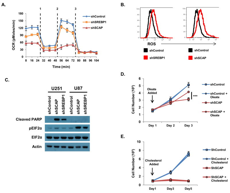
The sterol regulatory element-binding proteins (SREBP) are key transcriptional regulators of lipid metabolism and cellular growth. It has been proposed that SREBP signaling regulates cellular growth through its ability to drive lipid biosynthesis. Unexpectedly, we find that loss of SREBP activity inhibits cancer cell growth and viability by uncoupling fatty acid synthesis from desaturation. Integrated lipid profiling and metabolic flux analysis revealed that cancer cells with attenuated SREBP activity maintain long-chain saturated fatty acid synthesis, while losing fatty acid desaturation capacity. We traced this defect to the uncoupling of fatty acid synthase activity from stearoyl-CoA desaturase 1 (SCD1)-mediated desaturation. This deficiency in desaturation drives an imbalance between the saturated and monounsaturated fatty acid pools resulting in severe lipotoxicity. Importantly, replenishing the monounsaturated fatty acid pool restored growth to SREBP-inhibited cells. These studies highlight the importance of fatty acid desaturation in cancer growth and provide a novel mechanistic explanation for the role of SREBPs in cancer metabolism.


Conflict of interest statement

Disclosure of Potential Conflicts of Interest:

Dr. Paul Mischel has served as a scientific advisor to Celgene on the mTOR kinase inhibitor program. No other potential conflicts of interest were disclosed.

Figures

Figure 1
Figure 1. Cancer cells maintain a sterol sensitive, SCAP-dependent lipogenic program regardless of heightened PI3K/AKT signaling
(A, B) SREBP and lipogenic gene expression determined by q-PCR in U87 and U251 glioma cells cultured in complete media with 1% serum and treated with (A) 25μM 25-hydroxycholesterol or (B) 10 μM fatostatin for 24 h. (C) SREBP and lipogenic gene expression from U87 cells stably expressing shSREBP1, shSREBP2 or shSCAP cultured in complete media with 1% serum for 24 h. (D) Growth curve or (E) cell cycle analysis of U87 cells stably expressing shSREBP1, shSREBP2 or shSCAP U87 cells cultured in complete media with 10% serum. Frequency of cells in G1 indicated in plot. Cell cycle plots are representative of N=3 experiments. *p<0.05, **p<0.01, ***p<0.001.
Figure 2
Figure 2. SREBP1 regulates tumor growth
(A) Tumor area (mm2) of subcutaneous U87 tumors expressing shSREBP1, shSREBP2 or shControl on day 21. (B) Tumor area (mm2) of subcutaneous U87 tumors expressing shSREBP1, shSCAP or shControl on day 19 post-implantation. (C) Tumor area (mm2) subcutaneous wild-type U87 xenograft on day 21 post-implantation treated with fatostatin (5mg/kg or 30mg/kg) or vehicle i.p. every 3 days. p values indicated on plots.
Figure 3
Figure 3. The SCAP/SREBP signaling axis is not required to maintain cholesterol homeostasis in glioma cells
(A) GC/MS determination of total cellular cholesterol content of U87 or U251 glioma cells stably expressing shSREBP1, shSREBP2 or shSCAP as indicated cultured in complete media and 1% serum for 48 h. (B) Experimental schematic for determination of de novo lipid biosynthesis using 50% mixture of U-13C-glucose. (C) Percentage of cellular cholesterol derived from de novo synthesis in U87 or U251 cells stably expressing shSREBP1 or shSCAP cells as indicated and cultured in a 50% mixture of U-13C-glucose and 1% serum for 48 h. (D) Percentage of cellular cholesterol derived from de novo synthesis in U87 and U251 glioma cells cultured in complete media and 1% serum for 48 h. Cultures were treated with SREBP inhibitor compound 24 (25μM) or vehicle. (E) Total cellular cholesterol content of U87 and U251 cells treated with Compound 24 (25μM) as described above for 48 hours. *p<0.05, **p<0.01, ***p<0.001.
Figure 4
Figure 4. Loss of SREBP results in an increase total saturated long chain fatty acids
(A, B) Analysis of total saturated (16:0 and 18:0) and monounsaturated (16:1 and 18:1) long chain fatty acids (FAs) from U87 cells stably expressing shSREBP1 or shSCAP. Cell were cultured in complete media and 1% serum for 48 h. (C) Determination of the 16:0 to 16:1 and 18:0 to 18:1 ratios in shControl, shSREBP1 or shSCAP cells. (D) Determination of indicated FAs from U87 cells cultured in complete media and 1% serum for 48 h. Additionally, cultures were treated with vehicle or compound 24 (10 or 25μM) as indicated. (E) Ratio of 16:0 to 16:1, 18:0 to 18:1 and 20:0 to 20:1 from U87 and U251 cells treated with compound 24 (25μM) for 48 h.
Figure 5
Figure 5. SREBP activity is required to maintain de novo synthesis of monounsaturated fatty acids
(A) Determination of newly synthesized saturated and monounsaturated fatty acids (FAs) from U87 cells stably expressing shSREBP1 or shSCAP cultured in complete media containing a 50% mixture of U-13C-glucose and 1% serum for 48 h. (B) Percentage of indicated FAs derived from de novo synthesis in U87 and U251 glioma cells cultured in complete media containing a 50% mixture of U-13C-glucose and 1% serum for 48 h. In addition, cultures were treated with vehicle or compound 24 (25μM) as indicated. (C) The ratio of indicated FAs from U87 cells stably expressing mature SREBP1 (mSREBP1a), SREBP2 (mSREBP2) or vector control cultured in complete media and 10% serum for 48 h. (D) The ratio of indicated FAs from subcutaneous shSREBP1, shSCAP or shControl U87 xenograft harvested on day 19 post-implantation.
Figure 6
Figure 6. SREBP signaling is required to protect cells from lipotoxicity
(A) Oxygen consumption rate (OCR) of U87 SREBP or SCAP knockdown cells in basal state, and in response to sequential treatment with (1) oligomycin (ATPase inhibitor), (2) FCCP (uncoupling agent) and (3) rotenone/myxothiazol (mitochondrial blocker). (B) Flow cytometric analysis of cellular ROS levels in shControl, shSREBP1, or shSCAP cells. (C) Immunoblots assessing phospho- and total EIF2α, cleaved-PARP, and actin from U87 or U251 shControl, shSREBP1, or shSCAP cells as indicated. (D) Growth curve of shControl or shSCAP U87 cells cultured with 275 μM oleate conjugated to BSA or BSA vehicle. (E) Growth curve of shControl or shSCAP U87 cells cultured with MBCD-cholesterol (1μg/mL) or vehicle.
Figure 7
Figure 7. Loss of SCD1 phenocopies loss of SREBP1 in cancer cells
(A) Growth curve of U87 cells stably expressing shControl or shSCD1 cultured in complete media and 1% serum. (B) Ratio of indicated saturated to monounsaturated long chain FAs determined by GC/MS in control and SCD1 knockdown U87 cells cultured in complete media and 1% serum for 48 h. (C) Flow cytometric analysis of cellular ROS levels in control or shSCD1cells cultured as above. (D) Immunoblots assessing FASN, SCD1, phospho- and total EIF2α, and cleaved-PARP from U87 control or shSCD1cells cultured in complete media and 1% serum for 24 h. (E) A model for the mechanism by which SREBPs protect from lipotoxicity. Under low sterol or monounsaturated FA conditions, SREBPs are transcriptionally active coordinating de novo saturated FA production with de saturation through the regulation of SCD1. Likewise, de novo cholesterol biosynthesis is driven by the upregulation of genes involved in the mevalonate pathway. (F) When SCAP or SREBP1 are inhibited, cells compensate for the loss of de novo cholesterol synthesis by increased scavenging of cholesterol or decreased efflux in an SREBP independent manner. Importantly, SREBP-inhibited cells maintain saturated fatty acid production, but cannot desaturate these products due to a loss of SCD1 activity. Uncoupling saturated FA synthesis from desaturation alters the ratios of these FAs resulting in profound lipotoxicity.

References

    1. Hanahan D, Weinberg RA. Hallmarks of cancer: the next generation. Cell. 2011;144:646–74. - PubMed
    1. Warburg O. On the origin of cancer cells. Science. 1956;123:309–14. - PubMed
    1. Gatenby RA, Gillies RJ. Why do cancers have high aerobic glycolysis? Nat Rev Cancer. 2004;4:891–9. - PubMed
    1. DeBerardinis RJ, Lum JJ, Hatzivassiliou G, Thompson CB. The biology of cancer: metabolic reprogramming fuels cell growth and proliferation. Cell Metab. 2008;7:11–20. - PubMed
    1. DeBerardinis RJ, Mancuso A, Daikhin E, Nissim I, Yudkoff M, Wehrli S, et al. Beyond aerobic glycolysis: transformed cells can engage in glutamine metabolism that exceeds the requirement for protein and nucleotide synthesis. Proc Natl Acad Sci U S A. 2007;104:19345–50. - PMC - PubMed

Publication types

LinkOut - more resources