. 2013 Apr 15;73(8):2551-62.
doi: 10.1158/0008-5472.CAN-12-4108. Epub 2013 Feb 25.

Endocrine fibroblast growth factor FGF19 promotes prostate cancer progression

Affiliations

Endocrine fibroblast growth factor FGF19 promotes prostate cancer progression

Shu Feng et al. Cancer Res. .

Abstract

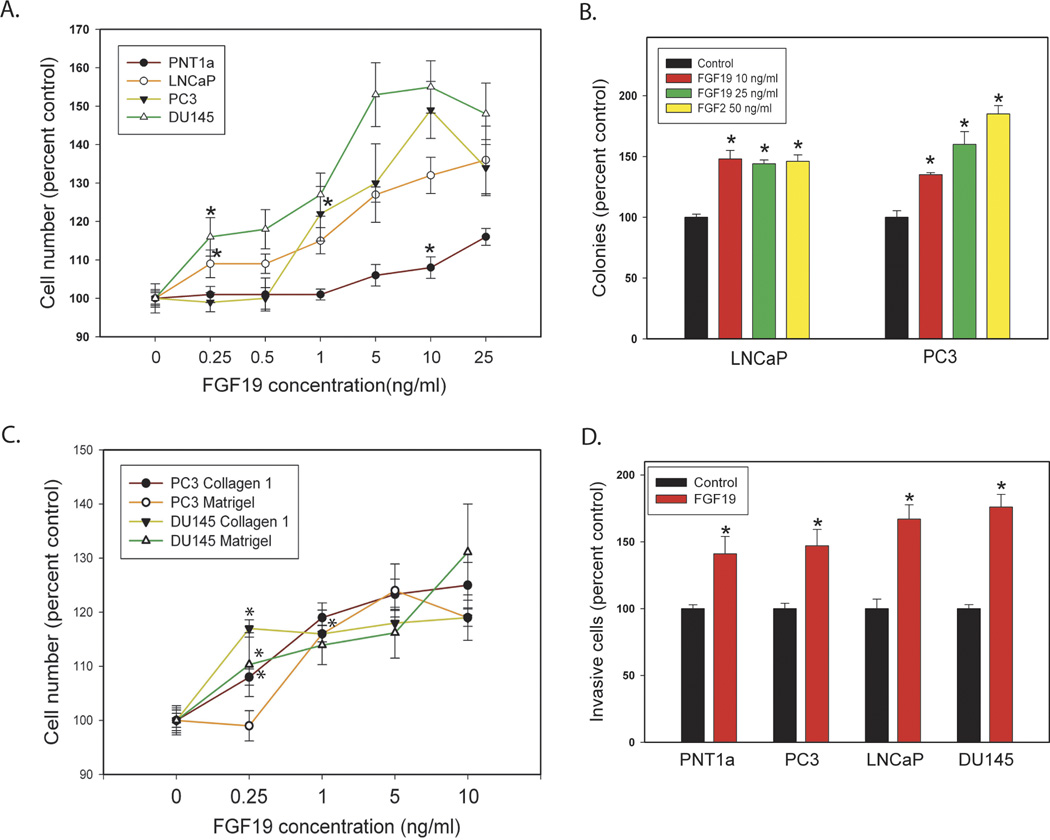
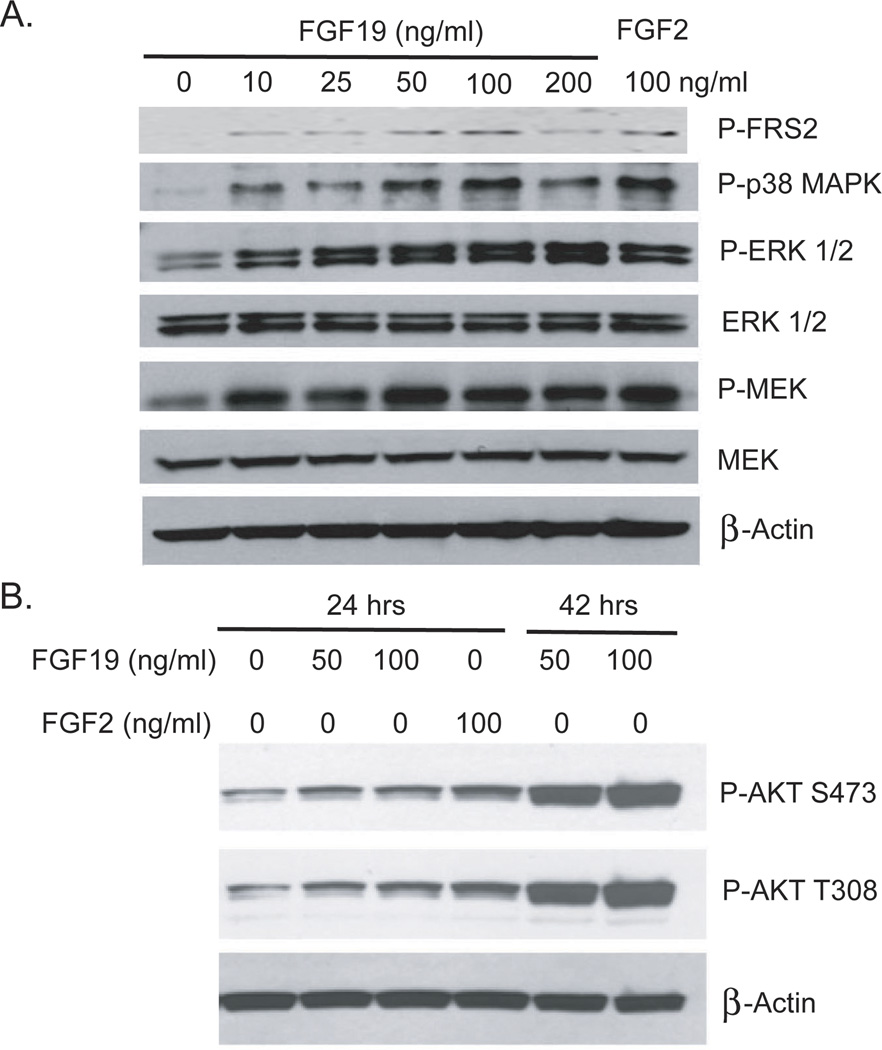
Prostate cancer is the most common visceral malignancy and the second leading cause of cancer deaths in US men. There is broad evidence that fibroblast growth factor (FGF) receptors are important in prostate cancer initiation and progression, but the contribution of particular FGFs in this disease is not fully understood. The FGF family members FGF19, FGF21, and FGF23 comprise a distinct subfamily that circulate in serum and act in an endocrine manner. These endocrine FGFs require α-Klotho (KL) and/or β-Klotho (KLB), two related single-pass transmembrane proteins restricted in their tissue distribution, to act as coreceptors along with classic FGF receptors (FGFR) to mediate potent biologic activity. Here we show that FGF19 is expressed in primary and metastatic prostate cancer tissues, where it functions as an autocrine growth factor. Exogenous FGF19 promoted the growth, invasion, adhesion, and colony formation of prostate cancer cells at low ligand concentrations. FGF19 silencing in prostate cancer cells expressing autocrine FGF19 decreased invasion and proliferation in vitro and tumor growth in vivo. Consistent with these observations, KL and/or KLB were expressed in prostate cancer cells in vitro and in vivo, raising the possibility that additional endocrine FGFs may also exert biologic effects in prostate cancer. Our findings support the concept that therapies targeting FGFR signaling may have efficacy in prostate cancer and highlight FGF19 as a relevant endocrine FGF in this setting.


Conflict of interest statement

The authors disclose no potential conflicts of interest.

Figures

Figure 1
Figure 1. FGF19 and Klotho co-receptors are expressed in prostate and PCa cell lines
A. FGF19 expression was determined by Q-RT-PCR using RNAs from the indicated cell lines. PNT1a and LNCaP were below the reliable detection limit of this assay. B. Expression of KL, KLB and FGFR4 in PNT1a and PCa cell lines by RT-PCR. HPRT is control for reverse transcription.
Figure 2
Figure 2. Biological effects of exogenous FGF19 on prostate and PCa cell lines
A. FGF19 induced proliferation. FGF19 was added to cultures of cells in serum free medium at the indicated concentration and cell number analyzed at either 48 or 72 hrs depending on the cell line. B. Colony formation in soft agar. FGF19, FGF2 or vehicle was added to cells in soft agar at the indicated concentration and colonies counted after 14 days. C. FGF19 induced cell adhesion. Numbers of cells adherent to collagen 1 or Matrigel were determined at the indicated FGF19 concentration as described in Materials and Methods. D. Invasion through Matrigel. Cell invasion though Matrigel was determined as described in Materials and Methods. Data is expressed relative to vehicle only control (100%). Mean +/− standard error of the mean (SEM) for triplicates are shown for all experiments. Asterisks indicate statistically significant differences. For A and C, the first statistically significant concentration for each cell line relative to control is indicated by an asterisk and all differences at higher concentrations are significant but asterisks are omitted for clarity.
Figure 3
Figure 3. FGF19 activates MAP kinase and AKT pathways in PCa cells
A. DU145 cells were serum starved for 24 hours and stimulated with the indicated concentration of FGF19 or FGF2 for 15 minutes. Cell lysates were prepared and Western blotting performed as described in Materials and Methods to assess activation of MAP kinase signaling. B. LNCaP cells were serum starved for 24 hours and then treated with the indicated concentration of FGF19 or FGF2 for either 24 or 42 hours. Cell lysates were prepared and Western blotting performed as described in Materials and Methods to assess activation of AKT.
Fig 4
Fig 4. Biological affects of FGF19 knockdown on PCa cells
A, B. FGF19 was transiently knocked down using two different shRNAs and mRNA levels evaluated by Q-RT-PCR. Proliferation and invasion were evaluated on parallel plates as described in Materials and Methods. A: DU145 cells; B: PC3 cells. C. FGF19 was stably knocked down using two different shRNAs. Protein expression was determined using an FGF19 ELISA. Proliferation and invasion were evaluated on parallel plates as described in Materials and Methods. For A–C, experimental data is expressed relative to vector control cells (100%). The mean +/− standard deviation is shown. D. PC3 cells with knockdown of FGF19 (FGF19 sh1, n=18) or vector controls (n=14) were inoculated subcutaneously on day 1. Calculated tumor volume is shown at intervals from day 13 to day 32. Mean +/− SEM is shown. Statistically significant differences from controls are indicated by asterisks for all experiments.
Figure 5
Figure 5. FGF19 is expressed in human PCa tissues
A. Quantitative RT-PCR for FGF19 mRNA expression was performed on 63 benign and 73 primary PCa samples. Relative expression after normalization to HPRT is shown (mean +/− SEM). The difference in FGF19 expression between benign and cancer samples was statistically significant (p=.025, t-test). B. Immunohistochemistry was performed as described in Materials and Methods. Upper left: Medium power view (200×) of primary PCa. Arrow shows normal epithelial cells. Upper right (200×) and lower left (100×): metastatic PCa lesions with strong FGF19 expression. Lower right: metastatic lesion with no FGF19 expression on same tissue microarray (100×).
Figure 6
Figure 6. Expression of Klotho co-receptor mRNAs in human prostate and PCa tissues
A. RT-PCR for KL and KLB mRNA in laser captured benign and cancer epithelium. HPRT is a reverse transcription control. B. KL mRNA levels across multiple tissue types (including prostate), for both cancer (upper panel) and normal (lower panel) tissues. Upper panel: BR: breast; CO: colon; KI: kidney; LU: lung; OV: ovarian; UT: uterine; PR: prostate C. Relative expression of KLB in normal/benign prostate tissue (“N”) versus prostate tumors (“T”), for four independent profiling datasets (surveyed using Oncomine).
Figure 7
Figure 7. Expression of Klotho protein by immunohistochemistry
Immunohistochemistry was performed as described in Materials and Methods. A. Kidney (positive control; 100×). B, Prostate cancer (100×); Cystic atrophy is present on the left. C. Prostate cancer (200×). Note membranous staining pattern. D. Normal epithelium with moderate staining (200×).

References

    1. Itoh N, Ornitz DM. Fibroblast growth factors: from molecular evolution to roles in development, metabolism and disease. J Biochem. 2011;149:121–130. - PMC - PubMed
    1. Harmer NJ, Pellegrini L, Chirgadze D, Fernandez-Recio J, Blundell TL. The crystal structure of fibroblast growth factor (FGF) 19 reveals novel features of the FGF family and offers a structural basis for its unusual receptor affinity. Biochemistry. 2004;43:629–640. - PubMed
    1. Kurosu H, Kuro OM. The Klotho gene family as a regulator of endocrine fibroblast growth factors. Mol Cell Endocrinol. 2009;299:72–78. - PubMed
    1. Fu L, John LM, Adams SH, Yu XX, Tomlinson E, Renz M, et al. Fibroblast growth factor 19 increases metabolic rate and reverses dietary and leptin-deficient diabetes. Endocrinology. 2004;145:2594–2603. - PubMed
    1. Choi M, Moschetta A, Bookout AL, Peng L, Umetani M, Holmstrom SR, et al. Identification of a hormonal basis for gallbladder filling. Nat Med. 2006;12:1253–1255. - PubMed

Publication types

MeSH terms