Review
. 2013 Jul;54(7):1761-75.
doi: 10.1194/jlr.R030833. Epub 2013 Feb 24.

Vitamin A and retinoid signaling: genomic and nongenomic effects

Affiliations
Review

Vitamin A and retinoid signaling: genomic and nongenomic effects

Ziad Al Tanoury et al. J Lipid Res. 2013 Jul.

Abstract

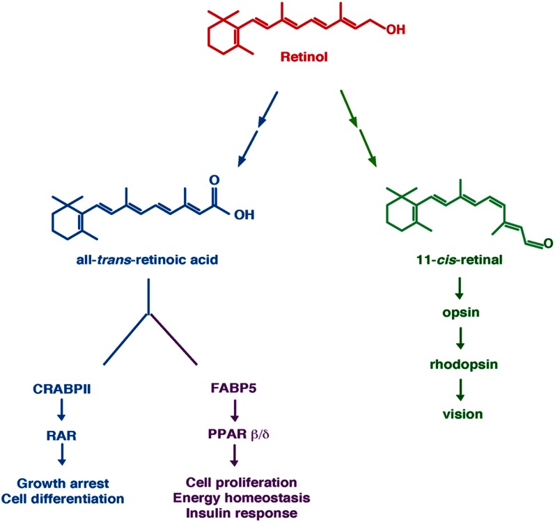
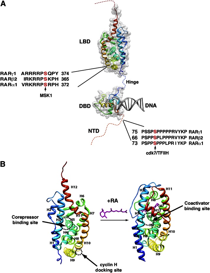
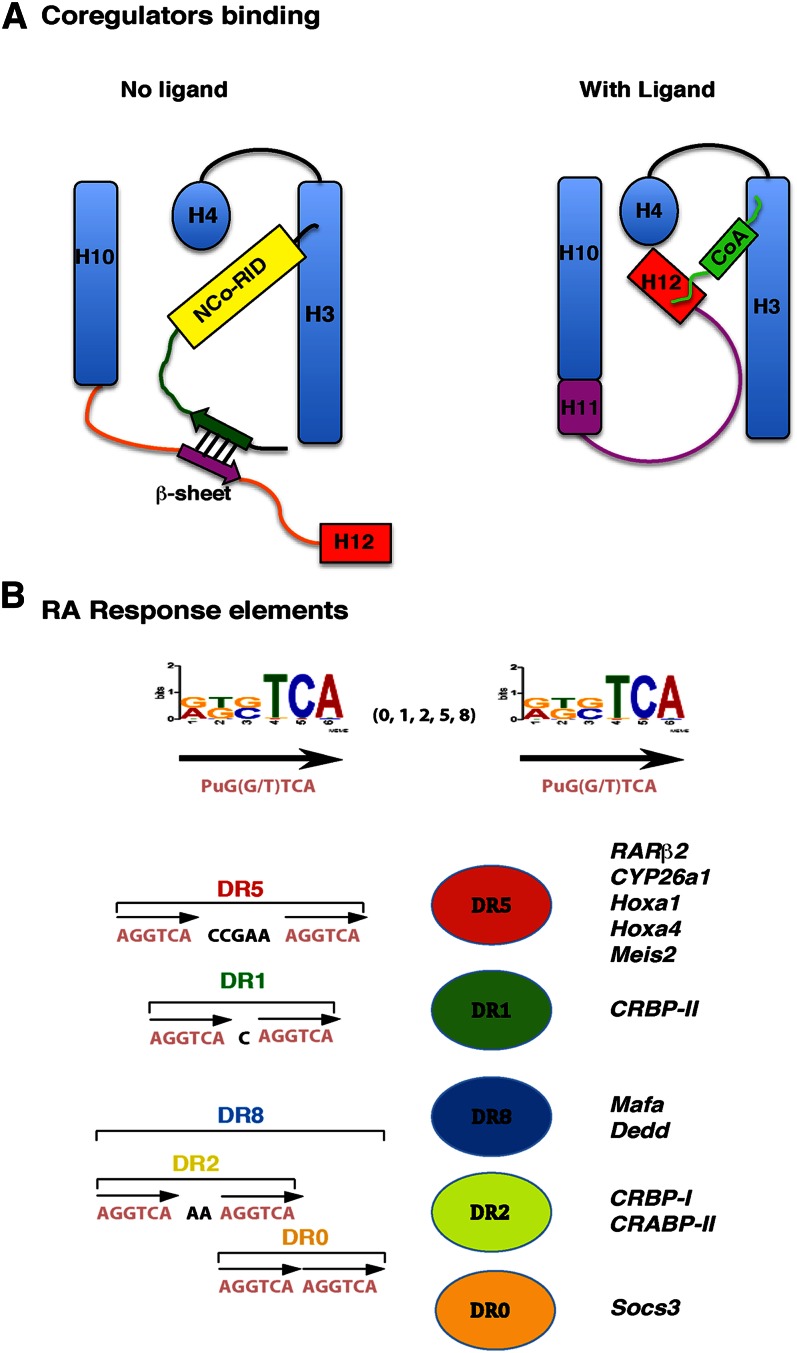
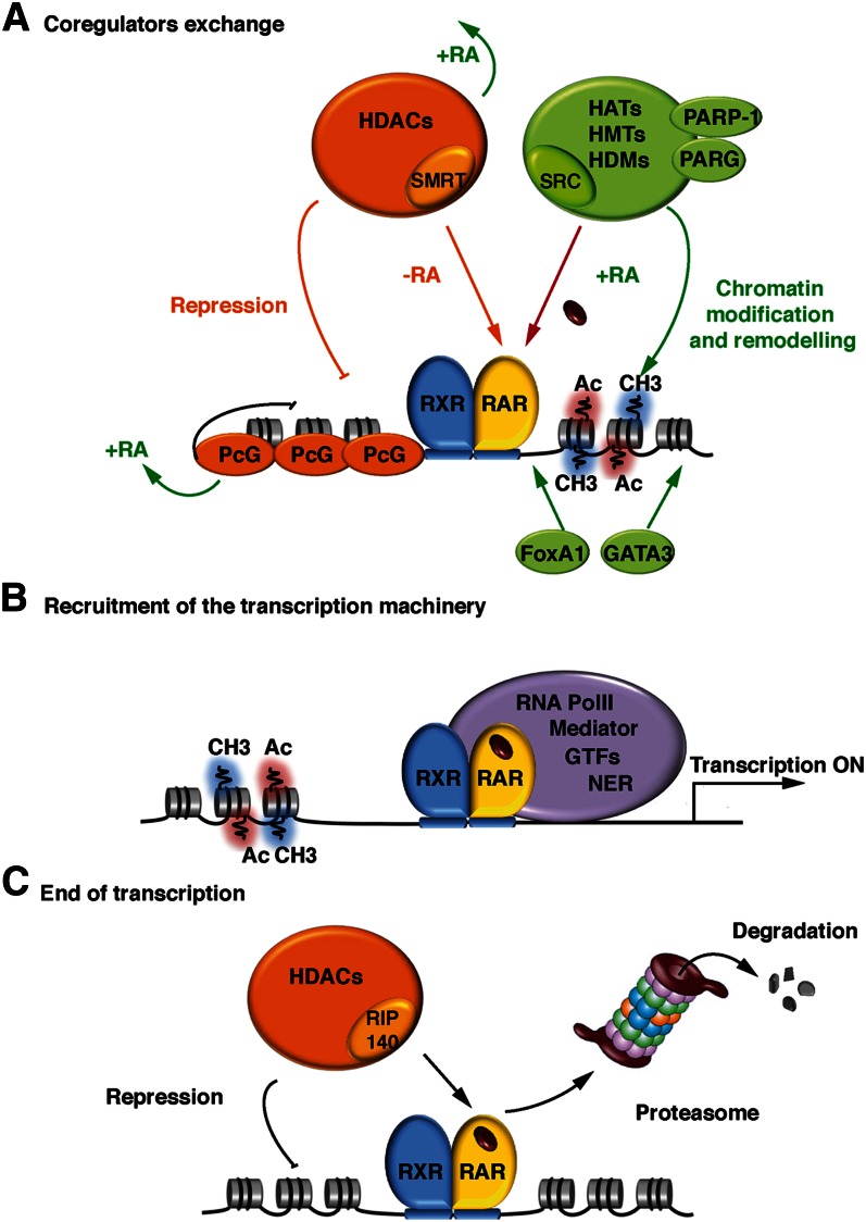
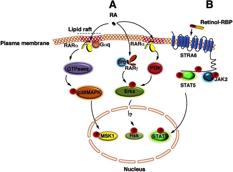
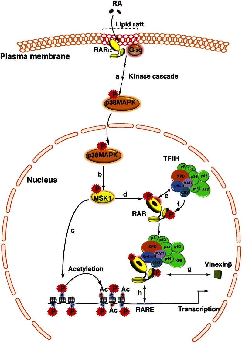
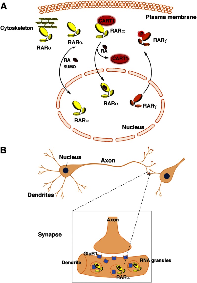
Vitamin A or retinol is arguably the most multifunctional vitamin in the human body, as it is essential from embryogenesis to adulthood. The pleiotropic effects of vitamin A are exerted mainly by one active metabolite, all-trans retinoic acid (atRA), which regulates the expression of a battery of target genes through several families of nuclear receptors (RARs, RXRs, and PPARβ/δ), polymorphic retinoic acid (RA) response elements, and multiple coregulators. It also involves extranuclear and nontranscriptional effects, such as the activation of kinase cascades, which are integrated in the nucleus via the phosphorylation of several actors of RA signaling. However, vitamin A itself proved recently to be active and RARs to be present in the cytosol to regulate translation and cell plasticity. These new concepts expand the scope of the biologic functions of vitamin A and RA.

Keywords: kinase cascade; nuclear receptor; retinoic acid receptor; retinoid X receptor; transcription.


Figures

Fig. 1.
Fig. 1.
Retinol and its two main metabolites, atRA and 11cRAL. AtRA can bind the intracellular lipid binding proteins CRABPII or FBP5. Binding to CRABPII channels RA to RARs, which then regulate genes involved in cell growth and differentiation. In contrast, binding to FABP5 channels RA to PPARβ/δ, which controls the expression of other subsets of genes involved in energy homeostasis and insulin response. 11cRAL serves as a cofactor for rhodopsin and is critical for vision. Adapted from Ref. .
Fig. 2.
Fig. 2.
RAR structure. (A) RARs depict a domain organization with an unstructured NTD and two well-structured domains: a central DBD and a C-terminal LBD. The phosphorylation sites located in the NTD and the LBD are shown. (B) Structural changes induced upon RA binding. The crystal structures of the unliganded RXRα and liganded RARγ LBDs are shown. Helices are represented as ribbons and labeled from H1 to H12. The binding domains for corepressors/coactivators and for cyclin H are shown. Adapted from Protein Data Bank 1lbd and 2lbd.
Fig. 3.
Fig. 3.
Coregulator binding surfaces and RA response elements. (A) Schematic models for the recruitment of corepressors and coactivators. Adapted from Ref. . (B) The classical retinoid response elements are composed of a direct repeat of the motif 5′-Pu G (G/T) TCA spaced by 0 (DR0), 1 (DR1), 2 (DR2), 5 (DR5), or 8 (DR8) base pairs. DR8 comprises three half-sites with DR2 and DR0 spacing. Some RARE-associated genes are shown.
Fig. 4.
Fig. 4.
Coregulator exchange at RXR/RAR heterodimers. (A) In the absence of ligand, RARα/RXR heterodimers bound to DNA are associated with corepressor complexes. Upon ligand binding, the corepressors dissociate, allowing the recruitment of coactivators and large complexes with enzymatic activities that decompact repressive chromatin. (B) When chromatin is decompacted, the transcriptional machinery, consisting of the Mediator, RNA PolII, the general transcription factors (GTF), and the nuclear excision repair (NER) factors, is recruited to the promoter, resulting in the initiation of transcription. (C) Transcription ends with the recruitment of nonconventional coactivators, such as RIP140, associated to large complexes with chromatin-repressing activity and/or through the degradation of RARs by the ubiquitin-proteasome system.
Fig. 5.
Fig. 5.
Extranuclear and nontranscriptional effects of RA and vitamin A. (A) A subpopulation of RARα is present in membrane lipid rafts and initiates cascades of kinase activations upon RA binding. In various epithelial and fibroblast cells, in response to RA, RARα localized in lipid rafts interacts with Gαq proteins and activates Rho-GTPases, p38MAPK, and MSK1. However, in neuronal cells, in response to RA, RARα activates Erks through the activation of the PI3K/Akt pathway. Whether Erks activate downstream effectors is unknown, but RSK2 would be an interesting candidate. Strikingly, in neuronal cells, RA has been also shown to activate Erks via RARγ in association with the Src kinase. (B) Vitamin A bound to RBP binds to the extracellular moiety of Stra6 and triggers the phosphorylation of its cytosolic domain. Phosphorylated STRA6 recruits and activates JAK2, which in turns phosphorylates STAT5. Then STAT5 translocates to the nucleus to regulate gene expression.
Fig. 6.
Fig. 6.
Crosstalk between the RA-activated p38MAPK pathway and the expression of RAR target genes. In response to RA, p38MAPK is activated (a), and then translocates into the nucleus and phosphorylates MSK1 (b). Activated MSK1 phosphorylates histones (c) and RARα at a serine located in the LBD (d). Subsequent to conformational changes, the cyclin H subunit of the CAK subcomplex of TFIIH is recruited to an adjacent domain (e), allowing the formation of a RARα/TFIIH complex and the phosphorylation of the NTD by the cdk7 kinase (f). In the case of the RARγ subtype, phosphorylation of the NTD promotes the dissociation of coregulators, such as vinexinß (g). Finally, phosphorylated RARα is recruited to response elements located in the promoter of target genes (h).
Fig. 7.
Fig. 7.
Unconventional presence of RARs out of the nucleus. (A) In certain cell types (Sertoli and hepatic stellate cells), RARs are sequestered in the cytosol upon phosphorylation, sumoylation, or interaction with proteins such as CART1 or cytoskeleton proteins. (B) RARα is present in the dentrites of neuronal cells, where it is associated to mRNA granules and controls translation.

References

    1. Harrison E. H. 2012. Mechanisms involved in the intestinal absorption of dietary vitamin A and provitamin A carotenoids. Biochim. Biophys. Acta. 1821: 70–77 - PMC - PubMed
    1. Clagett-Dame M., DeLuca H. F. 2002. The role of vitamin A in mammalian reproduction and embryonic development. Annu. Rev. Nutr. 22: 347–381 - PubMed
    1. Clagett-Dame M., Knutson D. 2011. Vitamin A in reproduction and development. Nutrients. 3: 385–428 - PMC - PubMed
    1. Altucci L., Gronemeyer H. 2001. The promise of retinoids to fight against cancer. Nat. Rev. Cancer. 1: 181–193 - PubMed
    1. McKenna N. J. 2012. EMBO retinoids 2011: mechanisms, biology and pathology of signaling by retinoic acid and retinoic acid receptors. Nucl. Recept. Signal. 10: e003. - PMC - PubMed

Publication types