. 2012;4(2):109-18.

Dilatation of the ascending aorta in patients with congenitally bicuspid aortic valves

Affiliations

Dilatation of the ascending aorta in patients with congenitally bicuspid aortic valves

F Robicsek et al. HSR Proc Intensive Care Cardiovasc Anesth. 2012.

Abstract

Introduction: The cause of ascending aortic dilatation occurring in patients with congenitally bicuspid aortic valves was investigated.

Methods: Flow patterns through human aortic roots with congenitally bicuspid aortic valves as well as through porcine constricted aortas were studied in a left heart simulator. Vibration was recorded as a measure of turbulence in the post-stenotic segment. Histological changes in fetal aortas with isolated congenitally bicuspid aortic valves were compared to fetal aortas with congenitally bicuspid aortic valves and hypoplastic left hearts, as well as to normal fetal aortas with tricuspid aortic valves.

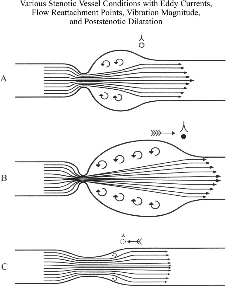
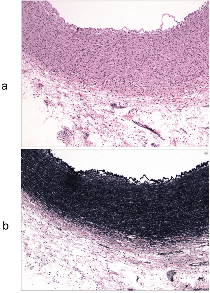
Results: Congenitally bicuspid aortic valves were anatomically stenotic even in the absence of pressure gradients and without history of relevant symptoms. Histology of the aortic wall in isolated fetal congenitally bicuspid aortic valves was similar to that of fetal aortas with normal tri-leaflet aortic valves, but was abnormal if congenitally bicuspid aortic valves was associated with other cardiovascular anomalies. Flow studies revealed that turbulence and vibration in the post-stenotic aortic segments generated by the stenosis were proportional to the degree of the narrowing.

Conclusions: Congenitally bicuspid aortic valves are inherently stenotic, asymmetrical, generate turbulence and vibration. This not only leads to early failure but also to injury of the ascending aortic wall and ascending aortic dilatation. The more progressive form of ascending aortic dilatation occurs in patients where congenitally bicuspid aortic valves is combined with other inborn anomalies and may require a radical procedure (replacement).

Keywords: aortic aneurysm; bicuspid aortic valve.


Conflict of interest statement

Conflict of interest None declared.

Figures

Figure 1
Figure 1
Three congenitally bicuspid aortic roots obtained from young individuals who had no symptoms of heart disease and died of non-cardiac causes shown in full diastole (a) and in full systole (b). None of the roots had any measurable trans-valvular pressure gradients. Note the morphological stenosis of various degrees in every specimen. Casts obtained in full diastole (c) shows creases and wrinkles in full diastole.
Figure 2
Figure 2
Valve C. Ultrasound image showing asymmetrical, elliptical and narrow (57% of the annulus area) leaflet opening.
Figure 3
Figure 3
2D ultrasound images of flow through a vascular stenosis show:
  1. The initial jet flow through the stenosis.

  2. The development of a recirculation zone.

  3. The region of reattachment to the aortic wall.

Figure 4
Figure 4
Extension of "functional" stenosis past the post-stenotic area (a) and formation of eddy-currents indicated by residual dye in a hydraulic model. Stenosis with post-stenotic dilatation (c) also shows extended "functional stenosis and increase in eddy-currents and turbulence" (from Robicsek F, et al: The post-stenotic dilatation of great vessels Acta Med Scand 1954; 155: 481-484).
Figure 5
Figure 5
Symmetrical opening (shaded, clover-shaped area showing maximal leaflet opening) of the trileaflet aortic valves with three even sinuses and leaflets expanding beyond the circumference of the annulus (dotted reversed cone) and the asymmetrical and incomplete (shaded ellipse) opening of the congenitally bicuspid aortic valve with two uneven sinuses. In the latter, the flow-jet impacts the aortic wall. TAV = trileaflet aortic valves ; CBAV = congenitally bicuspid aortic valve
Figure 6
Figure 6
Vibration recordings at 1, 2, 3, and 5 cm from the stenosis. Strongest signal occurs at a frequency of 110 Hz. Vibration is higher at 1 and 2 cm from the stenosis when the pressure gradient is 38 mmHg. Vibration is higher at 3 and 5 cm when the gradient is 56 mmHg.
Figure 7
Figure 7
Different degrees of aortic stenoses, formation of eddy currents, flow reattachment regions and different magnitudes of vibration, leading to development of poststenotic dilatation.
Figure 8
Figure 8
Digital re-creation of the flow pattern through bicuspid aortic root and aortic arch.
Figure 9
Figure 9
Shear-stress values measured in an aortic arch with a bicuspid aortic valve. Note the high values on the right-lateral aspect of the ascending aorta.
Figure 10
Figure 10
Ascending aorta of a fetus with normal tri-leaflet aortic valve. Note the orderly arrangement of smooth muscle cells of the media, thin intima and diffuse, homogeneous elastin staining throughout the entire thickness of the media. 2.5 X magnification.
  1. Hematoxylin eosin stain.

  2. Movat pentachrome stain.

Figure 11
Figure 11
Ascending aorta of a fetus with congenitally bicuspid aortic valve as the only cardiovascular abnormality. It represents a histological picture similar to those of a fetal aorta with a normal tri-leaflet aortic valve. 10 X magnification.
  1. Hematoxylin eosin stain.

  2. Movat pentachrome stain.

Figure 12
Figure 12
Ascending aortic wall of a fetus with congenitally bicuspid aortic valve and hypoplastic left heart syndrome. Note the relative thickening of the aortic wall and focally disordered architecture on both the H&E and elastin stains in an area of relative stenosis of the aorta. 10 X magnification.
  1. Hematoxylin eosin stain.

  2. Movat pentachrome stain.

Figure 13
Figure 13
Ascending aorta of a 5 y.o. boy with congenitally bicuspid valve as the only cardiovascular anomaly. Note the intimal damage in the aortic root caused by turbulent flow (a) and the disintegration of the elastic elements in the ascending aortic wall (b) 25X magnification. Movat pentachrome stain.

References

    1. Roberts W C. The congenitally bicuspid aortic valve. A study of 85 autopsy cases. Am J Cardiol. 1970;26:72–83. - PubMed
    1. Roberts W C. The structure of the aortic valve in clinically isolated aortic stenosis: an autopsy study of 162 patients over 15 years of age. Circulation. 1970;42:91–97. - PubMed
    1. Larson E W, Edwards W D. Risk factors for aortic dissection: a necropsy study of 161 cases. Am J Cardiol. 1984;53:849–855. - PubMed
    1. Stewart W J, King M E, Gilliam L D. et al. Prevalence of aortic valve prolapse with bicuspid aortic valve and its relation to aortic regurgitation: a cross-sectional echocardiographic study. Am J Cardiol 1984; 54: 1277-82. 1984;54:1277–1282. - PubMed
    1. Kuralay E, Demirkilic U, Ozal E. et al. Surgical approach to ascending aorta in bicuspid aortic valve. J Card Surg 2003; 18: 173-80. 2003;18:173–180. - PubMed

LinkOut - more resources