. 2013 May 15;591(10):2523-40.
doi: 10.1113/jphysiol.2012.243659. Epub 2013 Feb 25.

Glutamatergic modulation of synaptic-like vesicle recycling in mechanosensory lanceolate nerve terminals of mammalian hair follicles

Affiliations

Glutamatergic modulation of synaptic-like vesicle recycling in mechanosensory lanceolate nerve terminals of mammalian hair follicles

Robert W Banks et al. J Physiol. .

Abstract

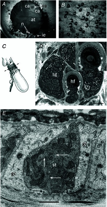
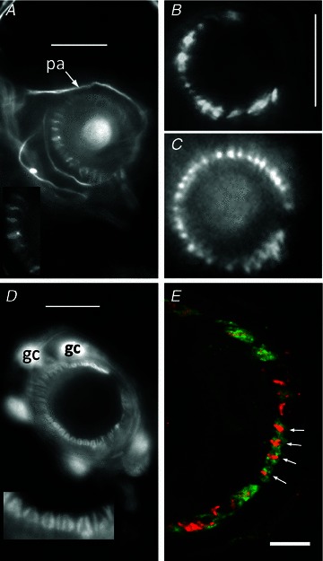
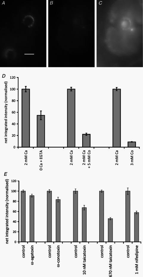
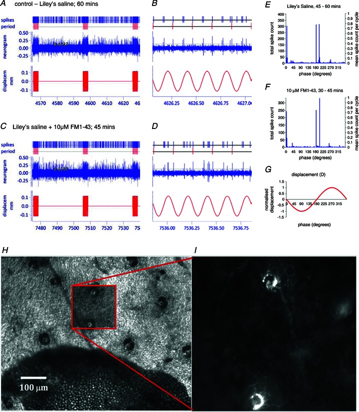
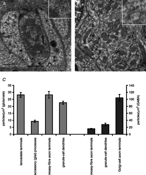
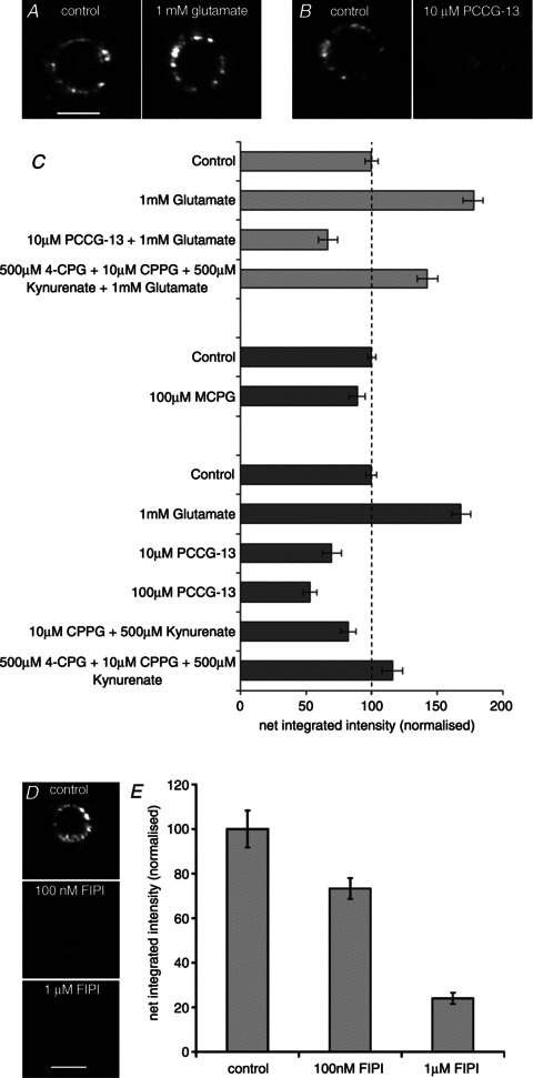
Our aim in the present study was to determine whether a glutamatergic modulatory system involving synaptic-like vesicles (SLVs) is present in the lanceolate ending of the mouse and rat hair follicle and, if so, to assess its similarity to that of the rat muscle spindle annulospiral ending we have described previously. Both types of endings are formed by the peripheral sensory terminals of primary mechanosensory dorsal root ganglion cells, so the presence of such a system in the lanceolate ending would provide support for our hypothesis that it is a general property of fundamental importance to the regulation of the responsiveness of the broad class of primary mechanosensory endings. We show not only that an SLV-based system is present in lanceolate endings, but also that there are clear parallels between its operation in the two types of mechanosensory endings. In particular, we demonstrate that, as in the muscle spindle: (i) FM1-43 labels the sensory terminals of the lanceolate ending, rather than the closely associated accessory (glial) cells; (ii) the dye enters and leaves the terminals primarily by SLV recycling; (iii) the dye does not block the electrical response to mechanical stimulation, in contrast to its effect on the hair cell and dorsal root ganglion cells in culture; (iv) SLV recycling is Ca(2+) sensitive; and (v) the sensory terminals are enriched in glutamate. Thus, in the lanceolate sensory ending SLV recycling is itself regulated, at least in part, by glutamate acting through a phospholipase D-coupled metabotropic glutamate receptor.


Figures

Figure 1
Figure 1. Lanceolate nerve ending in a hair follicle of mouse pinna labelled with FM1-43, showing the annulus used to define a region of interest (ROI) for quantitative analysis of fluorescence intensity
Epifluorescence image. The diameter of the annulus outer circle was set at 30 μm in all experiments. The inner circle diameter was set to ensure that the annulus enclosed all terminals analysed. This was either at 10 μm, as shown here, or at 12.5 μm and kept constant throughout a particular experiment. An annular ROI, rather than a circle, best approximated the area containing terminal labelling and also excluded from the analysis the diffuse autofluorescent spot often found at the base of the hair shaft.
Figure 2
Figure 2. Hair follicles of the anterior skin of the mouse pinna
A, the mouse pinna preparation pinned, anterior skin face down, in a Sylgard-lined dish filled with Liley's solution, and set up for electrophysiological recording. The whole posterior skin has been removed, as well as a large area of elastic cartilage and adipose tissue (at) from the cleared area (ca). By folding the cleared area back, access was gained to the hair shafts, allowing 2 or 3 within the vibrated area (va) to be mechanically displaced by a fire-polished glass capillary (not shown). The nerves (n) are branches of the mandibular division of the trigeminal and are set up for differential recording of the neurogram using recording (re) and indifferent (ie) suction electrodes. B, bright-field image of mouse pinna skin viewed from the dermal side, showing several hair follicles. The bases of the hair shafts are clearly seen; each shaft is partly surrounded by a sebaceous gland that appears dark. Scale bar indicates 100 μm. C, diagram of the structure and location of the innervation of a hair follicle. The lanceolate ending consists of the group of terminals forming the palisade-like structure immediately below the lobular sebaceous gland (from Bannister, 1976). The dashed line indicates the typical plane of section for subsequent images for fluorescence and light microscopy. D, semi-thin (1 μm) cross-section through a hair follicle (hf) at the level of the sebaceous gland (sg) and lanceolate ending, as indicated in C. The lanceolate ending surrounds the follicle, and terminals appear as dark structures alternating between lighter accessory cells, shown in greater detail in the inset. Mouse pinna, Toluidine Blue; scale bar (main image) indicates 20 μm. E, electron micrograph of an ultrathin cross-section through a lanceolate ending, showing a single, darkly stained, sensory terminal (st) almost completely enclosed by pale-staining glial cell (gc) processes. Note the numerous 50-nm-diameter vesicle profiles in the terminal axoplasm (white arrows). Mouse pinna; scale bar indicates 0.5 μm.
Figure 3
Figure 3. Immunohistochemical and genetic identification of the sensory terminals and glial cells of lanceolate nerve endings
A, anti-neurofilament protein (NFP)-like immunoreactivity is localized in structures identified as preterminal axons (pa) and sensory terminals in a mouse pinna follicle. Several terminals are shown enlarged in the inset. Epifluorescence; scale bar indicates 10 μm. B, structures identified as sensory terminals also react strongly with anti-synapsin I antibody. Mouse pinna, epifluorescence; scale bar indicates 20 μm. C, synaptopHluorin shows the expression of the v-SNARE synaptobrevin in the lanceolate terminals in a very similar pattern to NFP and synaptophysin. Mouse pinna, epifluorescence; scale as in B. D, anti-S-100 antibody, in contrast, labels paired structures identified as glial cells (gc) and their processes in a mouse pinna follicle. Pairing of the processes is particularly apparent in the enlarged inset and is distinct from the unpaired processes seen in A. Epifluorescence; scale bar indicates 10 μm. E, a follicle from rat pinna double labelled with antibodies against synaptophysin (red) and S-100 (green). Where the ending is precisely orthogonal within the section (white arrows), individual red profiles can be seen clearly to be almost entirely enclosed by paired green profiles, identified as sensory terminals and glial cell processes, respectively. Laser-scanning confocal microscopy; scale bar indicates 5 μm.
Figure 4
Figure 4. FM1-43 labelling of lanceolate endings is Ca2+ dependent
AC, 5 mm Co2+ has a strong inhibitory effect on labelling; epifluorescence. A, control (2 mm Ca2+); scale bar indicates 20 μm. B, 2 mm Ca2++ 5 mm Co2+, imaged in the same conditions as A. C, the same as B, with the brightness of the image greatly increased to show the low level of labelling present. D, histograms summarizing the quantitative data on effects of the following permutations: removal of external Ca2+ (left two columns); the addition of 5 mm Co2+ (middle two columns); and the replacement of external Ca2+ with 3 mm Co2+ (right two columns). Histograms show means ± SEM. (Statistics for the removal of external Ca2+: 2 pairs of preparations, each of 10 replicates, between preparations, F1,39= 38.3, P < 0.001; between pairs, F1= 10.3, P < 0.01; statistics for 5 mm Co2+: 4 pairs, 10 replicates, between preparations, F1,79= 474.2, P < 0.001; between pairs, F3= 1.85, n.s.; and statistics for replacing Ca2+/Mg2+ with 3 mm Co2+: 4 pairs, 10 replicates, between preparations, F1,79= 1020, P < 0.001; between pairs, F3= 0.15, n.s.). E, in part, the Ca2+ dependence of FM1-43 labelling of lanceolate endings is due to L-type channels, as shown by the differential effects of peptide blockers and nifedipine. Histograms (means ± SEM) summarize the quantitative data from separate experiments on the effects (left to right) of the P/Q-type channel blocker ω-agatoxin IVA, the N-type channel blocker ω-conotoxin GVIA, the L-type channel and SK channel blocker taicatoxin, and the L-type channel blocker nifedipine. [Statistics are as follows: (i) ω-agatoxin IVA: 4 pairs, 10 replicates, between preparations, F1,79= 7.28, P < 0.01; between pairs, F3= 4.48, P < 0.01; (ii) ω-conotoxin GVIA: 4 pairs, 10 replicates, between preparations, F1 79= 13.97, P < 0.001; between pairs, F3= 8.3, P < 0.001; (iii) taicatoxin,10 nm, Student's t= 5.18, P < 0.001 for 71 d.f.; 670 nm, 4 pairs, 10 replicates, between preparations F1,79= 436.6, P < 0.001; between pairs, F3= 9.33, P < 0.001; and (iv) nifedipine, Student's t = 6.01, P < 0.001].
Figure 5
Figure 5. Evidence that little, if any, FM1-43 labelling of lanceolate endings involves permeation of the mechanosensory channel
A, histograms (means ± SEM) summarize the quantitative data from separate experiments on the effects of different levels of external [Ca2+]. There is no, or only weak, inhibition of FM1-43 labelling in the presence of elevated external [Ca2+]. (Statistics for 3 quadruple preparations, 10 replicates, between preparations, F3,119= 26.1, P < 0.001; between quadruples, F2= 5.11, P < 0.01; Fisher's test vs. control, 0 Ca2+/3 mm Mg2+, P < 0.01; 5 mm Ca2+, n.s.; and 10 mm Ca2+, P < 0.05). B, lanceolate endings labelled with FM1-43 destain spontaneously when placed in standard Liley's fluid (control) and more rapidly when placed in Liley's fluid containing 3 nmα-latrotoxin (latrotoxin) to stimulate exocytosis. Epifluorescence; scale bar indicates 20 μm. C, the time-dependent loss of fluorescence can be fitted with single-exponent curves. The rate constant in the presence of latrotoxin is more than twice that of the control. Statistical comparison at individual times: Student's t, P < 0.01 at 15 min and P < 0.05 the rest.
Figure 6
Figure 6. Hair follicle afferent responses are highly phasic and unaffected by 10 μm FM1-43
A and C, samples lasting a little over 60 s, including 3 × 3 s periods of sinusoidal displacement of a small number of hair shafts, taken from the continuously recorded neurogram from a single pinna after approximately 60 min in standard Liley's solution (A) and after a further 45 min in Liley's solution with 10 μm FM1-43 (C). In each case, a 1 s interval from the last period of sinusoidal stimulation is shown on an expanded time scale to the right of the sample (B and D). Recordings were made with a 1401micro running Spike2 (Cambridge Electronic Design). Custom script files were written to mark individual events that crossed a threshold set by the horizontal cursor, for offline analysis. E and F, cycle histograms in 3 deg bins constructed from approximately 15 min sections of the neurogram, samples of which are given in AD, showing the phase relationships of responses of presumed lanceolate endings to mechanical stimulation of 2–4 hair shafts by sinusoidal displacement of a glass probe, shown in G. Note the marked phase locking in both histograms. E, the last change of standard Liley's solution prior to the addition of FM1-43 (406 complete sinusoidal displacements, ending with those in sample A). F, the last change of Liley's solution containing 10 μm FM1-43 (336 complete sinusoidal displacements, ending with those in sample D). G, normalized probe displacement; the amplitude was approximately 100 μm (see AD). H and I, part of a pinna preparation at the end of the electrophysiological experiment above showing (H) several hair follicles visible in bright-field illumination in the area cleared down to the dermis of the anterior skin. Dark, often bilobed shapes are sebaceous glands. I, enlarged view of the area marked by the box in H imaged with epifluorescence, showing two lanceolate endings labelled with FM1-43. Hair shafts were accessed by folding the free edge of the pinna over to bring the epidermis of the anterior skin uppermost for mechanical stimulation. Similar results were found in two further preparations.
Figure 7
Figure 7. Lanceolate sensory terminals are enriched in glutamate
A and B, electron micrographs of thin sections of a sensory lanceolate terminal (st) with enclosing glial cells (gc; A) and of a cerebellar cortical synaptic glomerulus (B) immunogold labelled to show glutamate-like immunoreactivity. A portion of each is enlarged in the insets, showing gold particles more clearly. The cerebellar glomerulus includes a mossy fibre terminal (mf) and several granule cell dendrites (d). Scale bars (main images) indicate 0.5 μm. C, histograms (means ± SEM) summarizing the quantitative assessment of glutamate-like immunoreactivity (light grey; left) and GABA-like immunoreactivity (dark grey; right). No GABA-like immunoreactivity was detected in lanceolate terminals or glial cells. (Statistics for glutamate, F3,240= 42.6; Fisher's test vs. lanceolate terminals: accessory (glial) cells, P < 0.01; mossy fibre terminals, n.s.; and granule cells, P < 0.01).
Figure 8
Figure 8. FM1-43 labelling of lanceolate sensory terminals is modulated by glutamate acting through a phospholipase-D-linked metabotropic glutamate receptor (mGluR)
A, FM1-43 labelling is enhanced in the presence of external glutamate. B, FM1-43 labelling is reduced in the presence of PCCG-13, a specific blocker of a non-canonical phospholipase-D-linked mGluR. In A and B, scale bar indicates 20 μm. C, histograms (means ± SEM) summarizing the quantitative data on the effects of external glutamate, alone or in the presence of various ionotropic and mGluR blockers (light grey; top) and, in a separate set of experiments, the effects of the blockers alone (dark grey; bottom). PCCG-13 is much more potent than blockers of the canonical glutamate receptors. [Statistics: (i) glutamate and blockers: 2 quadruple preparations, 7 replicates, between preparations, F3,55= 55.6, P < 0.001; between quadruples, F1= 2.61, n.s., Fisher's test P < 0.01, for all possible comparisons; (ii) MCPG alone: 2 double preparations, 22 replicates, between preparations, F1,87, n.s.; (iii) PCCG-13 and blockers of ionotropic glutamate receptor and mGluRs alone: F5,115= 36.2, Fisher's test, 10 μm PCCG-13, P < 0.05 and 100 μm PCCG-13, P < 0.01; all other blockers, n.s.]. D, FM1-43 labelling is reduced in the presence of FIPI, an inhibitor of phospholipase D. Scale bar indicates 20 μm. E, histograms (means ± SEM) summarizing the quantitative data showing that FIPI caused a dose-dependent decrease in FM1-43 labelling. (Statistics: F2,150= 24.1, Fisher's test, all comparisons, P < 0.01). A, B and D, epifluorescence.

References

    1. Albani-Torregrossa S, Attucci S, Marinozzi M, Pellicciari R, Moroni F, Pellegrini-Giampietro DE. Antagonist pharmacology of metabotropic glutamate receptors coupled to phospholipase D activation in adult rat hippocampus: focus on (2R,1′S,2′R,3′S)-2-(2′-carboxy-3′-phenylcyclopropyl) glycine versus 3,5-dihydroxyphenylglycine. Mol Pharmacol. 1999;55:699–707. - PubMed
    1. Andres KH. Über die Feinstruktur des Rezeptoren an Sinushaaren. Z Zellforsch Microsc Anat. 1966;75:339–365. - PubMed
    1. Banks RW. The distribution of static γ-axons in the tenuissimus muscle of the cat. J Physiol. 1991;442:489–512. - PMC - PubMed
    1. Banks RW, Bewick GS, Reid B, Richardson C. Evidence for activity-dependent modulation of sensory-terminal excitability in spindles by glutamate release from synaptic-like vesicles. Adv Exp Med Biol. 2002;508:13–18. - PubMed
    1. Bannister LH. Sensory terminals of peripheral nerves. In: Landon DH, editor. The Peripheral Nerve. London: Chapman and Hall; 1976. pp. 369–463.

Publication types