Review
. 2013 Feb 21:7:25.
doi: 10.3389/fncir.2013.00025. eCollection 2013.

Structural plasticity of climbing fibers and the growth-associated protein GAP-43

Affiliations
Review

Structural plasticity of climbing fibers and the growth-associated protein GAP-43

Giorgio Grasselli et al. Front Neural Circuits. .

Abstract

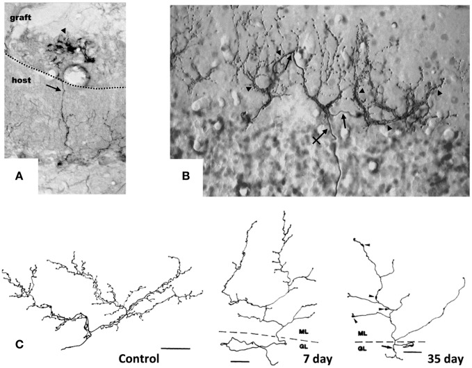
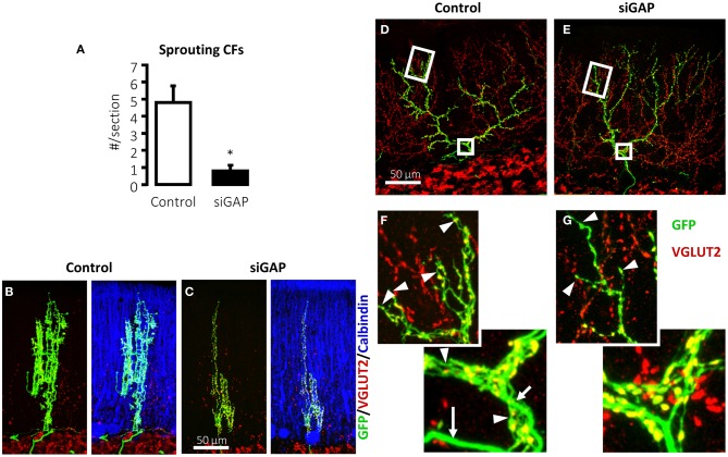
Structural plasticity occurs physiologically or after brain damage to adapt or re-establish proper synaptic connections. This capacity depends on several intrinsic and extrinsic determinants that differ between neuron types. We reviewed the significant endogenous regenerative potential of the neurons of the inferior olive (IO) in the adult rodent brain and the structural remodeling of the terminal arbor of their axons, the climbing fiber (CF), under various experimental conditions, focusing on the growth-associated protein GAP-43. CFs undergo remarkable collateral sprouting in the presence of denervated Purkinje cells (PCs) that are available for new innervation. In addition, severed olivo-cerebellar axons regenerate across the white matter through a graft of embryonic Schwann cells. In contrast, CFs undergo a regressive modification when their target is deleted. In vivo knockdown of GAP-43 in olivary neurons, leads to the atrophy of their CFs and a reduction in the ability to sprout toward surrounding denervated PCs. These findings demonstrate that GAP-43 is essential for promoting denervation-induced sprouting and maintaining normal CF architecture.

Keywords: GAP-43; atrophy; branching; climbing fiber; sprouting.


Figures

Figure 1
Figure 1
Target-dependent structural plasticity of CFs. (A) Sprouting of adult CFs 7 days following transplantation of embryonic cerebellum on to the surface of the adult cerebellar cortex. The sprouts (arrows) innervate PCs in the graft (arrowheads). Dotted line: host-graft border (Rossi et al., , unpublished). (B) CF sprouting and innervation of adjacent PCs previously denervated by a partial lesion of the IO, shown 1 year after the lesion on a sagittal plane. From 1 olivo-cerebellar axon (crossed arrow), a CFs is formed with collateral branches (arrows) giving rise to new CF-like structures (arrowheads) innervating the adjacent PCs (Rossi et al., 1991b). (C) A control CF and atrophic modification induced in CFs 7 and 35 days after deletion of PCs. CFs were reconstructed using a camera lucida. GL, granular layer; ML, molecular layer. Scale bar: 30 μm, 25 μm, and 25 μm, respectively, for control, 7 and 35 days (Rossi et al., 1993). CFs were labeled by PHA-l axon tracing, PCs by anti-calbindin immunostaining.
Figure 2
Figure 2
Silencing of GAP-43 in CFs prevents its sprouting and induces atrophy. (A) Sprouting in GFP-positive CFs, induced by a sub-total lesion of the IO, is dramatically reduced to nearly null levels in rats treated with GAP-43-silencing vectors (siGAP) 3 weeks before the lesion compared with controls as observed on coronal sections (N = 3 and 5 animals, respectively; *p < 0.05; mean ± SEM). (B,C) The total extension of CFs that are still able to grow sprouts following GAP-43 silencing was also significantly reduced as assessed on coronal sections. (D,E) Representative confocal images of rat CFs in sagittal sections under normal conditions 3 weeks after treatment with control or silencing vectors. (F,G) Details of the most distal segment and first main branching point of CFs shown in (D,E), showing a reduction in number and length of tendrils and consequent decrease in the density of varicosities. Arrows: thick axonal stalks; arrowheads: examples of tendrils [GFP, green; VGLUT2, red; calbindin, blue; modified from Grasselli et al. (2011)].

Similar articles

Cited by

References

    1. Aigner L., Arber S., Kapfhammer J. P., Laux T., Schneider C., Botteri F., et al. (1995). Overexpression of the neural growth-associated protein GAP-43 induces nerve sprouting in the adult nervous system of transgenic mice. Cell 83, 269–278 10.1016/0092-8674(95)90168-X - DOI - PubMed
    1. Allegra Mascaro A. L., Sacconi L., Pavone F. S. (2010). Multi-photon nanosurgery in live brain. Front. Neuroenergetics 2:21 10.3389/fnene.2010.00021 - DOI - PMC - PubMed
    1. Benedetti F., Montarolo P. G., Strata P., Tosi L. (1983). Collateral reinnervation in the olivocerebellar pathway in the rat, in Birth Defects Original Article Series, eds Haber P.-P. J., Hashim B., Giuffrida-Stella Am G. (New York, NY: Alan Liss, Inc.), 461–464 - PubMed
    1. Benowitz L. I., Rodriguez W. R., Neve R. L. (1990). The pattern of GAP-43 immunostaining changes in the rat hippocampal formation during reactive synaptogenesis. Brain Res. Mol. Brain Res. 8, 17–23 - PubMed
    1. Biewenga J. E., Schrama L. H., Gispen W. H. (1996). Presynaptic phosphoprotein B-50/GAP-43 in neuronal and synaptic plasticity. Acta Biochim. Pol. 43, 327–338 - PubMed

LinkOut - more resources