. 2013;8(2):e56316.
doi: 10.1371/journal.pone.0056316. Epub 2013 Feb 18.

The mitogen-activated protein kinase p38α regulates tubular damage in murine anti-glomerular basement membrane nephritis

Affiliations

The mitogen-activated protein kinase p38α regulates tubular damage in murine anti-glomerular basement membrane nephritis

Ralf Müller et al. PLoS One. 2013.

Abstract

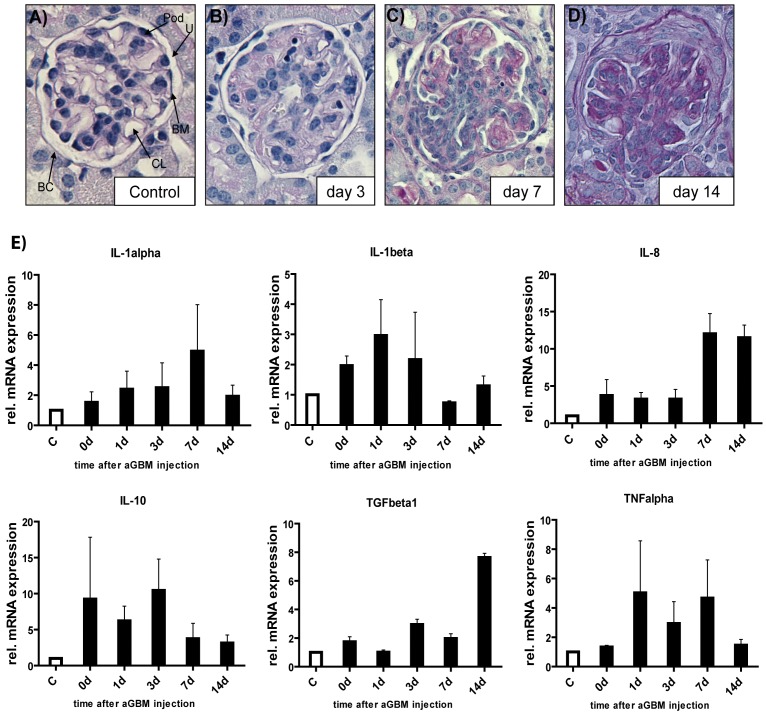
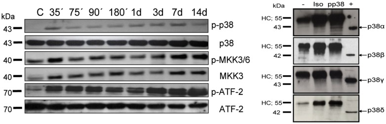
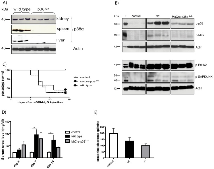
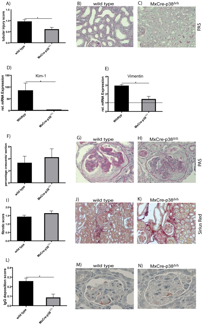
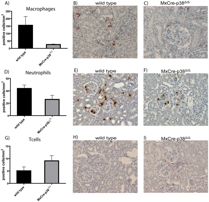
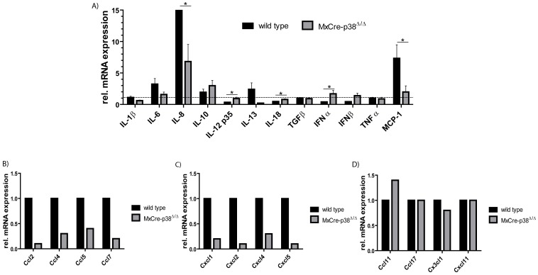
p38 mitogen-activated protein kinase (MAPK) is thought to play a central role in acute and chronic inflammatory responses. Whether p38MAPK plays a pathogenic role in crescentic GN (GN) and which of its four isoforms is preferentially involved in kidney inflammation is not definitely known. We thus examined expression and activation of p38MAPK isoforms during anti-glomerular basement membrane (GBM) nephritis. Therefore, p38α conditional knockout mice (MxCre-p38α(Δ/Δ)) were used to examine the role of p38α in anti-GBM induced nephritis. Both wild type and MxCre-p38α(Δ/Δ) mice developed acute renal failure over time. Histological examinations revealed a reduced monocyte influx and less tubular damage in MxCre-p38α(Δ/Δ) mice, whereas glomerular crescent formation and renal fibrosis was similar. Likewise, the levels of pro- and anti-inflammatory cytokines such as TNF, IL-1 and IL-10 were similar, but IL-8 was even up-regulated in MxCre-p38α(Δ/Δ) mice. In contrast, we could detect strong down-regulation of chemotactic cytokines such as CCL-2, -5 and -7, in the kidneys of MxCre-p38α(Δ/Δ) mice. In conclusion, p38α is the primary p38MAPK isoform expressed in anti-GBM nephritis and selectively affects inflammatory cell influx and tubular damage. Full protection from nephritis is however not achieved as renal failure and structural damage still occurs.


Conflict of interest statement

Competing Interests: The authors have declared that no competing interests exist.

Figures

Figure 1
Figure 1. Functionality of the anti- GBM nephritis model and differential cytokine activation.
Periodic acid-Schiff reagent (PAS) stain of a control kidney (A) and kidneys affected by crescentic GN over time are shown. BC = Bowmańs capsule, CL = capillary loop, BM = basement membrane, Pod = podocyte, U = urinary space. Development of full crescents occurs within 14 days (B–D). Mice were sacrificed at indicated days (original magnification 20×). (E) Expression analyses of pro- (TNFα, IL-1, IL-8) and anti-inflammatory (IL-10, TGFβ1) cytokines by qPCR in kidneys of control mice (white bars) and anti GBM-IgG treated mice (black bars). RNA was isolated from whole kidney lysates. Data are the mean value ± SEM (n = 4 for each time point).
Figure 2
Figure 2. Selective activation of p38α during anti- GBM nephritis.
(A) p38MAPK phosphorylation during anti-GBM induced nephritis was investigated at early (35 min) and late (14 days) time points after disease induction using phospho-specific antibodies (left graph). Control lane shows no p38 phosphorylation before injection of anti- GBM antibodies. (B) Co-Immunoprecipitation (IP) was used to determine which p38 isoform is activated during anti- GBM nephritis. Kidney tissue was retrieved three days after induction of glomerulonephritis. IP was performed with buffer only (−) or with kidney lysates and anti-phospho p38MAPK antibody, or with kidney lysates using an isotype-matched control antibody (Iso). IPs and positive control lysate (+) were separated by SDS-PAGE, blotted onto nitrocellulose and probed with specific antibodies against p38 isoforms (arrows). HC: heavy chain of the precipitating antibody (right graph).
Figure 3
Figure 3. Effects of conditional p38α deletion on anti-GBM nephritis.
(A) p38MAPKα deletion is depending on tissue and reaches from 50–100%. (B) Western blot experiments reveal significant reduction of p38 phosphorylation in kidneys of MxCre-p38αΔ/Δ transgenic mice at day 14 after induction of anti-GBM nephritis. Downstream activation of MK2 is also strongly reduced. Three representative animals are shown in each group. (C) Survival of wild type and MxCre-p38αΔ/Δ mice during anti-GBM nephritis shows no difference (controls n = 10, continuous line; wild type n = 16, spotted line; MxCre-p38αΔ/Δ mice n = 18, dashed line). (D) Serum urea levels in wild-type (day 14: mean 66.61±18.33 mg/dl; n = 9) and MxCre-p38αΔ/Δ mice (day 14: mean 58.43±10.46 mg/dl, p = ns vs. wild-type; n = 9). (E) Creatinine clearance decreases in both wild type (134.4±29.70; n = 8) and MxCre-p38αΔ/Δ (97.79±16.15; n = 8) mice.
Figure 4
Figure 4. Deletion of p38α ameliorates tubular but not glomerular damage during anti-GBM nephritis.
(A) Semi-quantitative scoring of tubular damage. p38α deletion significantly diminishes tubular damage (mean score 0.6±0.1, p<0.05 vs. control; n = 7) as compared to wild type mice (mean score 0.9±0.01; n = 7). Tubules of wild type mice (B) are dilated, whereas tubules of MxCre-p38αΔ/Δ (C) mice are still tightly packed. (D) KIM-1 mRNA level is dramatically increased in wild type mice (wild type 84.23±32.57; n = 4 vs. p38αΔ/Δ 2.675±1.248; n = 4) indicating high tubular damage. (E) Vimentin shows clear upregulation of its mRNA in wild type mice (2.900±0.1732; n = 4) whereas it is reduced nearly to the baseline level in MxCre-p38αΔ/Δ mice (1.375±0.3497; n = 4). Dashed line indicates RNA base level of control mice. (F–H) Analysis of crescent formation during anti GBM nephritis. Similar amounts of glomeruli revealed crescent formation in wild type (n = 6) and MxCre-p38αΔ/Δ mice (n = 6) (wild type: mean 3.3±1.2% vs. MxCre-p38αΔ/Δ: 4.2±1.5, p = ns). (I–K) Fibrotic tissue remodelling occurred in both wild type (n = 7) and MxCre-p38αΔ/Δ mice (n = 7) (wild-type: mean 1.409±0.1132 vs MxCre-p38αΔ/Δ: 1.615±0.1617, p = ns). Sirius red staining was performed 14 days after induction of anti-GBM nephritis. (L–M) p38α deletion affects the immune response to sheep IgG by decreased murine IgG depositions in the glomeruli.
Figure 5
Figure 5. Recruitment of leukocytes to kidneys in anti-GBM nephritis is p38α-dependent.
(A–C) F4/80 staining to detect macrophages clearly reveals a prominent infiltration in the kidneys of wild-type mice (mean 155.4±58.6 cells/mm2), whereas MxCre-p38αΔ/Δ mice show dramatically reduced macrophage numbers (mean 25.7±3.3 cells/mm2, p<0.05 vs. wild type). (D–F) Wild type mice show prominent neutrophil infiltration (mean 43.8±5.5 cells/mm2). There is a similar infiltration but less severe in MxCre-p38αΔ/Δ mice (mean 26.2±6.6 cells/mm2, p<0.05 vs. wild type). (G–I) In contrast to the other lymphocytes the number of T cells is not different among wild type (mean 5.0±1.5 cells/mm2) and MxCre-p38αΔ/Δ mice (mean 9.1±2.1 cells/mm2, p = ns vs. wild-type). Staining was performed 14 days after induction of anti-GBM nephritis (n = 7/group).
Figure 6
Figure 6. Inflammatory gene expression during anti-GBM nephritis is partially p38α dependent.
qPCRs from renal tissue of wild-type and MxCre-p38αΔ/Δ mice 14 days after injection with anti-GBM antibodies. There is no difference in the mRNA expression of pro-inflammatory cytokines like TNF, IL-1β and IL-6. Furthermore there is a significant regulation of IL-8, IL-12 and IL-18. There are no significant changes in anti-inflammatory cytokines like IL-10, IL-13 and TGFβ1. MCP-1 is down-regulated in MxCre-p38αΔ/Δ mice. n = 4/group measured in duplicates (A). Leukocyte infiltration is regulated by corresponding chemokines: Chemokines participating in (B) macrophage and (C) neutrophil recruitment are remarkably downregulated in MxCre-p38αΔ/Δ mice. (D) Chemotactic regulation of T cells is unaffected in MxCre-p38αΔ/Δ mice. In each group the RNA of five mice was pooled and then analyzed by RT-PCR with a RT2 Profiler™ PCR Array kit. mRNA expression was normalized to beta-actin of wildtype mice.

References

    1. Cobb BS, Payne DM, Reynolds AB, Parsons JT (1991) Regulation of the oncogenic activity of the cellular src protein requires the correct spacing between the kinase domain and the C-terminal phosphorylated tyrosine (Tyr-527). Mol Cell Biol 11: 5832–5838. - PMC - PubMed
    1. Ip YT, Davis RJ (1998) Signal transduction by the c-Jun N-terminal kinase (JNK)–from inflammation to development. Curr Opin Cell Biol 10: 205–219. - PubMed
    1. New L, Han J (1998) The p38 MAP kinase pathway and its biological function. Trends Cardiovasc Med 8: 220–228. - PubMed
    1. Pearson G, Robinson F, Beers Gibson T, Xu BE, Karandikar M, et al. (2001) Mitogen-activated protein (MAP) kinase pathways: regulation and physiological functions. Endocr Rev 22: 153–183. - PubMed
    1. Johnson GL, Lapadat R (2002) Mitogen-activated protein kinase pathways mediated by ERK, JNK, and p38 protein kinases. Science 298: 1911–1912. - PubMed

Publication types

MeSH terms

Substances

LinkOut - more resources