

Experimental respiratory Marburg virus haemorrhagic fever infection in the common marmoset (Callithrix jacchus)
- PMID: 23441639
- PMCID: PMC3607144
- DOI: 10.1111/iep.12018
Experimental respiratory Marburg virus haemorrhagic fever infection in the common marmoset (Callithrix jacchus)
Abstract
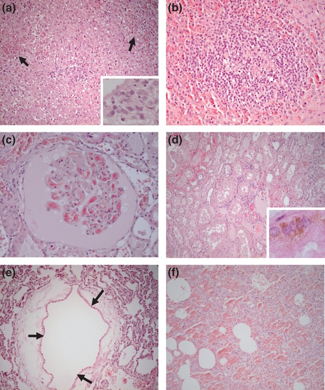
Marburg virus causes a highly infectious and lethal haemorrhagic fever in primates and may be exploited as a potential biothreat pathogen. To combat the infection and threat of Marburg haemorrhagic fever, there is a need to develop and license appropriate medical countermeasures. To determine whether the common marmoset (Callithrix jacchus) would be an appropriate model to assess therapies against Marburg haemorrhagic fever, initial susceptibility, lethality and pathogenesis studies were performed. Low doses of virus, between 4 and 28 TCID50 , were sufficient to cause a lethal, reproducible infection. Animals became febrile between days 5 and 6, maintaining a high fever before succumbing to disease between 8 and 11 days postchallenge. Typical signs of Marburg virus infection were observed including haemorrhaging and a transient rash. In pathogenesis studies, virus was isolated from the animals' lungs from day 3 postchallenge and from the liver, spleen and blood from day 5 postchallenge. Early signs of histopathology were apparent in the kidney and liver from day 3. The most striking features were observed in animals exhibiting severe clinical signs, which included high viral titres in all organs, with the highest levels in the blood, increased levels in liver function enzymes and blood clotting times, decreased levels in platelets, multifocal moderate-to-severe hepatitis and perivascular oedema.
© 2013 Crown copyright. International Journal of Experimental Pathology © 2013 International Journal of Experimental Pathology.
Figures
Similar articles
-
Experimental Respiratory Infection of Marmosets (Callithrix jacchus) With Ebola Virus Kikwit.J Infect Dis. 2015 Oct 1;212 Suppl 2:S336-45. doi: 10.1093/infdis/jiv371. Epub 2015 Jul 23. J Infect Dis. 2015. PMID: 26209682
-
A small nonhuman primate model for filovirus-induced disease.Virology. 2011 Nov 25;420(2):117-24. doi: 10.1016/j.virol.2011.08.022. Epub 2011 Sep 28. Virology. 2011. PMID: 21959017 Free PMC article.
-
Aerosol exposure to the angola strain of marburg virus causes lethal viral hemorrhagic Fever in cynomolgus macaques.Vet Pathol. 2010 Sep;47(5):831-51. doi: 10.1177/0300985810378597. Vet Pathol. 2010. PMID: 20807825
-
Marburg haemorrhagic fever in returning travellers: an overview aimed at clinicians.Clin Microbiol Infect. 2019 Apr;21S:e28-e31. doi: 10.1111/1469-0691.12673. Epub 2015 Jun 22. Clin Microbiol Infect. 2019. PMID: 24816494 Review.
-
Tissue and cellular tropism, pathology and pathogenesis of Ebola and Marburg viruses.J Pathol. 2015 Jan;235(2):153-74. doi: 10.1002/path.4456. J Pathol. 2015. PMID: 25297522 Review.
Cited by
-
Toxicological Evaluation of a Probiotic-Based Delivery System for P8 Protein as an Anti-Colorectal Cancer Drug.Drug Des Devel Ther. 2021 Nov 27;15:4761-4793. doi: 10.2147/DDDT.S319930. eCollection 2021. Drug Des Devel Ther. 2021. PMID: 34866901 Free PMC article.
-
Recent advances in marburgvirus research.F1000Res. 2019 May 21;8:F1000 Faculty Rev-704. doi: 10.12688/f1000research.17573.1. eCollection 2019. F1000Res. 2019. PMID: 31131088 Free PMC article. Review.
-
In vivo manipulation of γ9(+) T cells in the common marmoset (Callithrix Jacchus) with phosphoantigen and effect on the progression of respiratory melioidosis.PLoS One. 2013 Sep 30;8(9):e74789. doi: 10.1371/journal.pone.0074789. eCollection 2013. PLoS One. 2013. PMID: 24098670 Free PMC article.
-
A novel nonhuman primate model for influenza transmission.PLoS One. 2013 Nov 14;8(11):e78750. doi: 10.1371/journal.pone.0078750. eCollection 2013. PLoS One. 2013. PMID: 24244352 Free PMC article.
-
Comparative Pathology and Pathogenesis of African Swine Fever Infection in Swine.Front Vet Sci. 2020 May 19;7:282. doi: 10.3389/fvets.2020.00282. eCollection 2020. Front Vet Sci. 2020. PMID: 32509811 Free PMC article. Review.
References
-
- Alves DA, Glynn AR, Steele KE, et al. Aerosol exposure to the angola strain of Marburg virus causes lethal viral hemorrhagic Fever in cynomolgus macaques. Vet. Pathol. 2010;47:831–851. - PubMed
-
- Bancroft JD, Gamble M. Theory and Practice of histological techniques. 6th edn. Elsevier Health Sciences: Churchill Livingston Elsevier; 2008.
-
- Baskerville A, Bowen ET, Platt GS, McArdell LB, Simpson DI. The pathology of experimental Ebola virus infection in monkeys. J Pathol. 1978;125:131–138. - PubMed
Publication types
MeSH terms
Substances
LinkOut - more resources
Full Text Sources
Other Literature Sources
