. 2013 Feb 25:10:23.
doi: 10.1186/1742-4690-10-23.

Evidence for IFNα-induced, SAMHD1-independent inhibitors of early HIV-1 infection

Affiliations

Evidence for IFNα-induced, SAMHD1-independent inhibitors of early HIV-1 infection

Caroline Goujon et al. Retrovirology. .

Abstract

Background: Type I interferon (IFN) treatment of some cells, including dendritic cells, macrophages and monocytic THP-1 cells, restricts HIV-1 infection and prevents viral cDNA accumulation. Sterile alpha motif and HD domain protein 1 (SAMHD1), a dGTP-regulated deoxynucleotide triphosphohydrolase, reduces HIV-1 infectivity in myeloid cells, likely by limiting dNTPs available for reverse transcription, and has been described as IFNα-inducible. Myeloid cell infection by HIV-1 is enhanced by HIV-2/SIVSM Vpx, which promotes SAMHD1 degradation, or by exogenous deoxyribonucleoside (dN) addition.

Findings: SAMHD1 expression was not substantially influenced by IFNα treatment of monocyte-derived macrophages or THP-1 cells. The contributions of SAMHD1 to the inhibition of HIV-1 infectivity by IFNα were assessed through the provision of Vpx, exogenous dN addition, or via RNAi-mediated SAMHD1 knock-down. Both Vpx and dN efficiently restored infection in IFNα-treated macrophages, albeit not to the levels seen with these treatments in the absence of IFNα. Similarly using differentiated THP-1 cells, the addition of Vpx or dNs, or SAMHD1 knock-down, also stimulated infection, but failing to match the levels observed without IFNα. Neither Vpx addition nor SAMHD1 knock-down reversed the IFNα-induced blocks to HIV-1 infection seen in dividing U87-MG or THP-1 cells. Therefore, altered SAMHD1 expression or function cannot account for the IFNα-induced restriction to HIV-1 infection seen in many cells and cell lines.

Conclusion: IFNα establishes an anti-HIV-1 phenotype in many cell types, and appears to accomplish this without potentiating SAMHD1 function. We conclude that additional IFNα-induced suppressors of the early stages of HIV-1 infection await identification.


Figures
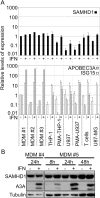
Figure 1
Figure 1
SAMHD1 is induced poorly by IFNα in monocyte-derived macrophages. A. 1 to 2 × 10⁶ monocyte-derived macrophages (MDMs) or primary CD4+ T cells (T cells), PMA-treated or dividing THP-1 and U937 cells, or U87-MG cells were incubated (or not) with 1000 U/ml IFNα for 24 h prior to total RNA extraction. cDNA was synthesized and the relative levels of SAMHD1 expression (upper panel), as well as two well-known ISGs, ISG15 and APOBEC3A (lower panel), were analyzed by RT-qPCR and the data were normalized to both GAPDH and β-actin expression. The graph shows the data obtained from 3 independent donors for MDMs (#1 #2 #3), and the mean values of relative expression in each cell type, obtained from 3 independent repetitions with cell lines or 3 different donors for CD4+ T cells. B. Western blot analysis. MDMs from 2 different donors were treated or not with 1000 U/ml IFNα for 24 h (donor #4), or for 8, 24 and 48 h (donor #5) before cell lysis. Protein levels of SAMHD1 and APOBEC3A (A3A) were determined, and tubulin served as a loading control.
Figure 2
Figure 2
Vpx-VLPs or exogenous dN treatment greatly enhance HIV-1 infection in IFNα-treated MDMs. A. VSV-G pseudotyped HIV-1 derived GFP reporter virus was produced in 293T cells and used to infect control or IFNα-treated MDMs at different MOIs (0.08 to 10, as determined in 293T cells), in the presence or the absence of either Vpx-VLP or 0.5 mM or 2.5 mM deoxyribonucleosides (dN). Levels of infection were monitored using flow cytometry to measure the percentage of cells expressing GFP, and mean values from 4 independent experiments (using cells from 4 different donors) are shown. Notably, treatment of MDMs with the higher concentration of dN changed their morphology, with the cells becoming rounder and less adherent. We conclude that experiments using high dN doses must be analysed with caution. B. MDMs were treated or not with IFNα for 24 h and subsequently incubated or not with Ctrl-VLPs, Vpx-VLPs or dN (at 0.5 or 2.5 mM as indicated) for 16 h before lysis. Whole cell dATP was quantified using a single nucleotide quantification assay. Mean values from 4 independent experiments (using cells from 4 different donors) are shown. C. Western blot analysis. Cell lysates from MDMs (donor #4) were harvested 24 h post IFNα and Vpx-VLP treatment, and SAMHD1 expression was analysed by western blot; Hsp90 served as a loading control.
Figure 3
Figure 3
Vpx-VLPs, exogenous dN treatment or SAMHD1 knock-down do not rescue the IFNα-induced block to HIV-1 infection in THP-1 cells. A. and B. VSV-G pseudotyped HIV-1 derived GFP reporter virus was produced in 293T cells and used to infect control or IFNα-treated PMA-differentiated THP-1 cells (A), or dividing THP-1 cells (B) at different MOIs (0.08 to 10), in the presence or the absence of either Vpx-VLP or 0.5 mM deoxyribonucleosides (dN). Levels of infection were monitored using flow cytometry to measure the percentage of cells expressing GFP, and mean values from 3 independent experiments are shown. C. Western blot analysis. Cell lysates from THP-1 cells were harvested 24 h post IFNα and Vpx-VLP treatment, and SAMHD1 expression was analysed by western blot; tubulin served as a loading control. D. THP-1 cells expressing a control shRNA or shRNA targeting SAMHD1 were differentiated with PMA or not in the presence or absence of IFNα for 24 h. Cells were infected with 3 different doses of VSV-G pseudotyped GFP reporter virus for 48 h, and GFP positive cells were enumerated by flow cytometry. The data are representative of 3 independent experiments with two independent shRNAs against SAMHD1. E. Western blot analysis of parallel samples from D. Protein levels of SAMHD1 and APOBEC3A (A3A) were determined and tubulin served as a loading control.
Figure 4
Figure 4
SAMHD1 is induced by IFNα in U87-MG cells, but Vpx triggered degradation does not rescue IFNα-mediated HIV-1 suppression. A. U87-MG cells were treated for 24 h with IFNα, or not, and incubated with no VLPs, Vpx-VLPs, or control VLPs (Ctrl-VLPs) and then infected with increasing amounts of GFP reporter virus (MOI 0.008 to 1), and GFP expression monitored as before. The mean values from 3 independent experiments are shown. B. Western blot analysis of a parallel samples from A. Protein levels of SAMHD1 were determined and tubulin served as a loading control.

References

    1. Goujon C, Malim MH. Characterization of the alpha interferon-induced postentry block to HIV-1 infection in primary human macrophages and T cells. J Virol. 2010;84:9254–9266. doi: 10.1128/JVI.00854-10. - DOI - PMC - PubMed
    1. Cheney KM, McKnight A. Interferon-alpha mediates restriction of human immunodeficiency virus type-1 replication in primary human macrophages at an early stage of replication. PLoS One. 2010;5:e13521. doi: 10.1371/journal.pone.0013521. - DOI - PMC - PubMed
    1. Baca-Regen L, Heinzinger N, Stevenson M, Gendelman HE. Alpha interferon-induced antiretroviral activities: restriction of viral nucleic acid synthesis and progeny virion production in human immunodeficiency virus type 1-infected monocytes. J Virol. 1994;68:7559–7565. - PMC - PubMed
    1. Meylan PR, Guatelli JC, Munis JR, Richman DD, Kornbluth RS. Mechanisms for the inhibition of HIV replication by interferons-alpha, -beta, and -gamma in primary human macrophages. Virology. 1993;193:138–148. doi: 10.1006/viro.1993.1110. - DOI - PubMed
    1. Goujon C, Jarrosson-Wuilleme L, Bernaud J, Rigal D, Darlix JL, Cimarelli A. With a little help from a friend: increasing HIV transduction of monocyte-derived dendritic cells with virion-like particles of SIV(MAC) Gene Ther. 2006;13:991–994. doi: 10.1038/sj.gt.3302753. - DOI - PubMed

Publication types

MeSH terms

Substances