. 2013 Mar 1;73(5):1635-46.
doi: 10.1158/0008-5472.CAN-12-3349. Epub 2013 Feb 26.

HER2 drives luminal breast cancer stem cells in the absence of HER2 amplification: implications for efficacy of adjuvant trastuzumab

Affiliations

HER2 drives luminal breast cancer stem cells in the absence of HER2 amplification: implications for efficacy of adjuvant trastuzumab

Suthinee Ithimakin et al. Cancer Res. .

Abstract

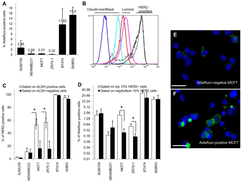
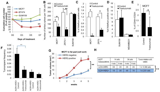
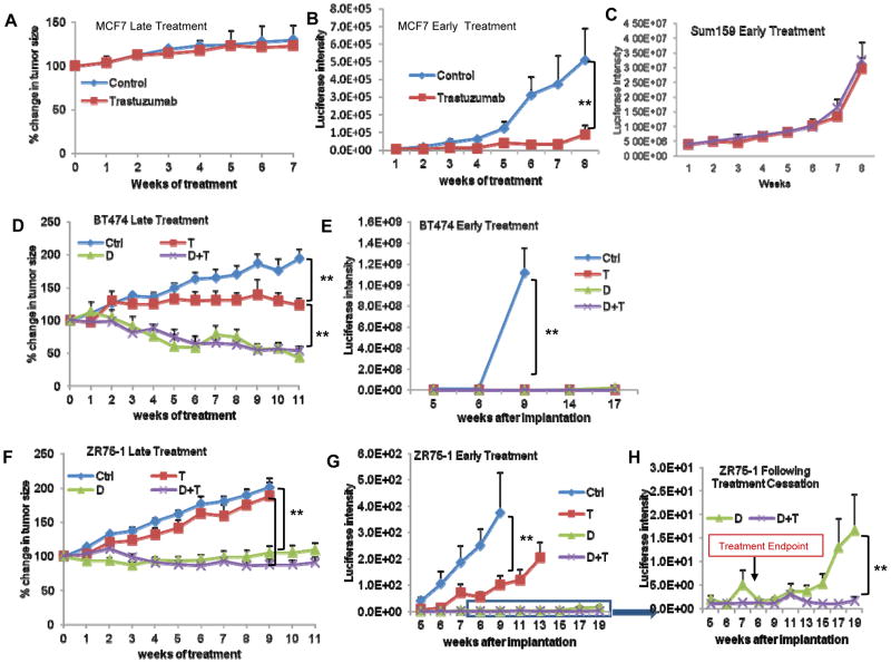
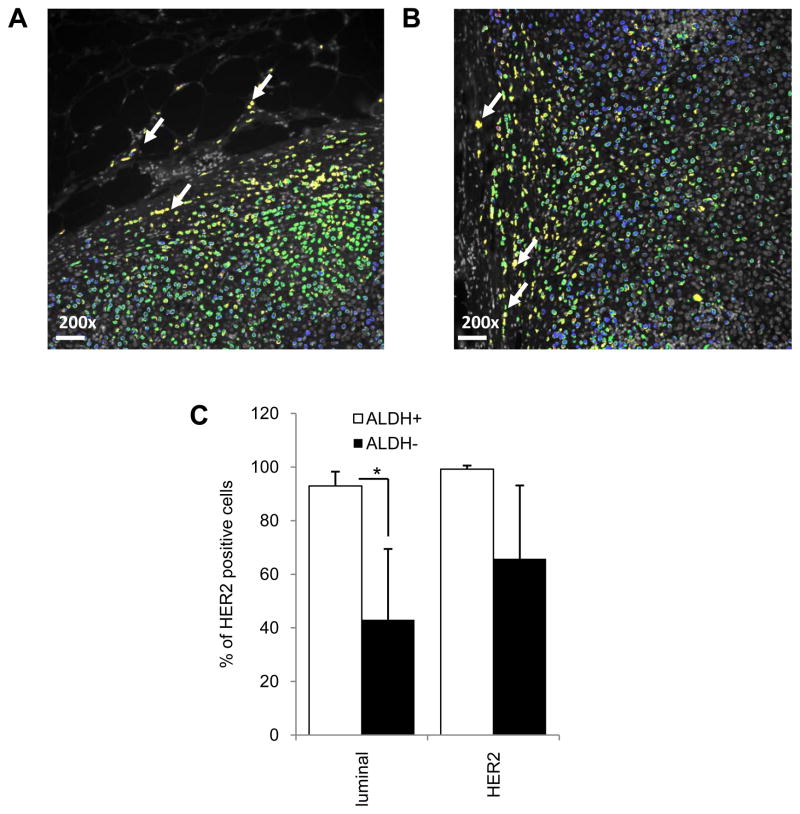
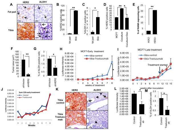
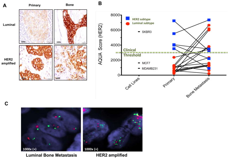
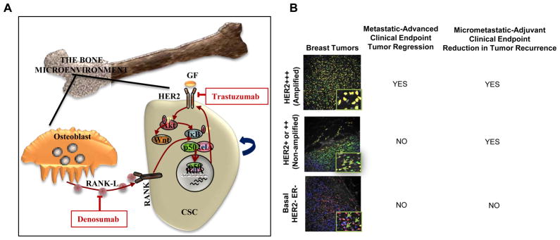
Although current breast cancer treatment guidelines limit the use of HER2-blocking agents to tumors with HER2 gene amplification, recent retrospective analyses suggest that a wider group of patients may benefit from this therapy. Using breast cancer cell lines, mouse xenograft models and matched human primary and metastatic tissues, we show that HER2 is selectively expressed in and regulates self-renewal of the cancer stem cell (CSC) population in estrogen receptor-positive (ER(+)), HER2(-) luminal breast cancers. Although trastuzumab had no effects on the growth of established luminal breast cancer mouse xenografts, administration after tumor inoculation blocked subsequent tumor growth. HER2 expression is increased in luminal tumors grown in mouse bone xenografts, as well as in bone metastases from patients with breast cancer as compared with matched primary tumors. Furthermore, this increase in HER2 protein expression was not due to gene amplification but rather was mediated by receptor activator of NF-κB (RANK)-ligand in the bone microenvironment. These studies suggest that the clinical efficacy of adjuvant trastuzumab may relate to the ability of this agent to target the CSC population in a process that does not require HER2 gene amplification. Furthermore, these studies support a CSC model in which maximal clinical benefit is achieved when CSC targeting agents are administered in the adjuvant setting. Cancer Res; 73(5); 1635-46. ©2012 AACR.


Conflict of interest statement

Disclosure of potential conflicts of interest

M. S. Wicha has financial holdings in OncoMed Pharmaceuticals, receives research support from Dompe and MedImmune, serves on the scientific advisory board of VERISTEM.

H. Korkaya receives research support from MedImmune.

D. F. Hayes has received research support from Pfizer, Novartis and Veridex and holds stock option for his role on the scientific advisory board for OncImmune.

Figures

Figure 1
Figure 1
High HER2 protein expression correlates with increased levels of ALDH activity in luminal breast cancer cell lines. A, The percentages of ALDH positivity as assessed by the Aldefluor assay in basal/claudin-low (SUM159, MDA-MB231), luminal (MCF7, ZR75-1) and HER2-amplified (BT474, SKBR3) cells. B, HER2 expression by FACS of the same cell lines demonstrated low HER2 expression (basal/claudin-low (dark and light blue)), moderate HER2 protein (luminal (red and pink)) and high levels of HER2 expression (amplified (light and dark grey)). C, FACS analysis of Aldefluor positive and negative cells demonstrated enrichment for HER2 expression in Aldefluor positive cells. D, FACS analysis of the top 10% of HER2-positive and bottom 10% HER2-negative cells demonstrated enrichment for Aldefluor positivity within HER2-positive cells. Increased HER2 expression correlated with increased Aldefluor activity where * is denoted, p<0.05. E–F, Immunofluorescence demonstrating increased HER2 expression in Aldefluor-positive compared to Aldefluor-negative MCF7 cells. Scale bar 50μm.
Figure 2
Figure 2
Trastuzumab targets CSCs in luminal breast cancer cells. A, MTT assay in 2-D culture demonstrated inhibition of cell growth by trastuzumab in BT474, HER2 amplified but not in MCF7 or SUM159 cells that do not display HER2 amplification. B, Tertiary tumorsphere formation of MCF7, ZR75-1 and SUM159 cells was analyzed after treatment of primary spheres with trastuzumab (at 21μg/ml) and two serial passages were performed in the absence of trastuzumab. Trastuzumab treatment significantly reduced the number of tertiary tumorspheres of luminal MCF7 and ZR75-1 cells but had no effect on basal/claudin-low Sum159 cells. C, Trastuzumab reduced the percent of Aldefluor-positive cells in luminal cell lines, D, but had no effect on basal/claudin-low cell lines. E, Trastuzumab significantly reduced the proportion of CD44+/CD24− in MCF7 cells. F, Trastuzumab significantly reduced the sphere formation in ALDH+HER2+ but had no significant effect onALDH+HER2− cells. G, MCF7 HER2-positive and HER2-negative cell populations were FACS sorted and 10,000 cells were then injected into mammary fat pads of NOD-SCID mice. HER2-positive cells produced larger tumors compared to HER2 negative cells as assessed by luciferase imaging. H, HER2 expression in ALDH-positive MCF7 cells demonstrated a significantly higher frequency of tumor initiating cells as compared to the ALDH-positive MCF7 cells without HER2 expression. (*p<0.05, **p<0.01).
Figure 3
Figure 3
Effects of trastuzumab on mouse tumor xenograft depend on the timing of administration. Luciferase expressing MCF7, BT474 and ZR75-1 cells were grown in mouse fat pads, and tumor size was measured with calipers or determined by luciferase intensity. In the late treatments, control (Ctrl), trastuzumab (T) and/or docetaxel (D) were administered over 6 weeks starting at 4 weeks post inoculation. In early treatments, the treatment started on the day of inoculation. A–B, MCF7 tumor xenografts were inhibited when trastuzumab treatment was administered early but not late. C, The Sum159 cells (basal/claudin-low breast cancer cells) showed no difference between control animals and trastuzumab treated even when treatments initiated at early time. D–E, Both trastuzumab and docetaxel inhibited growth of BT474 xenografts when administered in late or early setting. F–G, Trastuzumab inhibited ZR75-1 tumor xenograft growth when administered early but not at late setting. H, Following the cessation of treatment, tumors recurred in mice treated with docetaxel alone but did not recur in those treated with docetaxel plus trastuzumab. (**p<0.01).
Figure 4
Figure 4
HER2 and ALDH are co-expressed in primary luminal breast tumor cells. A–B, Cells co-expressing HER2 (green) and ALDH1 (red) in human breast carcinoma cells identified by cytokeratin 8 (blue) are located (yellow; white arrows) at invasive edges of primary mammary carcinomas in FFPE sections. C, Shown here are average HER2-positive cells in ALDH1-positive versus -negative populations analyzed by AQUA. Scale bar 50μm, as the original images were taken at 200X magnification. (*p<0.05, **p<0.01).
Figure 5
Figure 5
HER2 and ALDH1 expression are increased in MCF7 cells growing in the bone microenvironment. A, Representative images demonstrate an increase in HER2 and ALDH1 expression in MCF7 cells grown in mouse tibia compared to mammary fat pads. B–C, Quantitation of HER2 expression and ALDH1 expression was evaluated by counting positive cells by IHC. D, MCF7 cells co-cultured in vitro with human osteoblasts expressed increased levels of HER2 and this effect is inhibited by denosumab. E, HER2 and RANK expression in MCF7 cells was analyzed and the percent RANK expressing cells was assessed for the top 10% and bottom 10% of HER2 expressing cells. F, MCF7 cells with HER2 knockdown generated smaller tumors with G, fewer ALDH1-positive cells compared to tumors generated from MCF7 control cells as determined by IHC. H, Trastuzumab effectively inhibited luciferase expressing MCF7 cell growth as measured by luciferase expression in mouse tibias when administered in early setting but I, had no effect on established tumors in tibia (late setting, arrow indicates the initiation of trastuzumab). J, Trastuzumab had no effect on basal/claudin-low Sum159 cells introduced into tibia even when administered in the early setting. K–L–M, Trastuzumab reduced the expression of HER2 and ALDH1 in MCF7 cells grown in mouse tibia and L, resulted in smaller tumors with M, fewer ALDH1 positive cells than tumors generated in the absence of trastuzumab treatment. (*p<0.05, **p<0.01).
Figure 6
Figure 6
HER2 expression is increased in bone metastases compared to matched primary tumors in luminal breast cancers. A, HER2 expression by IHC score was examined in bone metastases as compared to matched breast primary tumor in patients with luminal and HER2 amplified breast cancers (additional examples in Supplementary Figure 6). B, HER2 expression was quantitated by AQUA score of 19 matched bone metastasis and primary breast cancers. 12 of 14 (87%) luminal breast cancers (red) demonstrated significant increase in HER2 expression in bone metastasis compared to matched primary breast tumors (95% confidence interval: 57–98%). All cases of HER2 intrinsic subtype had high score except for two patients that received trastuzumab treatment (blue line falls below the clinical threshold line). Cell lines MDA-MB231 (basal/claudin-low), MCF7 (luminal) and SKBR3 (HER2 amplified) were used to determine the high and low levels of HER2. C, Fluorescence in situ hybridization (FISH) assay for HER2 amplification shows two normal copies of the HER2 gene in representative bone metastasis sample of a patient with luminal breast cancer, while bone metastasis sample from a patient with HER2-amplified primary tumor as a positive control show multiple copies of the HER2 gene.
Figure 7
Figure 7
Regulation of breast CSCs by the bone microenvironment. A, Osteoblasts generated RANK-L activates the RANK receptor on breast CSCs activating NF-κB and HER2 expression. HER2 signaling further activates NF-κB generating a positive feedback loop. B, Clinical benefit of trastuzumab in advance setting as assessed by tumor regression is limited to tumors with HER2 gene amplification where HER2 is expressed in both bulk and CSC populations. In contrast, in the adjuvant setting, the clinical benefit of trastuzumab extends to luminal breast cancers in which HER2 is selectively expressed in the CSC population at sites of bone micrometastases. Tumors that are truly negative for HER2 do not benefit from trastuzumab in either setting.

References

    1. Slamon DJ, Clark GM, Wong SG, Levin WJ, Ullrich A, McGuire WL. Human breast cancer: correlation of relapse and survival with amplification of the HER-2/neu oncogene. Science. 1987;235:177–82. - PubMed
    1. Vogel CL, Cobleigh MA, Tripathy D, Gutheil JC, Harris LN, Fehrenbacher L, et al. Efficacy and safety of trastuzumab as a single agent in first-line treatment of HER2-overexpressing metastatic breast cancer. J Clin Oncol. 2002;20:719–26. - PubMed
    1. Slamon DJ, Leyland-Jones B, Shak S, Fuchs H, Paton V, Bajamonde A, et al. Use of chemotherapy plus a monoclonal antibody against HER2 for metastatic breast cancer that overexpresses HER2. N Engl J Med. 2001;344:783–92. - PubMed
    1. Vogel CL, Cobleigh MA, Tripathy D, Gutheil JC, Harris LN, Fehrenbacher L, et al. First-line Herceptin monotherapy in metastatic breast cancer. Oncology. 2001;61 (Suppl 2):37–42. - PubMed
    1. Mass RD, Press MF, Anderson S, Cobleigh MA, Vogel CL, Dybdal N, et al. Evaluation of clinical outcomes according to HER2 detection by fluorescence in situ hybridization in women with metastatic breast cancer treated with trastuzumab. Clin Breast Cancer. 2005;6:240–6. - PubMed

Publication types