. 2013 Feb 26:13:90.
doi: 10.1186/1471-2407-13-90.

Promoter hypomethylation, especially around the E26 transformation-specific motif, and increased expression of poly (ADP-ribose) polymerase 1 in BRCA-mutated serous ovarian cancer

Affiliations

Promoter hypomethylation, especially around the E26 transformation-specific motif, and increased expression of poly (ADP-ribose) polymerase 1 in BRCA-mutated serous ovarian cancer

Fang-Fang Bi et al. BMC Cancer. .

Abstract

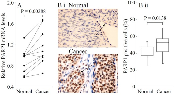
Background: Poly (ADP-ribose) polymerase 1 (PARP1) overexpression plays a critical role in ovarian cancer progression and the clinical development of PARP1 inhibitors to treat BRCA-mutated ovarian cancer has advanced rapidly. However, the mechanism regulating PARP1 expression remains unknown. Alterations in gene expression mediated by promoter methylation are being increasingly recognized and have frequently been reported in ovarian cancer. We therefore investigated the methylation status of the PARP1 promoter region and its correlation with PARP1 expression in BRCA-mutated ovarian cancer.

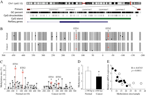
Methods: DNA from BRCA-mutated serous ovarian cancer samples and adjacent normal ovarian tissues were analyzed by bisulfite sequence using primers focusing on the CpG island in the promoter region of PARP1. Expression levels of PARP1 were assessed by immunohistochemistry and real-time PCR.

Results: Serous ovarian cancer tissues displayed decreased DNA methylation in the promoter region of PARP1 compared to normal tissue, and methylation intensity correlated inversely with PARP1 mRNA levels. More importantly, E26 transformation-specific (ETS) defined CpG sites were significantly less methylated in ovarian cancer samples.

Conclusions: These results indicate that hypomethylation of the promoter region, especially around the ETS motif might play a role in the upregulation of PARP1 expression in the progression of ovarian cancer.


Figures

Figure 1
Figure 1
Overexpression of PARP1 mRNA and protein in BRCA-mutated ovarian cancer. A, Relative PARP1 mRNA levels were measured by real-time quantitative PCR. B i, Sections were subjected to immunostaining for PARP1 in normal and ovarian cancer tissue. Arrow shows positive staining for PARP1 in the nuclei. B ii, Summary of the percentages of PARP1-positive cells from the measurements shown in (B i). Bar graphs show mean ± SD. Magnification is 400 × .
Figure 2
Figure 2
Hypomethylation of the ETS motif and promoter region of PARP1 in BRCA-mutated ovarian cancer. A, Location of PARP1 core promoter CpG sites. Genomic coordinates are shown, along with the primer-amplified fragments, GC percentage, location of individual CpG dinucleotides (dashes), CpG island (green bar), and the PARP1 RefSeq gene (exon 1 shown as a blue box and intron shown as an arrowed line). The arrow indicates the transcriptional direction. B, Changes of methylation patterns in the core promoter region of PARP1 (each group, n = 10). The circles correspond to CpG sites denoted by the black dashes in (A). Closed circles, methylation; open circles, unmethylation. Ten individual clones were sequenced for each sample. Red circles show a methylation of cytosine that located in a CpG within an ETS motif. C, Summary of the promoter DNA methylation pattern in ovarian cancer and normal tissues. The y-axis shows the mean methylation sites (each group n = 10). Red circles show a methylation of cytosine that located in a CpG within an ETS motif. D, Overall methylation percentage within the promoter region from ovarian cancer and normal tissues (each group n = 10). E, Correlation between the relative PARP1 mRNA levels and the total number of methylated sites for each sample. Open circles, ovarian cancer tissues. Closed circles, normal ovarian tissues (each group n = 10). * P < 0.05 ovarian cancer vs. normal tissues.

Similar articles

Cited by

References

    1. Siegel R, Naishadham D, Jemal A. Cancer statistics 2012. CA Cancer J Clin. 2012;62:10–29. doi: 10.3322/caac.20138. - DOI - PubMed
    1. Pruthi S, Gostout BS, Lindor NM. Identification and Management of Women With BRCA Mutations or Hereditary Predisposition for Breast and Ovarian Cancer. Mayo Clin Proc. 2010;85:1111–1120. doi: 10.4065/mcp.2010.0414. - DOI - PMC - PubMed
    1. Bryant HE, Schultz N, Thomas HD, Parker KM, Flower D, Lopez E, Kyle S, Meuth M, Curtin NJ, Helleday T. Specific killing of BRCA2-deficient tumours with inhibitors of poly(ADPribose) polymerase. Nature. 2005;434:913–917. doi: 10.1038/nature03443. - DOI - PubMed
    1. Farmer H, McCabe N, Lord CJ, Tutt AN, Johnson DA, Richardson TB, Santarosa M, Dillon KJ, Hickson I, Knights C, Martin NM, Jackson SP, Smith GC, Ashworth A. Targeting the DNA repair defect in BRCA mutant cells as a therapeutic strategy. Nature. 2005;434:917–921. doi: 10.1038/nature03445. - DOI - PubMed
    1. Barnett JC, Bean SM, Nakayama JM, Kondoh E, Murphy SK, Berchuck A. High poly (adenosine diphosphate-ribose) polymerase expression and poor survival in advanced-stage serous ovarian cancer. Obstet Gynecol. 2010;115:49–54. doi: 10.1097/AOG.0b013e3181c2d294. - DOI - PubMed

MeSH terms