. 2013 Mar 15;12(6):899-906.
doi: 10.4161/cc.23879. Epub 2013 Feb 26.

Centrobin regulates centrosome function in interphase cells by limiting pericentriolar matrix recruitment

Affiliations

Centrobin regulates centrosome function in interphase cells by limiting pericentriolar matrix recruitment

Jessie M Jeffery et al. Cell Cycle. .

Abstract

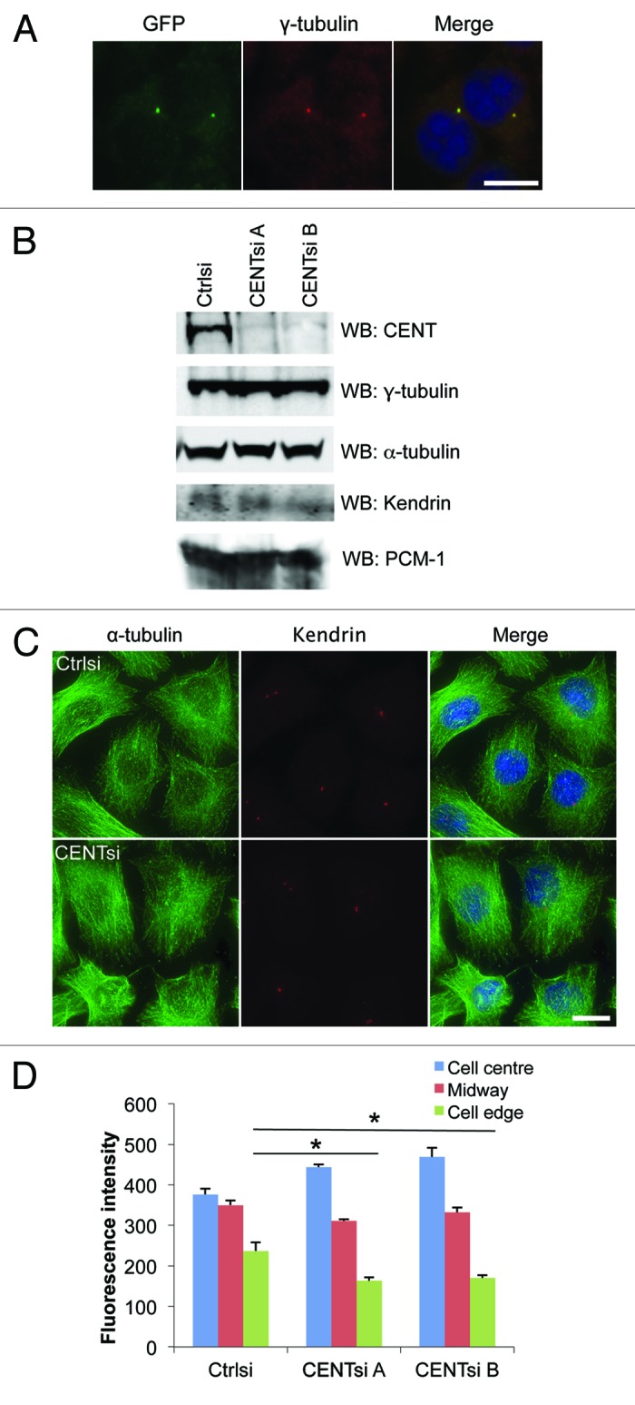
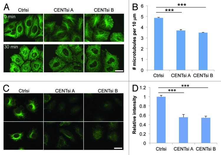
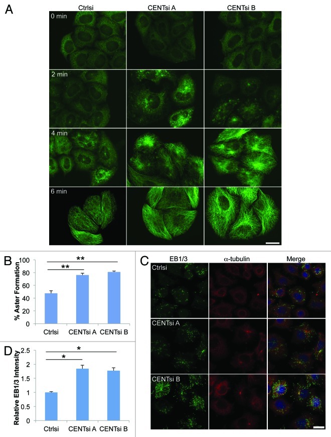
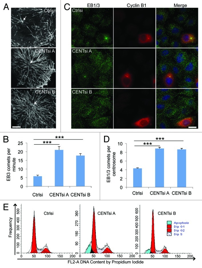
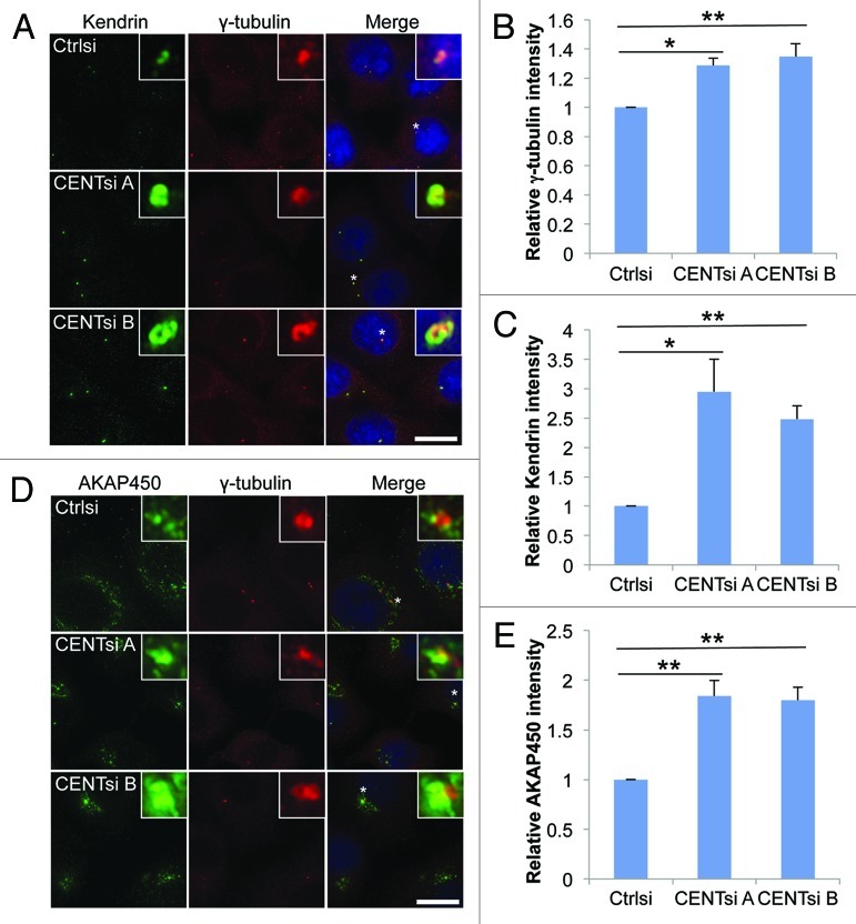
The amount of pericentriolar matrix at the centrosome is tightly linked to both microtubule nucleation and centriole duplication, although the exact mechanism by which pericentriolar matrix levels are regulated is unclear. Here we show that Centrobin, a centrosomal protein, is involved in regulating these levels. Interphase microtubule arrays in Centrobin-depleted cells are more focused around the centrosome and are less stable than the arrays in control cells. Centrobin-depleted cells initiate microtubule nucleation more rapidly than control cells and exhibit an increase in the number of growing microtubule ends emanating from the centrosome, while the parameters of microtubule plus end dynamics around the centrosome are not significantly altered. Finally, we show that Centrobin depletion results in the increased recruitment of pericentriolar matrix proteins to the centrosome, including γ-tubulin, AKAP450, Kendrin and PCM-1. We propose that Centrobin might regulate microtubule nucleation and organization by controlling the amount of pericentriolar matrix.

Keywords: centriole duplication; centrobin; microtubules; nucleation; pericentriolar matrix.


Figures

None
Figure 1. Centrobin-depleted cells exhibit a redistribution of the microtubule network and microtubule stability defect. (A) HeLa cells stably expressing GFP-Centrobin at near-endogenous levels were fixed and co-stained for GFP, γ-tubulin and DAPI. Scale bar represents 10 μm. (B–D) Control or Centrobin-depleted (CENTsi) HeLa cells were harvested for (B) western blotting with the indicated antibodies or (C) co-stained for α-tubulin, Kendrin and DAPI. Scale bar represents 20 μm. (D) The intensity of microtubule staining from (C) was measured at the densest region of microtubules near the cell center, at the cell edge closest to this region and midway between the two points by quantifying the intensity of a 2.14 μm square at each point. For each experiment, 50 cells were measured per condition. Error bars represent the standard error of the mean from three independent experiments. p-values are denoted as follows: *p < 0.05.
None
Figure 2. Centrobin-depleted cells exhibit a microtubule stability defect. (A) Control and Centrobin-depleted cells were treated with 10 μM nocodazole for 30 min and stained for α-tubulin. (B) Quantification of the average number of microtubules remaining per 10 μm after nocodazole treatment from (A). For each cell, a 10 μm line was drawn in five different positions around the cell parallel to the nearest cell membrane and the number of microtubules crossing this line counted. Twenty cells were measured for each condition, resulting in a total of 100 measurements per condition. (C) Control or Centrobin-depleted HeLa cells were stained for acetylated tubulin. (D) Quantification of relative acetylated tubulin intensity per cell from (C). For each experiment, 20 cells were measured per condition. Scale bars represent 20 μm. Error bars represent the standard error of the mean from three independent experiments. p-values are denoted as follows: ***p < 0.001.
None
Figure 3. Centrobin-depleted cells initiate microtubule nucleation more rapidly than control cells. HeLa cells were transfected with control or Centrobin siRNA, incubated for 48 h and placed on ice for 1 h. (A) Cells were returned to warm (37°C) media for the indicated times, fixed and stained for α-tubulin. (B–D) Cells were returned to warm media for 2 min and then fixed. (B) Cells were stained for α-tubulin and quantified for the percentage of cells with aster formation after 2 min. Asters were identified by using the thresholding tool in ImageJ. For each experiment, 100 cells were counted. (C) Cells were fixed and stained for α-tubulin, EB1/3 and DAPI. (D) Quantification of total EB1/3 intensity per cell from (C) relative to control cells. For each experiment, 50 cells were counted. Error bars represent the standard error of the mean from three independent experiments. p-values are denoted as follows: *p < 0.05, **p < 0.01. Scale bars represent 20 μm.
None
Figure 4. Centrobin-depleted cells exhibit increased microtubule nucleation. (A) HeLa cells were co-transfected with EB3-GFP and control or Centrobin siRNA. Maximum intensity projections of live cell image sequences of control and Centrobin-depleted cells. Images were collected every 0.5 sec, and the movies were 2 min in duration. Scale bar represents 10 μm. (B) Microtubule nucleation frequency from the centrosome as quantified from movies of EB3-GFP described in (A). A circle measuring 5 μm in diameter was drawn around the centrosome and comets originating in the circle were counted. (C) Control and Centrobin-depleted cells were co-stained with antibodies against Cyclin B1 and EB1/3. Scale bar represents 20 μm. (D) Quantification of the number of EB1/3 comets around the centrosome from fixed cells described in (C). The location of the centrosome was identified via 3D reconstruction of the cells then a maximum projection image generated and a circle measuring 5 μm in diameter was drawn around the centrosome. All the comets contained within the circle were counted. (E) Cell cycle profiles of control and Centrobin-depleted cells. Error bars represent the standard error of the mean from three independent experiments. For each of these experiments, 10 cells were measured per condition. p-values are denoted as follows: ***p < 0.001.
None
Figure 5. PCM accumulation at the centrosome is increased in Centrobin-depleted cells. Control or Centrobin-depleted cells were stained with the indicated antibodies: (A–C) γ-tubulin and Kendrin, (D and E) γ-tubulin and AKAP450. Insets are 8× magnification of centrosomes marked with asterisk. Intensity of the centrosomal staining was measured by drawing a 2.14 μm square around the centrosome and is graphed relative to control cells. Error bars represent the standard error of the mean from three independent experiments. For each experiment 20 cells were counted per condition. p-values are denoted as follows: *p < 0.05, **p < 0.01. Scale bars represent 20 μm.

Comment in

Similar articles

Cited by

References

    1. Kleylein-Sohn J, Westendorf J, Le Clech M, Habedanck R, Stierhof Y-D, Nigg EA. Plk4-induced centriole biogenesis in human cells. Dev Cell. 2007;13:190–202. doi: 10.1016/j.devcel.2007.07.002. - DOI - PubMed
    1. Carvalho-Santos Z, Machado P, Branco P, Tavares-Cadete F, Rodrigues-Martins A, Pereira-Leal JB, et al. Stepwise evolution of the centriole-assembly pathway. J Cell Sci. 2010;123:1414–26. doi: 10.1242/jcs.064931. - DOI - PubMed
    1. Song MH, Aravind L, Müller-Reichert T, O’Connell KF. The conserved protein SZY-20 opposes the Plk4-related kinase ZYG-1 to limit centrosome size. Dev Cell. 2008;15:901–12. doi: 10.1016/j.devcel.2008.09.018. - DOI - PMC - PubMed
    1. Bobinnec Y, Khodjakov A, Mir LM, Rieder CL, Eddé B, Bornens M. Centriole disassembly in vivo and its effect on centrosome structure and function in vertebrate cells. J Cell Biol. 1998;143:1575–89. doi: 10.1083/jcb.143.6.1575. - DOI - PMC - PubMed
    1. Zou CZ, Li J, Bai YJ, Gunning WT, Wazer DE, Band V, et al. Centrobin: a novel daughter centriole-associated protein that is required for centriole duplication. J Cell Biol. 2005;171:437–45. doi: 10.1083/jcb.200506185. - DOI - PMC - PubMed

Publication types

LinkOut - more resources