

Effect of extracellular pH on selectin adhesion: theory and experiment
- PMID: 23442851
- PMCID: PMC3552277
- DOI: 10.1016/j.bpj.2012.12.005
Effect of extracellular pH on selectin adhesion: theory and experiment
Abstract
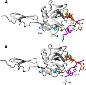
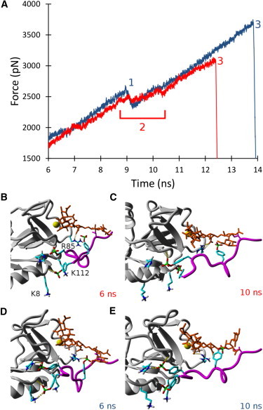
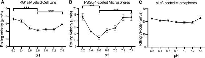
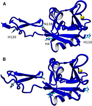
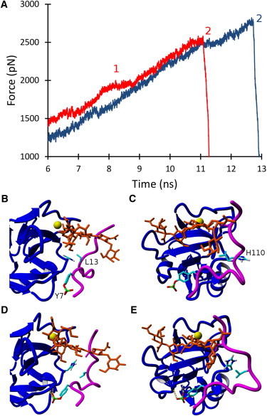
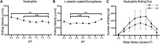
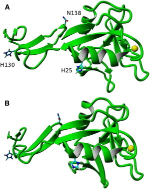
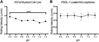
Selectins mediate circulatory leukocyte trafficking to sites of inflammation and trauma, and the extracellular microenvironments at these sites often become acidic. In this study, we investigated the influence of slightly acidic pH on the binding dynamics of selectins (P-, L-, and E-selectin) to P-selectin glycoprotein ligand-1 (PSGL-1) via computational modeling (molecular dynamics) and experimental rolling assays under shear in vitro. The P-selectin/PSGL-1 binding is strengthened at acidic pH, as evidenced by the formation of a new hydrogen bond (seen computationally) and the observed decrease in the rolling velocities of model cells. In the case of L-selectin/PSGL-1 binding dynamics, the binding strength and frequency increase at acidic pH, as indicated by the greater cell-rolling flux of neutrophils and slower rolling velocities of L-selectin-coated microspheres, respectively. The cell flux is most likely due to an increased population of L-selectin in the high-affinity conformation as pH decreases, whereas the velocities are due to increased L-selectin/PSGL-1 contacts. In contrast to P- and L-selectin, the E-selectin/PSGL-1 binding does not exhibit significant changes at acidic pH levels, as shown both experimentally and computationally.
Copyright © 2013 Biophysical Society. Published by Elsevier Inc. All rights reserved.
Figures
Similar articles
-
L- and P-selectins collaborate to support leukocyte rolling in vivo when high-affinity P-selectin-P-selectin glycoprotein ligand-1 interaction is inhibited.Am J Pathol. 2005 Mar;166(3):945-52. doi: 10.1016/S0002-9440(10)62314-0. Am J Pathol. 2005. PMID: 15743805 Free PMC article.
-
Threshold levels of fluid shear promote leukocyte adhesion through selectins (CD62L,P,E).J Cell Biol. 1997 Feb 10;136(3):717-27. doi: 10.1083/jcb.136.3.717. J Cell Biol. 1997. PMID: 9024700 Free PMC article.
-
Regulation of PSGL-1 interactions with L-selectin, P-selectin, and E-selectin: role of human fucosyltransferase-IV and -VII.J Biol Chem. 2005 Feb 18;280(7):5378-90. doi: 10.1074/jbc.M410899200. Epub 2004 Dec 3. J Biol Chem. 2005. PMID: 15579466
-
PSGL-1 function in immunity and steady state homeostasis.Immunol Rev. 2009 Jul;230(1):75-96. doi: 10.1111/j.1600-065X.2009.00797.x. Immunol Rev. 2009. PMID: 19594630 Review.
-
Selectins--potential pharmacological targets?Drug Discov Today. 2006 Nov;11(21-22):1034-40. doi: 10.1016/j.drudis.2006.09.004. Epub 2006 Sep 26. Drug Discov Today. 2006. PMID: 17055414 Review.
Cited by
-
Platelets in inflammation and atherogenesis.Front Immunol. 2015 Mar 6;6:98. doi: 10.3389/fimmu.2015.00098. eCollection 2015. Front Immunol. 2015. PMID: 25798138 Free PMC article. Review.
-
Glycopeptide analogues of PSGL-1 inhibit P-selectin in vitro and in vivo.Nat Commun. 2015 Mar 31;6:6387. doi: 10.1038/ncomms7387. Nat Commun. 2015. PMID: 25824568 Free PMC article.
-
Tumor Vasculature as an Emerging Pharmacological Target to Promote Anti-Tumor Immunity.Int J Mol Sci. 2023 Feb 23;24(5):4422. doi: 10.3390/ijms24054422. Int J Mol Sci. 2023. PMID: 36901858 Free PMC article. Review.
-
Modulation of selectin-mediated adhesion of flowing lymphoma and bone marrow cells by immobilized SDF-1.Int J Mol Sci. 2014 Aug 27;15(9):15061-72. doi: 10.3390/ijms150915061. Int J Mol Sci. 2014. PMID: 25167133 Free PMC article.
-
Stem cell enrichment with selectin receptors: mimicking the pH environment of trauma.Sensors (Basel). 2013 Sep 17;13(9):12516-26. doi: 10.3390/s130912516. Sensors (Basel). 2013. PMID: 24048341 Free PMC article.
References
-
- Ley K., Gaehtgens P., Rosen S.D. Lectin-like cell adhesion molecule 1 mediates leukocyte rolling in mesenteric venules in vivo. Blood. 1991;77:2553–2555. - PubMed
-
- Springer T.A. Traffic signals for lymphocyte recirculation and leukocyte emigration: the multistep paradigm. Cell. 1994;76:301–314. - PubMed
-
- Weston B.W., Hiller K.M., Cusack J.C., Jr. Expression of human α(1,3)fucosyltransferase antisense sequences inhibits selectin-mediated adhesion and liver metastasis of colon carcinoma cells. Cancer Res. 1999;59:2127–2135. - PubMed
-
- Geng Y., Marshall J.R., King M.R. Glycomechanics of the metastatic cascade: tumor cell-endothelial cell interactions in the circulation. Ann. Biomed. Eng. 2012;40:790–805. - PubMed
Publication types
MeSH terms
Substances
Grants and funding
LinkOut - more resources
Full Text Sources
Other Literature Sources
