. 2013 Feb 5;104(3):575-84.
doi: 10.1016/j.bpj.2012.11.3837.

Amphipathic lipid packing sensor motifs: probing bilayer defects with hydrophobic residues

Affiliations

Amphipathic lipid packing sensor motifs: probing bilayer defects with hydrophobic residues

Stefano Vanni et al. Biophys J. .

Abstract

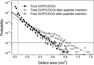
Sensing membrane curvature allows fine-tuning of complex reactions that occur at the surface of membrane-bound organelles. One of the most sensitive membrane curvature sensors, the Amphipathic Lipid Packing Sensor (ALPS) motif, does not seem to recognize the curved surface geometry of membranes per se; rather, it recognizes defects in lipid packing that arise from membrane bending. In a companion paper, we show that these defects can be mimicked by introducing conical lipids in a flat lipid bilayer, in agreement with experimental observations. Here, we use molecular-dynamics (MD) simulations to characterize ALPS binding to such lipid bilayers. The ALPS motif recognizes lipid-packing defects by a conserved mechanism: peptide partitioning is driven by the insertion of hydrophobic residues into large packing defects that are preformed in the bilayer. This insertion induces only minor modifications in the statistical distribution of the free packing defects. ALPS insertion is severely hampered when monounsaturated lipids are replaced by saturated lipids, leading to a decrease in packing defects. We propose that the hypersensitivity of ALPS motifs to lipid packing defects results from the repetitive use of hydrophobic insertions along the monotonous ALPS sequence.


Figures

Figure 1
Figure 1
The ALPS motif of ArfGAP1 binds to large DOPC/DOG liposomes. (a) CD spectra of the ArfGAP1 (amino acids 197–231) peptide in the absence (white dots) or presence (black dots) of sonicated DOPC liposomes (RH = 16.6 ± 8 nm). (b) Spectra of the peptide alone (white dots) or in the presence of large DOPC (RH = 115 ± 52 nm; black dots) or DOPC/DOG liposomes (85/15 mol/mol, RH = 108 ± 48 nm; gray dots). All experiments were performed at 25°C in 10 mM Tris pH 7.2 and 120 mM NaCl buffer. Peptide concentration: 25 μM; lipid concentration: 5 mM.
Figure 2
Figure 2
Insertion mechanism of the ALPS motif of ArfGAP1 in a DOPC/DOG bilayer. (a) Time evolution of the distance between the center of mass of residue F26 and the average glycerol level of lipid molecules. (b) Time evolution of the distance between the center of mass of residue F4 and the average glycerol level of lipid molecules. (c) Time evolution of the LJ energy between the protein and the lipid molecules. (d) Averaged time evolution of protein backbone density along the normal to the bilayer plane. (e) MD snapshot (t = 280 ns) of the ALPS motif of ArfGAP1 inserted into a DOPC/DOG bilayer. The peptide is shown in green in cartoon representation and the inserting phenylalanine residues (F26 and F4) are shown in van der Waals (vdW) representation. All data shown are taken from MD run c.
Figure 3
Figure 3
Colocalization between lipid packing defects and peptide insertion. (a) Top view of lipid molecules and packing defects in a representative snapshot from MD simulations of the ALPS motif of ArfGAP1 with a DOPC/DOG bilayer. Lipids are shown in surface representation with acyl chains in yellow and polar heads in gray. Packing defects are depicted in blue. The peptide is not shown. The apparent defects at the contour of the lipid bilayer are shown for clarity but are not considered as packing defects in the analysis. (b) Colocalization between the ALPS motif of ArfGAP1 and lipid packing defects. The peptide is shown in green in cartoon representation and the inserting phenylalanine residues (F26 and F4) are shown in vdW representation. DOG molecules are shown in licorice representation.
Figure 4
Figure 4
Role of packing defects in the insertion mechanism of hydrophobic residues. (a) Time evolution of the distance computed along the normal to the lipid bilayer between the center of mass of F26 and the average coordinate of the glycerol atoms (red) and time evolution of the size of the packing defect with the same coordinates in the membrane plane as F26 (black). (b–d) Upper panel: side view of the protein and lipid molecules in the proximity of a lipid packing defect at different times. In b, insertion does not take place; in c the residue is about to insert below the glycerol level; and in d the residue is fully inserted. Lower panel: localization of packing defects in the corresponding snapshots. The packing defect that is localized in close proximity to F26 is depicted in red.
Figure 5
Figure 5
Effect of peptide insertion on the size distribution of lipid packing defects. Histograms and exponential fit of defect areas in different conditions: pure DOPC/DOG bilayer (black dots) and DOPC/DOG bilayer after peptide insertion (white squares and white triangles). White squares: all packing defects in the leaflet where insertion takes place; white triangles: packing defects in the leaflet where insertion takes place after removal of defects that are occupied by the inserting peptide.
Figure 6
Figure 6
The ALPS motif of GMAP210 exhibits repeated hydrophobic insertions in DOPC/DOG bilayers. (a) Side view of an MD snapshot (after 1 μs) of the ALPS motif of GMAP210 inserted into a DOPC/DOG lipid bilayer. The peptide is shown in green in cartoon representation and the inserting hydrophobic residues (M1, W4, L5, L8, L16, L26, I30, F33, M37, and L38) are shown in vdW representation. (b) Top view of the colocalization between the hydrophobic insertions and geometrical lipid packing defects (blue). DOG molecules are shown in licorice representation. C- and N-terminal regions are explicitly indicated. A small defect below the N-terminus is not visible due to overlap with the N-terminus of the peptide.
Figure 7
Figure 7
Effect of packing defects on ALPS partitioning in the lipid bilayer. (a) Size distribution of packing defects for mixed DOPC/DOG (black) and pure DMPC (gray) bilayers. The ratio between the total area of lipid packing defects > 0.2 nm2 and the total surface area is 0.47% for DOPC/DOG and 0.19% for DMPC. (b) Total number of hydrophobic insertions in five independent MD simulations of 400 ns each for the ALPS motifs of ArfGAP1 and GMAP210 with DOPC/DOG (black) and DMPC (gray) bilayers. (Note that panel a is a simplified version of Fig. 5 in the companion article (23).

Comment in

Similar articles

Cited by

References

    1. Antonny B., Huber I., Cassel D. Activation of ADP-ribosylation factor 1 GTPase-activating protein by phosphatidylcholine-derived diacylglycerols. J. Biol. Chem. 1997;272:30848–30851. - PubMed
    1. Bigay J., Gounon P., Antonny B. Lipid packing sensed by ArfGAP1 couples COPI coat disassembly to membrane bilayer curvature. Nature. 2003;426:563–566. - PubMed
    1. McMahon H.T., Gallop J.L. Membrane curvature and mechanisms of dynamic cell membrane remodelling. Nature. 2005;438:590–596. - PubMed
    1. Antonny B. Mechanisms of membrane curvature sensing. Annu. Rev. Biochem. 2011;80:101–123. - PubMed
    1. Zimmerberg J., Kozlov M.M. How proteins produce cellular membrane curvature. Nat. Rev. Mol. Cell Biol. 2006;7:9–19. - PubMed

Publication types

LinkOut - more resources