. 2013 Feb 5;104(3):705-15.
doi: 10.1016/j.bpj.2012.12.003.

Modeling the assembly of the multiple domains of α-actinin-4 and its role in actin cross-linking

Affiliations

Modeling the assembly of the multiple domains of α-actinin-4 and its role in actin cross-linking

Timothy Travers et al. Biophys J. .

Abstract

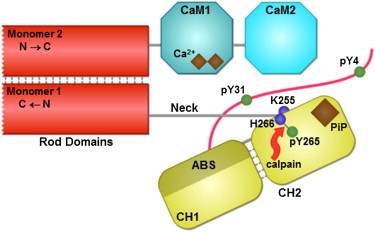
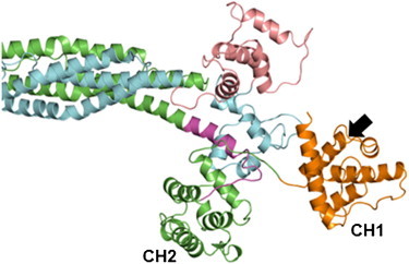
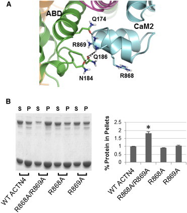
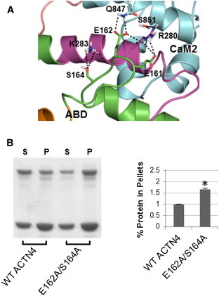
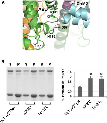
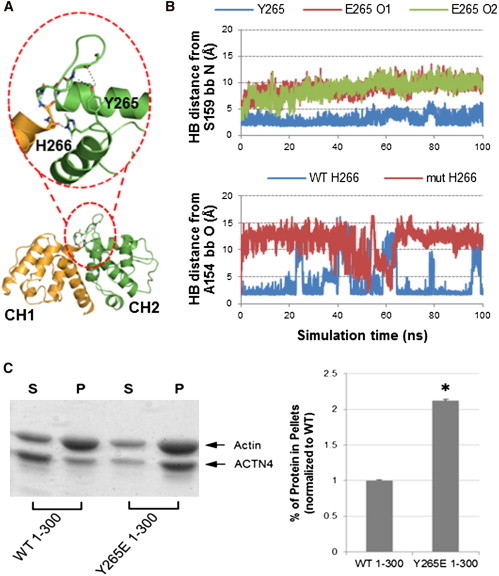
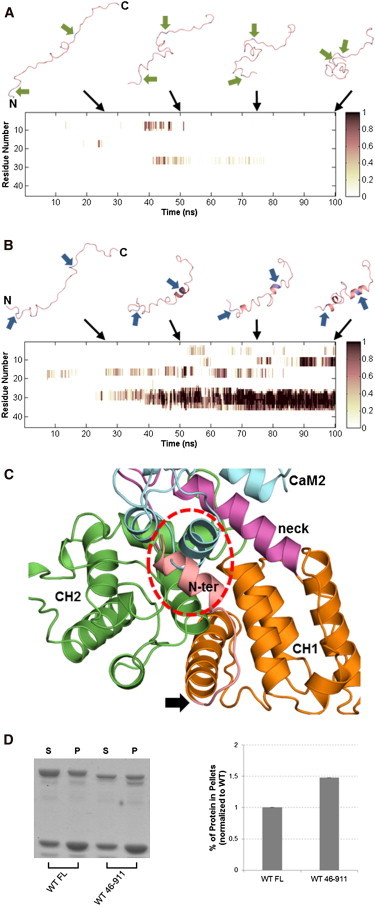
The assembly of proteins into multidomain complexes is critical for their function. In eukaryotic nonmuscle cells, regulation of the homodimeric actin cross-linking protein α-actinin-4 (ACTN4) during cell migration involves signaling receptors with intrinsic tyrosine kinase activity, yet the underlying molecular mechanisms are poorly understood. As a first step to address the latter, we validate here an atomic model for the ACTN4 end region, which corresponds to a ternary complex between the N-terminal actin-binding domain (ABD) and an adjacent helical neck region of one monomer, and the C-terminal calmodulin-like domain of the opposite antiparallel monomer. Mutagenesis experiments designed to disrupt this ternary complex confirm that its formation reduces binding to F-actin. Molecular dynamics simulations show that the phosphomimic mutation Y265E increases actin binding by breaking several interactions that tether the two calponin homology domains into a closed ABD conformation. Simulations also show a disorder-to-order transition in the double phosphomimic mutant Y4E/Y31E of the 45-residue ACTN4 N-terminal region, which can inhibit actin binding by latching both calponin homology domains more tightly. Collectively, these studies provide a starting point for understanding the role of external cues in regulating ACTN4, with different phenotypes resulting from changes in the multidomain assembly of the protein.


Figures

Figure 1
Figure 1
Domain architecture of ACTN4. Human ACTN4 (911 residues) forms antiparallel homodimers through binding of the spectrin rod domains. Monomer 1 shows an unstructured region; an ABD comprised of two CH domains (CH1 contains the ABS and CH2 has a putative PiP-binding site); and a neck region that connects CH2 to the first spectrin repeat of the rod domain. Monomer 2 shows the rod domain and C-terminal CaM-like domain (only CaM1 contains putative binding sites for calcium ions). Also shown are a target for m-calpain proteolysis between Y265 and H266, and residue K255 that has been implicated as a disease mutation site. The tyrosine residues Y4, Y31, and Y265 are phosphorylatable.
Figure 2
Figure 2
Neck-CaM2 complex homology model. (A) The helical neck is shown in surface representation with coloring based on side-chain hydrophobicity using the Eisenberg scale (52). Colors go from white to yellow, in the direction of increased side-chain hydrophobicity. CaM2 is shown in blue. (B) Comparison of our pairwise sequence alignment between titin repeat ZR7 and the neck region of ACTN4 (residues 272–294) with that suggested by Young and Gautel (residues 276–298) (8). Red boxes denote matching of neck residues with those in titin ZR7 that are in contact with CaM2 based on the solution structure in PDB 1H8B (23). Numbers in parentheses give the percent sequence similarity for the global alignment as computed by the EMBOSS (53) stretcher algorithm (using default parameters) at the EMBL-EBI website (http://www.ebi.ac.uk/Tools/psa/emboss_stretcher/). (C) Surface representation is of CaM2, colored according to electrostatic potential using APBS (54). The neck is shown in magenta, except for the three helical turns in yellow that contribute hydrophobic residues to the binding interface. Also shown are polar contacts that stabilize neck-CaM2, including K273-E870, R280-Q847, R280-S851, N288-Y878, and E292-R882. (D) Backbone root mean-square deviation (RMSD) plots of the neck region from 50-ns explicit solvent MD simulations of the neck-CaM2 complex. Blue curve shows that the simulation of the homology model based on our alignment was stable throughout the trajectory; the model based on the Y&G alignment (magenta curve) shows a large increase in backbone RMSD at around 34 ns into the simulation when the neck region twisted to a position similar to our homology model (green curve; RMSD relative to our model).
Figure 3
Figure 3
The ACTN4 neck fits in between the ABD and CaM2. (A) Surface representation shows the ABD-CaM2 complex from the closed ABD end of cryo-EM structure PDB 1SJJ (25). View of the neck (shown in cartoon only) is through its helical axis. (B) Interactions between the neck and CH2 of the ABD are predominantly polar in nature. Residues that form polar interactions are shown in sticks. Several nonpolar residues are also shown extending from three helical turns and away from CH2. These residues are part of the binding interface with CaM2.
Figure 4
Figure 4
Atomic model of six domains of ACTN4 linked by a novel ternary complex between ABD, neck, and CaM2 resolved from known pairwise interactions. The full model (shown in the center) was built from homology models to published structures (PDB IDs of known pairwise interactions shown surrounding the central model). Monomer 1 containing ABD is colored green, except for CH1 (orange) and the neck region (magenta). Monomer 2 containing the CaM-like domain is colored light blue, with CaM1 (pink) positioned on top of the ABD-neck-CaM2 ternary complex. The black arrow points to the ABS on CH1, whose access to F-actin is partially blocked by the rod domains in the closed ABD structure. For clarity, the disordered N-terminal is not shown.
Figure 5
Figure 5
Atomic model of open ABD in ACTN4. Same reference and color scheme as in Fig. 4. Except for the unbinding of CH1 and CH2, none of the other domains need to move to access the open ABD state. Note that this state is in an optimal position to bind F-actin because the other domains do not hinder the ABS (pointed to by black arrow) on CH1.
Figure 6
Figure 6
Experimental validation of residues R868/R869 that stabilize the ternary complex. (A) ABD-CaM2 interface: R869 of CaM2 makes HBs (dashed lines) with polar side chains from CH2. R868 can make these same H-bonds in the R869A mutant by turning toward the polar cluster. (B) Representative image (left) and quantitative results (right) of Coomassie Blue G-250 stained polyacrylamide gels for WT, R868A/R869A, R868A, and R869A constructs of full-length human ACTN4. Image analysis and quantitation of three experiments are shown in the plot, with the percent of ACTN4 that cosedimented with actin for each construct normalized relative to WT. Student’s t-test was performed to compare each mutant with WT. Asterisks denote p < 0.05 between the labeled mutant and WT. Error bars indicate ± SD.
Figure 7
Figure 7
Experimental validation of residues E162/S164 that stabilize the ternary complex. (A) ABD-neck-CaM2 interface: E161, E162, and S164 of the ABD interact with R280 and K283 of the neck and Q847 of CaM2 to keep the core ternary complex stable. (B) Representative image (left) and quantitative results of three experiments (right) of Coomassie Blue G-250 stained polyacrylamide gels (as in Fig. 6B) for WT and E162A/S164A constructs of full-length human ACTN4.
Figure 8
Figure 8
Experimental validation of residue H189 that stabilizes the ternary complex. (A) ABD-CaM2 interface: H189 of ABD interacts with D874 of CaM2. The other two residues (K181 and K193) of the putative PiP-binding site on CH2 are also shown, with neither of them able to make contacts with CaM2. (B) Representative image (left) and quantitative results of three experiments (right) of Coomassie Blue G-250 stained polyacrylamide gels (as in Fig. 6B) for WT, H189L, and ΔPBD (see text) constructs of full-length human ACTN4.
Figure 9
Figure 9
ACTN4 regulation by phosphorylation at Y265. (A) Crystal structure of CH1 (orange) and CH2 (green) from ACTN4 ABD (PDB 2R0O) (11). Zoomed area highlights HBs of Y265 and H266 with the backbone of the loop connecting the two CH domains. (B) HB distance plots between the S159 backbone and the side chain at residue 265 (top) and between the A154 backbone and H266 side chain (bottom) from 100-ns explicit solvent MD simulations. For the WT simulation (blue curves), these HBs are kept mostly stable throughout the trajectory. For the Y265E simulation (magenta and green curves), the HB to the S159 backbone is not formed with either carboxylate oxygen of Glu, whereas the HB involving H266 is broken for most of the trajectory. (C) Phosphorylation mimic at Y265 increases actin binding for the 1-300 construct. Representative image (left) and quantitative results of three experiments (right) of Coomassie Blue G-250-stained polyacrylamide gels (as in Fig. 6B) for WT and Y265E constructs of human ACTN4 1-300.
Figure 10
Figure 10
ACTN4 regulation by the N-terminal region. Helicity plots over 100-ns implicit solvent Langevin dynamics simulations of WT (A) and Y4E/Y31E (B) constructs of the 45-residue N-terminal region. Time points were averaged per residue over sequential 100-ps windows. Darker shades signify higher helical content. Above each plot are representative snapshots at 25, 50, 75, and 100 ns into each trajectory. The termini of both constructs are labeled for the 25-ns snapshots. The two arrows adjacent to each peptide (A) point to WT Y4 and Y31 residues, or (B) point to the phosphomimic mutations Y4E and Y31E. Both simulations were started with constructs in fully extended conformations. (C) Top-ranked docked model of the helical motif around Y31E and ACTN4 ABD superimposed with the core ternary complex. The circled area highlights a clash between the N-terminal helix and CaM2. Arrow points to where the N-terminal connects to CH1, indicating that this is a feasible docked model. (D) Removal of the N-terminal enhances binding to F-actin. Representative image (left) and quantitative results (right) of Coomassie Blue G-250-stained polyacrylamide gel for full-length (FL) and 46-911 truncation mutant constructs of human ACTN4. Image analysis and quantitation are shown in the plot, with percent protein in pellets for the mutant normalized relative to WT.

Similar articles

Cited by

References

    1. Otey C.A., Carpen O. Alpha-actinin revisited: a fresh look at an old player. Cell Motil. Cytoskeleton. 2004;58:104–111. - PubMed
    1. Sjöblom B., Salmazo A., Djinović-Carugo K. Alpha-actinin structure and regulation. Cell. Mol. Life Sci. 2008;65:2688–2701. - PMC - PubMed
    1. Dandapani S.V., Sugimoto H., Pollak M.R. Alpha-actinin-4 is required for normal podocyte adhesion. J. Biol. Chem. 2007;282:467–477. - PubMed
    1. Shao H., Wang J.H., Wells A. α-actinin-4 is essential for maintaining the spreading, motility and contractility of fibroblasts. PLoS ONE. 2010;5:e13921. - PMC - PubMed
    1. Kanchanawong P., Shtengel G., Waterman C.M. Nanoscale architecture of integrin-based cell adhesions. Nature. 2010;468:580–584. - PMC - PubMed

Publication types