. 2013:3:1293.
doi: 10.1038/srep01293.

Photothermal nanodrugs: potential of TNF-gold nanospheres for cancer theranostics

Affiliations

Photothermal nanodrugs: potential of TNF-gold nanospheres for cancer theranostics

Jingwei Shao et al. Sci Rep. 2013.

Abstract

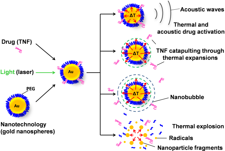
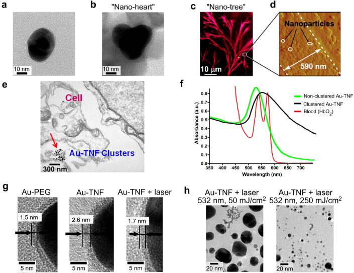
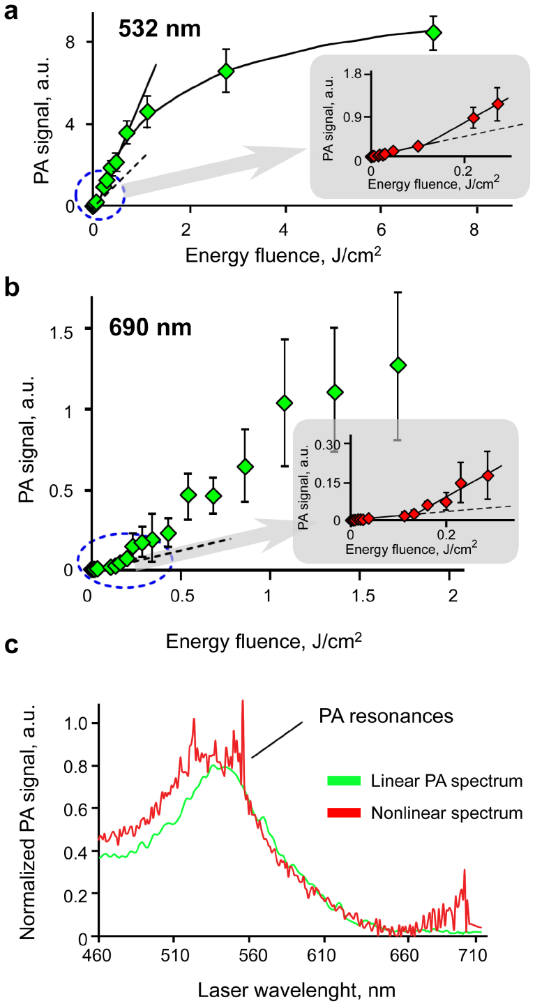
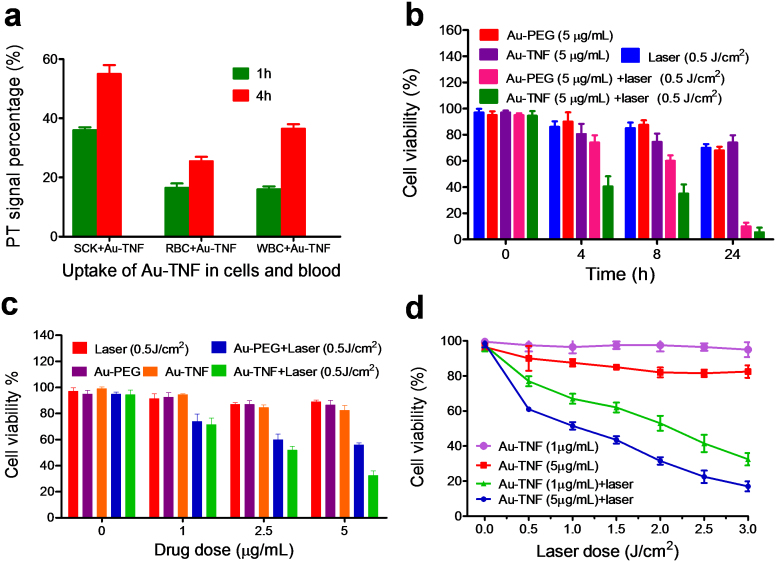
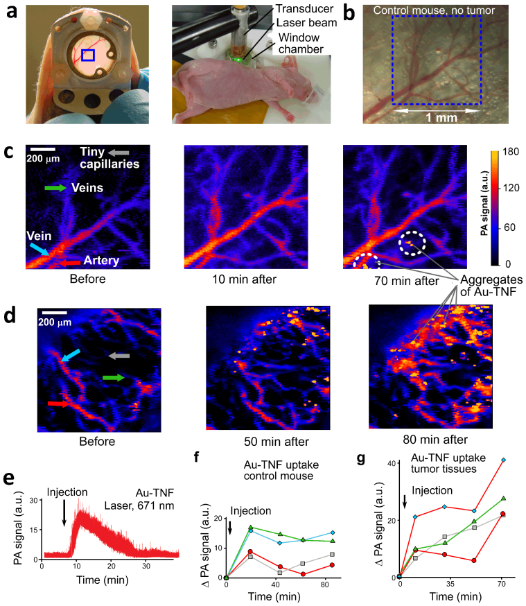
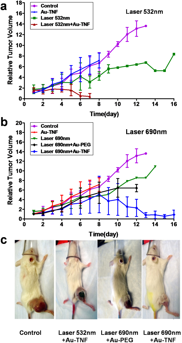
Nanotechnology has been extensively explored for drug delivery. Here, we introduce the concept of a nanodrug based on synergy of photothermally-activated physical and biological effects in nanoparticle-drug conjugates. To prove this concept, we utilized tumor necrosis factor-alpha coated gold nanospheres (Au-TNF) heated by laser pulses. To enhance photothermal efficiency in near-infrared window of tissue transparency we explored slightly ellipsoidal nanoparticles, its clustering, and laser-induced nonlinear dynamic phenomena leading to amplification and spectral sharpening of photothermal and photoacoustic resonances red-shifted relatively to linear plasmonic resonances. Using a murine carcinoma model, we demonstrated higher therapy efficacy of Au-TNF conjugates compared to laser and Au-TNF alone or laser with TNF-free gold nanospheres. The photothermal activation of low toxicity Au-TNF conjugates, which are in phase II trials in humans, with a laser approved for medical applications opens new avenues in the development of clinically relevant nanodrugs with synergistic antitumor theranostic action.


Figures

Figure 1
Figure 1. Principle of nanodrug action based on PT activation of TNF- gold nanosphere conjugates through controllable temperature increase, thermal expansion, acoustic nanobubble, and explosion phenomena.
Figure 2
Figure 2. Au-TNF nanoparticle characterization.
TEM imaging of (a) single nanoparticle and (b) of a small cluster of three nanoparticles (“nano-heart”). (c) Optical image of large nanoparticle cluster (“nano-tree”) in dried solution. (d) Atomic force microscopy of nanoparticle cluster fragment corresponding to a white square in (c). (e) Transmission electron microscopy (TEM) image of Au-TNF clusters in 2H11 cells, 24 h incubation. (f) Absorption spectra of blood, single, and clustered Au-TNF nanoparticles. (g) TNF release from PEG coating of Au-TNF during laser heating (wavelength, 532 nm; energy fluence, 50 mJ/cm2). (h) Nanoparticle fragmentation by high energy laser pulses.
Figure 3
Figure 3. Nonlinear PA dynamics in Au-TNF conjugates.
(a), (b) PA signal amplitudes as a function of laser energy fluence at laser wavelengths of 532 nm (a) and 690 nm (b). (c) Linear and nonlinear normalized PA spectra of Au-TNF suspension at laser energy fluence of 50 mJ/cm2 and 500 mJ/cm2. All PA experiments were performed in 1011 particles/mL Au-TNF solution (120 μm light path), laser beam diameter 50 μm.
Figure 4
Figure 4. In vitro cell-nanoparticle interaction and cytotoxicity of Au-TNF with pulsed laser.
(a) Cellular uptake of Au-TNF in SCK cancer cells and murine blood. (b) Cell viability after different therapeutic regimens. (c) Cell viability after photothermal therapy and graded drug concentration. (d) Cell viability after different laser energies for selected Au-TNF concentration. Results are expressed as the mean ±SD of three replicates of each treatment.
Figure 5
Figure 5. Au-TNF pharmacokinetics in murine model.
(a) Window chamber (left) and PA probe with ultrasound transducer (right). (b) Healthy mouse vasculature in window chamber. (c) PA imaging of healthy tissues before and after i.v. Au-TNF injection. (d) SCK tumor vasculature after i.v. Au-TNF injection. Local intensive PA signals correspond to nanoparticle clusters. (e) In vivo PA flow cytometry of Au-TNF nanoparticle kinetics in mouse ear microvessels. (f, g) Increase in PA signal after Au-TNF injection (data from panels c and d) for healthy (f) and tumor bearing (g) mice. Laser parameters for c and d: wavelength, 532 nm; energy fluence, 50 mJ/cm2; scanning step size, 10 μm. Laser parameters for e: 671 nm, 100 mJ/cm2.
Figure 6
Figure 6. In vivo anti-tumor effect of Au-TNF nanoparticles activated by pulsed laser at wavelength of 532 nm and 690 nm.
Relative tumor volume increase after treatment with laser alone, and laser with Au-TNF or Au-PEG at laser wavelength of 532 nm (a) or 690 nm (b) at 8 h after an i.v. injection of nanoparticles. (c) Tumor images of different mouse groups. Each group contained 5 mice.

References

    1. Agarwal A., Mackey M. A., El-Sayed M. A. & Bellamkonda R. V. Remote triggered release of doxorubicin in tumors by synergistic application of thermosensitive liposomes and gold nanorods. ACS Nano 5, 4919–4926 (2011). - PubMed
    1. Dreaden E. C. & El-Sayed M. A. Detecting and destroying cancer cells in more than one way with noble metals and different confinement properties on the nanoscale. Chem. Res. 45, 1854–65 (2012). - PMC - PubMed
    1. Farokhzad O. C. & Langer R. Impact of nanotechnology on drug delivery. ACS Nano 3, 16–20 (2009). - PubMed
    1. Ferrari M. Cancer nanotechnology: opportunities and challenges. Nat. Rev. Cancer 5, 161–171 (2005). - PubMed
    1. Koo O. M., Rubinstein I. & Onyuksel H. Role of nanotechnology in targeted drug delivery and imaging: a concise review. Nanomedicine: Nanotechnol, Biology and Medicine 1, 193–212 (2005). - PubMed

Publication types

MeSH terms