. 2012 Nov 27;13(12):15867-80.
doi: 10.3390/ijms131215867.

Impact of cyanidin-3-glucoside on glycated LDL-induced NADPH oxidase activation, mitochondrial dysfunction and cell viability in cultured vascular endothelial cells

Affiliations

Impact of cyanidin-3-glucoside on glycated LDL-induced NADPH oxidase activation, mitochondrial dysfunction and cell viability in cultured vascular endothelial cells

Xueping Xie et al. Int J Mol Sci. .

Abstract

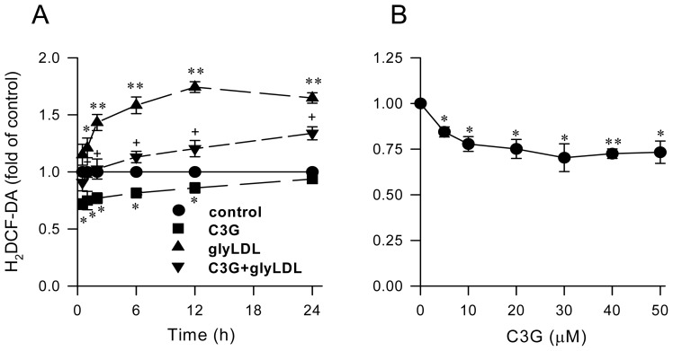
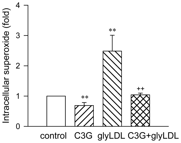
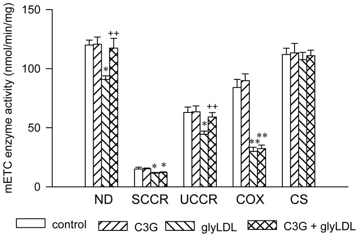
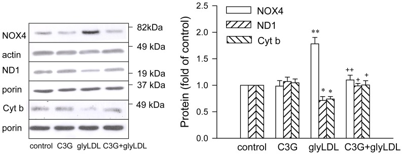
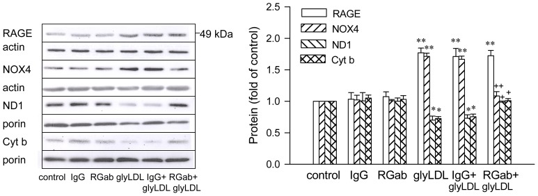
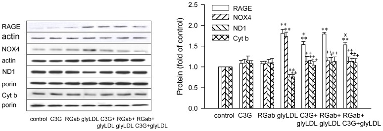
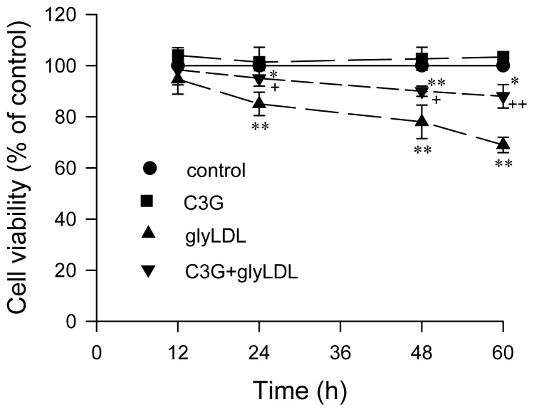
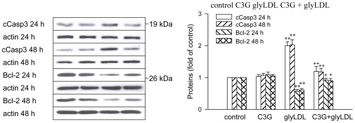
Elevated levels of glycated low density lipoprotein (glyLDL) are frequently detected in diabetic patients. Previous studies demonstrated that glyLDL increased the production of reactive oxygen species (ROS), activated NADPH oxidase (NOX) and suppressed mitochondrial electron transport chain (mETC) enzyme activities in vascular endothelial cells (EC). The present study examined the effects of cyanidin-3-glucoside (C3G), a type of anthocyanin abundant in dark-skinned berries, on glyLDL-induced ROS production, NOX activation and mETC enzyme activity in porcine aortic EC (PAEC). Co-treatment of C3G prevented glyLDL-induced upregulation of NOX4 and intracellular superoxide production in EC. C3G normalized glyLDL-induced inhibition on the enzyme activities of mETC Complex I and III, as well as the abundances of NADH dehydrogenase 1 in Complex I and cytochrome b in Complex III in EC. Blocking antibody for the receptor of advanced glycation end products (RAGE) prevented glyLDL-induced changes in NOX and mETC enzymes. Combination of C3G and RAGE antibody did not significantly enhance glyLDL-induced inhibition of NOX or mETC enzymes. C3G reduced glyLDL-induced RAGE expression with the presence of RAGE antibody. C3G prevented prolonged incubation with the glyLDL-induced decrease in cell viability and the imbalance between key regulators for cell viability (cleaved caspase 3 and B cell Lyphoma-2) in EC. The findings suggest that RAGE plays an important role in glyLDL-induced oxidative stress in vascular EC. C3G may prevent glyLDL-induced NOX activation, the impairment of mETC enzymes and cell viability in cultured vascular EC.


Figures

Figure 1
Figure 1
Effect of C3G on redox status in EC. PAEC were treated with vehicle (control), 30 μM C3G, 100 μg/mL of glyLDL or C3G + glyLDL for 0.5–24 h (A) or with 0–50 μM C3G for 30 min (B). Redox status was assessed by measuring fluorescence intensity in cells at 485/530 nm (excitation/emission) using a fluorescence microplate reader. Values were expressed in mean ± SD in fold of control (n = 3 independent experiments). *, **: p < 0.05 or 0.01 versus control; +: p < 0.05 versus glyLDL.
Figure 2
Figure 2
Effect of C3G on glyLDL-induced intracellular superoxide in EC. PAEC were treated with vehicle (control), C3G (30 μM), glyLDL (100 μg/mL) or C3G + gLDL for 2 h. Intracellular superoxide was measured using lucigenin method. Values were expressed in means ± SD in fold of control (n = 3 independent experiments). **: p < 0.01 versus control; ++: p < 0.01 versus glyLDL.
Figure 3
Figure 3
Effects of C3G on the activities of mitochondrial respiratory chain complex enzymes in glyLDL-treated EC. PAEC were treated with vehicle (control), 30 μM C3G, 100 μg/mL of glyLDL or C3G + glyLDL for 12 h. Activities of NADH dehydrogenase (ND), succinate cytochrome c redutase (SCCR), ubiquinol cytochrome c reductase (UCCR), cytochrome c oxidase (COX) and citrate synthase (CS) in EC were analyzed as described in the experimental section. Values were expressed in mean ± SD in nmol/min/mg protein (n = 3 independent experiments). *, **: p < 0.05 or 0.01 versus control; ++: p < 0.01 versus glyLDL.
Figure 4
Figure 4
Effects of C3G on glyLDL-induced changes in NOX4, ND1 and Cyt b content. PAEC were treated with vehicle (control), 30 μM C3G, 100 μg/mL of glyLDL or C3G + glyLDL for 12 h. Abundances of NOX4, ND1 and Cyt b were measured using Western blotting. Molecular weights of standards were marked beside the blots. Values were expressed in mean ± SD folds of control (n = 3 independent experiments). *, **: p < 0.05 or 0.01 versus control; +, ++: p < 0.05 or 0.01 versus glyLDL.
Figure 5
Figure 5
Effects of RAGE antibody on glyLDL-induced changes in RAGE, NOX4, ND1 and Cyt b content. PAEC were treated with vehicle (control), 10 μg/mL of rabbit IgG, 10 μg/mL of RAGE antibody (RGab), 100 μg/mL of glyLDL, IgG + glyLDL or RGab + glyLDL for 12 h. Abundances of NOX4, RAGE, ND1 and Cyt b were measured using Western blotting. Values were expressed in mean ± SD folds of control (n = 3 independent experiments). *, **: p < 0.05 or 0.01 versus control; +, ++: p < 0.05 or 0.01 versus glyLDL.
Figure 6
Figure 6
Effects of C3G and RAGE antibody on glyLDL-induced changes in RAGE, NOX4, ND1 and Cyt b content. PAEC were treated with vehicle (control), 30 μM C3G, 10 μg/mL of RAGE antibody (RGab), 100 μg/mL of glyLDL, C3G + glyLDL or RGab + C3G + glyLDL for 12 h. Abundances of NOX4, RAGE, ND1 and Cyt b were measured using Western blotting. Values were expressed in mean ± SD folds of control (n = 3 independent experiments). **: p < 0.01 versus control; +, ++: p < 0.05 or 0.01 versus glyLDL; x: p < 0.05 versus glyLDL plus RGab.
Figure 7
Figure 7
Effects of C3G and glyLDL on cell viability. PAEC were treated with vehicle (control), 30 μM C3G, 100 μg/mL of glyLDL or C3G + glyLDL for 12–60 h. Cell viability was measured using MTT assay as described in the experimental section. Values were expressed in means ± SD in percentage of control (n = 3 independent experiments). *, **: p < 0.05 or 0.01 versus control with corresponding incubation time; +, ++: p < 0.05 or 0.01 versus glyLDL.
Figure 8
Figure 8
Effects of C3G and glyLDL on regulators for cell viability. PAEC were treated with vehicle (control), 30 μM C3G, 100 μg/mL of glyLDL or C3G + glyLDL for 24 h or 48 h. Abundances of cleaved caspase 3 (cCasp3) and Bcl-2 were measured using Western blotting. Values were expressed in mean ± SD folds of control (n = 3 independent experiments). **: p < 0.01 versus control; +, ++: p < 0.05 or 0.01 versus glyLDL.

References

    1. Virella G., Lopes-Virella M.F. The Pathogenic Role of the Adaptive Immune Response to Modified LDL in Diabetes. Front. Endocrinol. (Lausanne) 2012;3:76. - PMC - PubMed
    1. Chao P.C., Huang C.N., Hsu C.C., Yin M.C., Guo Y.R. Association of dietary AGEs with circulating AGEs, glycated LDL, IL-1α and MCP-1 levels in type 2 diabetic patients. Eur. J. Nutr. 2010;49:429–434. - PubMed
    1. Zhao R., Shen G.X. Functional modulation of antioxidant enzymes in vascular endothelial cells by glycated LDL. Atherosclerosis. 2005;179:277–284. - PubMed
    1. Abid M.R., Kachra Z., Spokes K.C., Aird W.C. NADPH oxidase activity is required for endothelial cell proliferation and migration. FEBS Lett. 2000;4862:252–256. - PubMed
    1. Block K., Gorinm Y., Abboud H.E. Subcellular localization of Nox4 and regulation in diabetes. Proc. Natl. Acad. Sci. USA. 2009;106:14385–14390. - PMC - PubMed

Publication types