. 2013:4:1518.
doi: 10.1038/ncomms2504.

Greater efficiency of photosynthetic carbon fixation due to single amino-acid substitution

Affiliations
Free PMC article

Greater efficiency of photosynthetic carbon fixation due to single amino-acid substitution

Judith Katharina Paulus et al. Nat Commun. 2013.
Free PMC article

Abstract

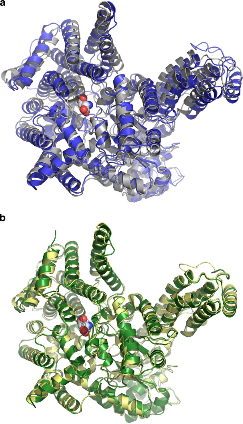
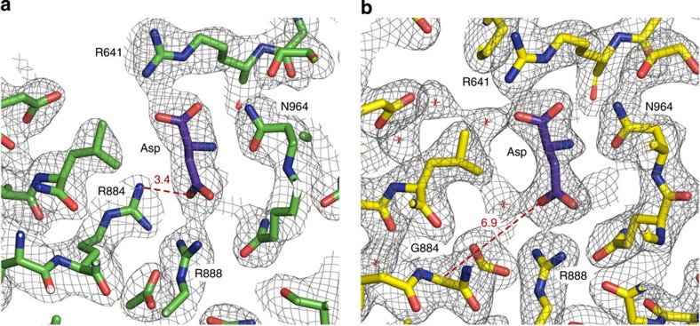
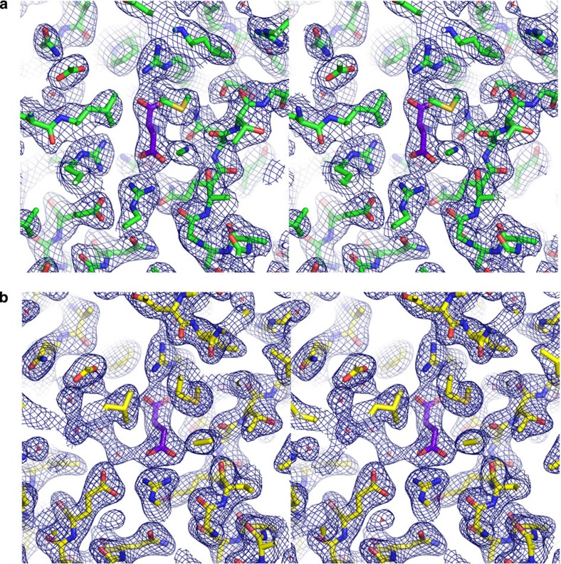
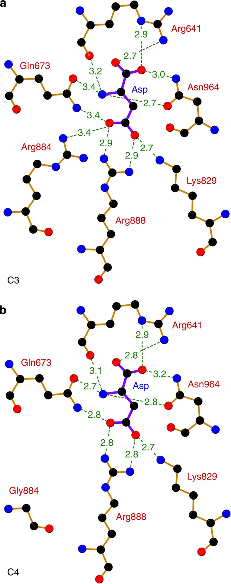
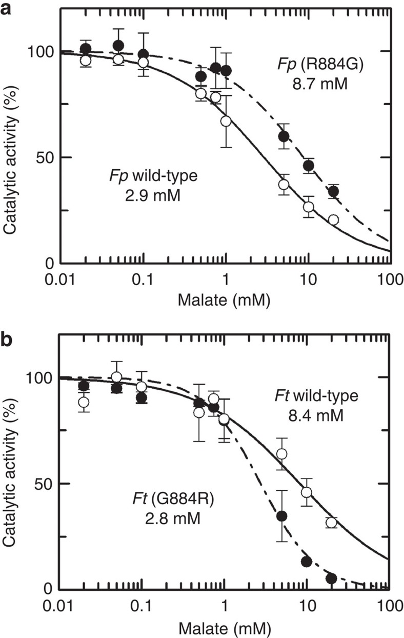
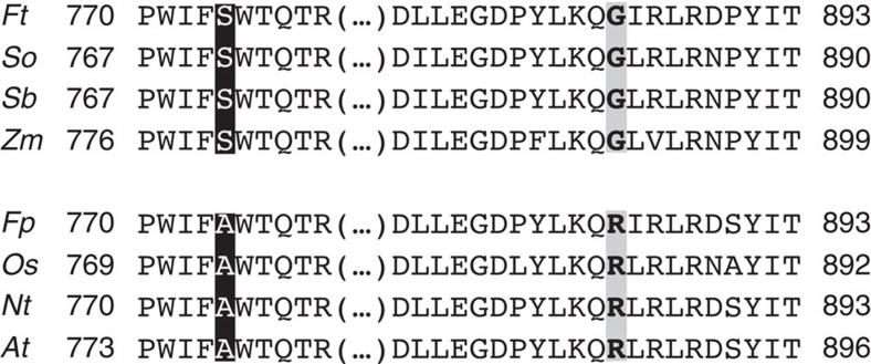
The C4-photosynthetic carbon cycle is an elaborated addition to the classical C3-photosynthetic pathway, which improves solar conversion efficiency. The key enzyme in this pathway, phosphoenolpyruvate carboxylase, has evolved from an ancestral non-photosynthetic C3 phosphoenolpyruvate carboxylase. During evolution, C4 phosphoenolpyruvate carboxylase has increased its kinetic efficiency and reduced its sensitivity towards the feedback inhibitors malate and aspartate. An open question is the molecular basis of the shift in inhibitor tolerance. Here we show that a single-point mutation is sufficient to account for the drastic differences between the inhibitor tolerances of C3 and C4 phosphoenolpyruvate carboxylases. We solved high-resolution X-ray crystal structures of a C3 phosphoenolpyruvate carboxylase and a closely related C4 phosphoenolpyruvate carboxylase. The comparison of both structures revealed that Arg884 supports tight inhibitor binding in the C3-type enzyme. In the C4 phosphoenolpyruvate carboxylase isoform, this arginine is replaced by glycine. The substitution reduces inhibitor affinity and enables the enzyme to participate in the C4 photosynthesis pathway.


Figures

Figure 1
Figure 1. Structural comparison of C3 PEPC and C4 PEPC monomers.
(a) Overlay of PEPC from E. coli (C3-type, grey) and Z. mays (C4-type, blue). The calculated root-mean-square deviaton (r.m.s.d.) of both monomers is 2.0 Å. (b) Superposition of PEPC from F. pringlei (C3-type, green) and F. trinervia (C4-type, yellow). The Flaveria PEPCs have a high overall topological similarity with a r.m.s.d. of 0.4 Å. Structures are shown as ribbons with the aspartate/malate-binding site on top. The bound inhibitors are shown as spheres representing van der Waals radii.
Figure 2
Figure 2. Comparison of the inhibitor-binding sites.
(a) C3 PEPC from F. pringlei. (b) C4 PEPC from F. trinervia. Arg884 in C3 PEPC functions as a clamp by providing an additional hydrogen bond for inhibitor binding, while Gly884 in C4 PEPC is more than 6 Å away from the inhibitor. Structures and 2FoFc electron densities at 1.2σ of the malate/aspartate-binding pockets.
Figure 3
Figure 3. Electron density maps and final model structures.
(a) C3 PEPC from F. pringlei, (b) C4 PEPC from F. trinervia. Shown are the refined 2FoFc electron density maps around the inhibitor-binding sites. The maps are contoured at 1.2σ, where σ represents the root mean square of the density fluctuation of the unit cell. The model structures are shown as sticks. Carbon atoms are coloured green, yellow and purple for F. pringlei PEPC, F. trinveria PEPC and the inhibitor Asp, respectively. Nitrogen atoms are coloured blue, oxygens red and sulphurs yellow.
Figure 4
Figure 4. Side by side comparison of the feedback inhibitor-binding site.
(a) Inhibitor-binding site of C3 PEPC (F. pringlei). (b) Inhibitor-binding site of C4 PEPC (F. trinervia). The distances between the inhibitor aspartate and amino acids of the binding pocket are given in Å. The estimated coordinate errors are 0.24 Å (F. pringlei C3 PEPC) and 0.29 Å (F. trinervia C4 PEPC). Residues Arg641, Lys829, Arg888 and Asn964 have been identified as the malate-binding motif. In C3 PEPC, Arg884 provides an additional hydrogen bond for inhibitor binding. In C4 PEPC, this residue is replaced by glycine, which is 6.9 Å away from the inhibitor molecule.
Figure 5
Figure 5. Determination of IC50 values for wild-type and mutant PEPC from F. pringlei (C3) and F. trinervia (C4).
(a) The mean activity values of F. pringlei wild-type C3 PEPC (open circles) and F. pringlei R884G mutant (solid circles) are plotted against the concentration of the feedback inhibitor malate. The calculated IC50 values are 2.9 mM for the wild-type (solid curve) and 8.7 mM for the R884G mutant C3 PEPC (dashed curve). (b) Plot of mean activity values of F. trinervia wild-type C4 PEPC (open circles) and F. trinervia G884R mutant (closed circles). IC50 values calculated from the experimental data are 8.4 mM for wild-type and 2.8 mM for the mutant C4 PEPC. S.d. was calculated from three independent experiments. Activities were measured in triplicates for each experiment.
Figure 6
Figure 6. Sequence alignment of C3 and C4 determining regions in various PEPCs.
Amino acids that determine C3/C4-specific function are highlighted. In the substrate-binding centre, Ala774 (Flaveria numbering) mediates C3 specificity, while Ser774 determines the increased kinetic efficiency of C4 PEPC (black boxes). Malate binding in the inhibitory site is controlled by Arg884 in the C3 enzyme, while the increased tolerance of the C4 PEPC towards the feedback inhibitor is mediated by Gly884 (grey boxes). The sequences are: Ft (F. trinervia UniProt No. P30694), So (Saccharum officinarum Q9FS96), Sb (Sorghum bicolor P15804), Zm (Zea mays P04711), Fp (F. pringlei GenBank CAA88829.1), Os (Oryza sativa subsp. indica Q84XH0), Nt (Nicotiana tabacum P27154) and At (Arabidopsis thaliana Q84VW9).

References

    1. Sage R. F.. The evolution of C4 photosynthesis. New Phytol. 161, 341–370 (2004). - PubMed
    1. Peterhänsel C. & Maurino V. G.. Photorespiration redesigned. Plant Physiol. 155, 49 (2011). - PMC - PubMed
    1. Sage R. F., Sage T. L. & Kocacinar F.. Photorespiration and the evolution of C4 photosynthesis. Annu. Rev. Plant Physiol. 63, 17.1–17.29 (2012). - PubMed
    1. Stitt M. & Heldt H. W.. Generation and maintenance of concentration gradients between the mesophyll and bundle sheath in maize leaves. Biochim. Biophys. Acta Bioenerg. 808, 400–414 (1985).
    1. Voznesenskaya E. V., Franceschi V. R., Kiirats O., Freitag H. & Edwards G. E.. Kranz anatomy is not essential for terrestrial C4 plant photosynthesis. Nature 414, 543–546 (2001). - PubMed

Publication types

MeSH terms