. 2013 May;93(5):771-80.
doi: 10.1189/jlb.1212647. Epub 2013 Feb 26.

Selenoprotein K is required for palmitoylation of CD36 in macrophages: implications in foam cell formation and atherogenesis

Affiliations

Selenoprotein K is required for palmitoylation of CD36 in macrophages: implications in foam cell formation and atherogenesis

Svenja Meiler et al. J Leukoc Biol. 2013 May.

Abstract

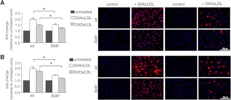
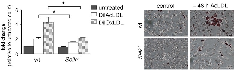
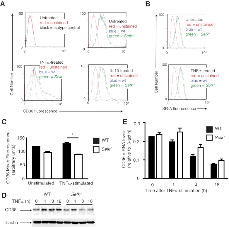
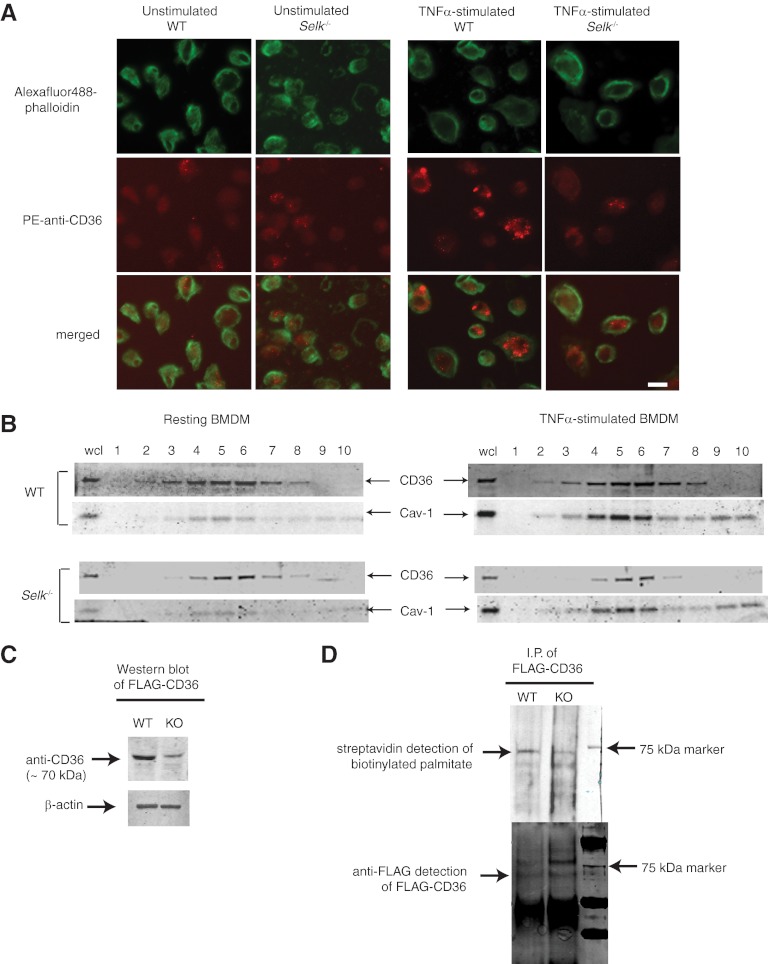
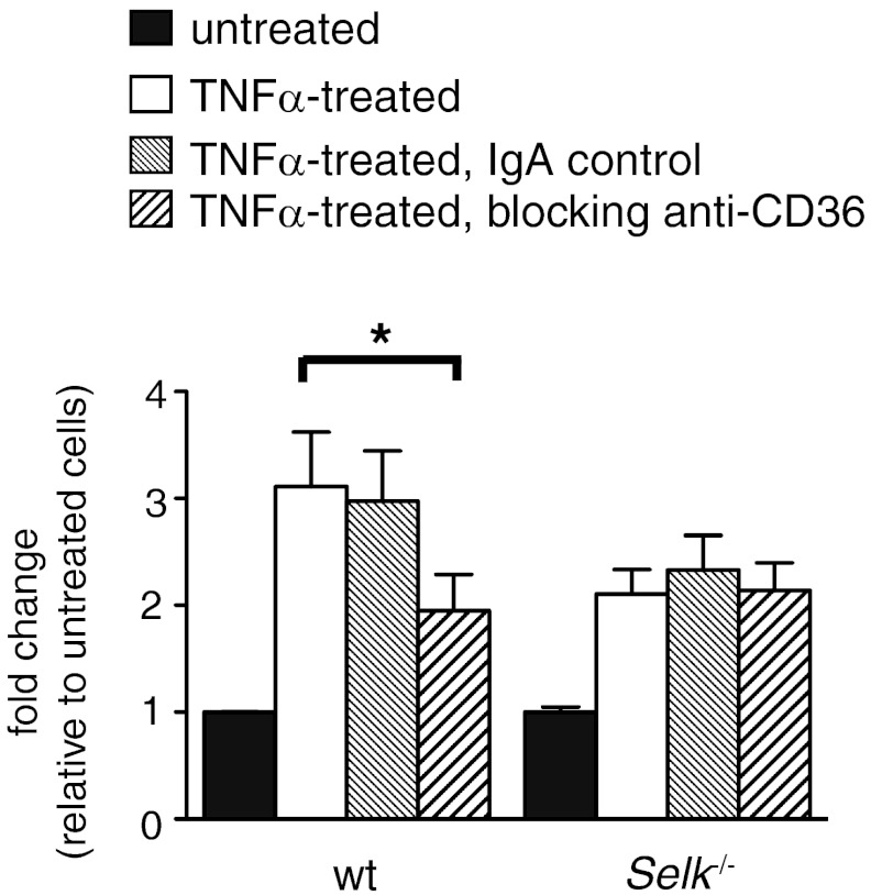
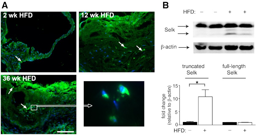
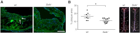
Selk is an ER transmembrane protein important for calcium flux and macrophage activation, but its role in foam cell formation and atherosclerosis has not been evaluated. BMDMs from Selk(-/-) mice exhibited decreased uptake of modLDL and foam cell formation compared with WT controls, and the differences were eliminated with anti-CD36 blocking antibody. CD36 expression was decreased in TNF-α-stimulated Selk(-/-) BMDMs compared with WT controls. Fluorescence microscopy revealed TNF-α-induced clustering of CD36 in WT BMDMs indicative of lipid raft localization, which was absent in Selk(-/-) BMDMs. Fractionation revealed lower levels of CD36 reaching lipid rafts in TNF-α-stimulated Selk(-/-) BMDMs. Immunoprecipitation showed that Selk(-/-) BMDMs have decreased CD36 palmitoylation, which occurs at the ER membrane and is crucial for stabilizing CD36 expression and directing its localization to lipid rafts. To assess if this phenomenon had a role in atherogenesis, a HFD was fed to irradiated Ldlr(-/-) mice reconstituted with BM from Selk(-/-) or WT mice. Selk was detected in aortic plaques of controls, particularly in macrophages. Selk(-/-) in immune cells led to reduction in atherosclerotic lesion formation without affecting leukocyte migration into the arterial wall. These findings suggest that Selk is important for stable, localized expression of CD36 in macrophages during inflammation, thereby contributing to foam cell formation and atherogenesis.


Figures

Figure 1.
Figure 1.. Selk−/− in macrophages decreases uptake of modLDL.
(A) WT or Selk−/− BMDMs were incubated with Dil-AcLDL or Dil-OxLDL, and levels of ingested, fluorescent modLDL determined by plate-based fluorimetry (bar graphs) and fluorescent microscopy (representative images). (B) BMDMs from WT or Selk−/− mice were treated with TNF-α for 4 h, followed by the addition of Dil-AcLDL or Dil-OxLDL, and uptake measured similar untreated BMDMs. Data represent mean ± se (n=3). *P < 0.05.
Figure 2.
Figure 2.. Selk−/− leads to decreased foam cell formation.
BMDMs from WT or Selk−/− mice were treated with TNF-α for 4 h, followed by incubation with AcLDL for 24 or 48 h. Levels of ingested AcLDL were determined by fluorescent microscopic evaluation of Oil Red O. Representative images are shown for lipid content at 48 h. Data represent mean ± se (n=3). *P < 0.05.
Figure 3.
Figure 3.. Selk−/− reduces TNF-α-induced surface expression of CD36.
(A) Flow cytometry was used to detect surface CD36 levels on BMDMs from WT (wt; blue) or Selk−/− (green) BMDMs with no treatment, TNF-α treatment, or IL-10 treatment. Several different time-points after TNF-α or IL-10 treatment were evaluated for effects on CD36 surface expression, and representative histograms are shown for 18 h poststimulation. (B) Flow cytometry was used to detect surface SR-A levels on BMDMs from WT (blue) or Selk−/− (green) BMDMs with no treatment or with TNF-α treatment. (C) Mean fluorescence for CD36 was measured by flow cytometry for WT or Selk−/− BMDMs, with or without TNF-α treatment for 3 h. (D) Western blot was performed on whole cell lysates from WT and Selk−/− BMDMs treated with TNF-α for increasing time to determine levels of CD36 protein. (E) Real-time PCR was used to evaluate CD36 mRNA levels normalized to β-actin mRNA in WT or Selk−/− BMDMs stimulated with TNF-α for increasing time. Data represent mean ± se (n=3). *P < 0.05.
Figure 4.
Figure 4.. Selk is required for palmitoylation and membrane clustering of CD36 during TNF-α stimulation.
(A) Unstimulated or TNF-α-stimulated WT and Selk−/− BMDMs were stained with PE anti-CD36 (red) and F-actin stained using AlexaFluor488-phalloidin (green). Staining was evaluated using confocal microscopy. Original scale bar: 10 μm. (B) Cell lysates from unstimulated or TNF-α-stimulated WT and Selk−/− BMDMs were separated into fractions by sucrose gradient centrifugation and proteins separated and analyzed by Western blot. CD36 was detected in several fractions and was particularly enriched in those fractions exhibiting the marker for lipid rafts, Cav-1. wcl, whole cell lysate. (C) Western blot was used to analyze expression levels of FLAG-CD36 in WT or Selk−/− [knockout (KO)] BMDMs with β-actin used as a loading control. (D) Metabolic labeling of palmitoylated FLAG-CD36 and coupling to biotin showed higher levels of palmitoylation in WT BMDMs compared with Selk−/− BMDMs (upper panel). As a control, immunoprecipitated (I.P.) FLAG-CD36 was detected by Western blot using anti-FLAG, and similar levels of protein were pulled down for WT and Selk−/− BMDMs (lower panel). Note that bands below the 75-kD marker band correspond to the palmitoylated FLAG-CD36 protein, whereas the bands above the 75-kD marker band likely represent glycosylated FLAG-CD36 that has not been palmitoylated.
Figure 5.
Figure 5.. Decreased uptake of modLDL by Selk−/− macrophages is a result of TNF-α-induced CD36 expression.
Untreated or TNF-α-treated WT and Selk−/− BMDMs were preincubated with anti-CD36 IgA to block this scavenger receptor or with a control IgA. Dil-AcLDL was then added for 4 h and levels of ingested Dil-AcLDL determined by plate-based fluorimetry. *P < 0.05.
Figure 6.
Figure 6.. Macrophage-associated Selk is detected in atherosclerotic plaques.
(A) Immunofluorescence microscopy was performed on aortic roots from Ldlr−/− mice fed a HFD for 2, 12, and 36 weeks. Representative images of Selk-positive cells (green) within the aortic intima and in the plaque. Nuclei (blue) were stained with DAPI (original scale bar: 100 μm). (B) Western blot analyses of aorta tissue from Ldlr−/− mice fed a normal chow or a HFD indicated higher levels of the lower band (12 kDa) that corresponds to truncated Selk, found predominantly in macrophages. *P < 0.05.
Figure 7.
Figure 7.. Decreased atherosclerotic plaque in Selk BMT mice.
(A) Selk-positive cells (green) detected in atherosclerotic plaques from mice reconstituted with WT BM versus mice reconstituted with Selk−/− BM. (B) Quantification of Oil Red O+ lipid depositions in the aorta (left) of Ldlr−/− mice reconstituted with WT BM or with Selk−/− BM (right; representative images). *P < 0.05.

References

    1. Roger V. L., Go A. S., Lloyd-Jones D. M., Adams R. J., Berry J. D., Brown T. M., Carnethon M. R., Dai S., de Simone G., Ford E. S.., et al. (2011) Heart disease and stroke statistics—2011 update: a report from the American Heart Association. Circulation 123, e18–e209 - PMC - PubMed
    1. Lloyd-Jones D., Adams R. J., Brown T. M., Carnethon M., Dai S., De Simone G., Ferguson T. B., Ford E., Furie K., Gillespie C.., et al. (2010) Heart disease and stroke statistics—2010 update: a report from the American Heart Association. Circulation 121, e46–e215 - PubMed
    1. Hansson G. K., Robertson A. K., Soderberg-Naucler C. (2006) Inflammation and atherosclerosis. Ann. Rev. Pathol. 1, 297–329 - PubMed
    1. Weber C., Zernecke A., Libby P. (2008) The multifaceted contributions of leukocyte subsets to atherosclerosis: lessons from mouse models. Nat. Rev. Immunol. 8, 802–815 - PubMed
    1. Hansson G. K., Libby P. (2006) The immune response in atherosclerosis: a double-edged sword. Nat. Rev. Immunol. 6, 508–519 - PubMed

Publication types