. 2013 Apr;41(8):4447-58.
doi: 10.1093/nar/gkt114. Epub 2013 Feb 26.

PML bodies provide an important platform for the maintenance of telomeric chromatin integrity in embryonic stem cells

Affiliations

PML bodies provide an important platform for the maintenance of telomeric chromatin integrity in embryonic stem cells

Fiona T M Chang et al. Nucleic Acids Res. 2013 Apr.

Abstract

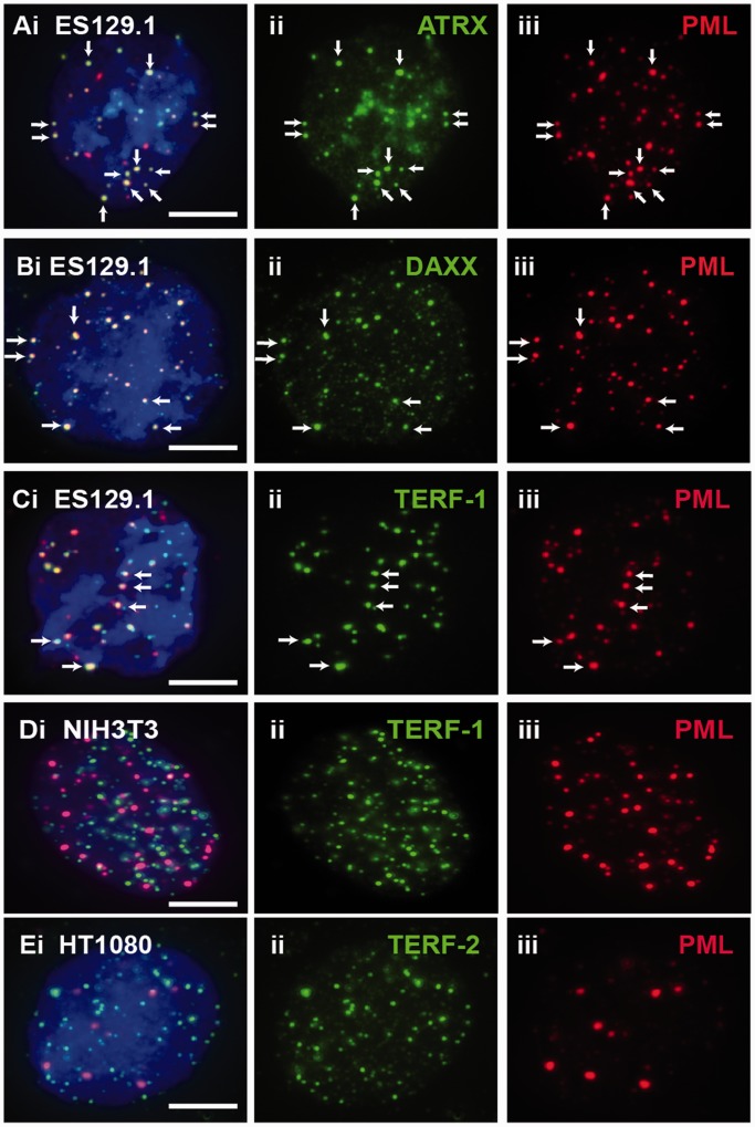
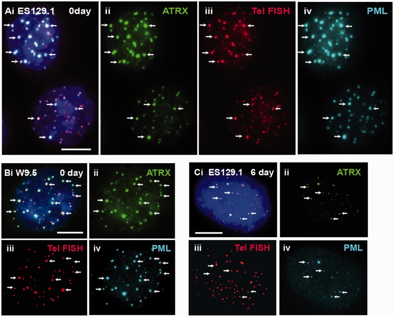
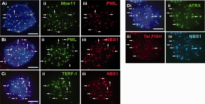
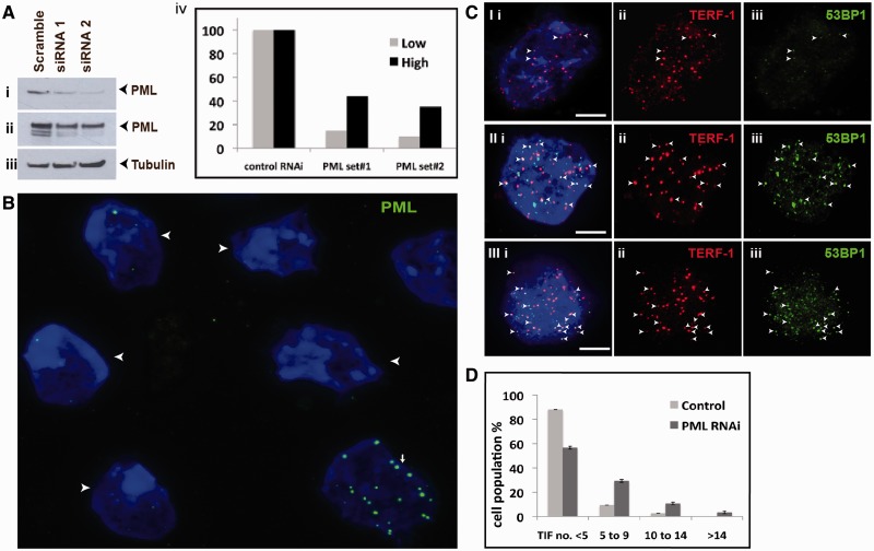
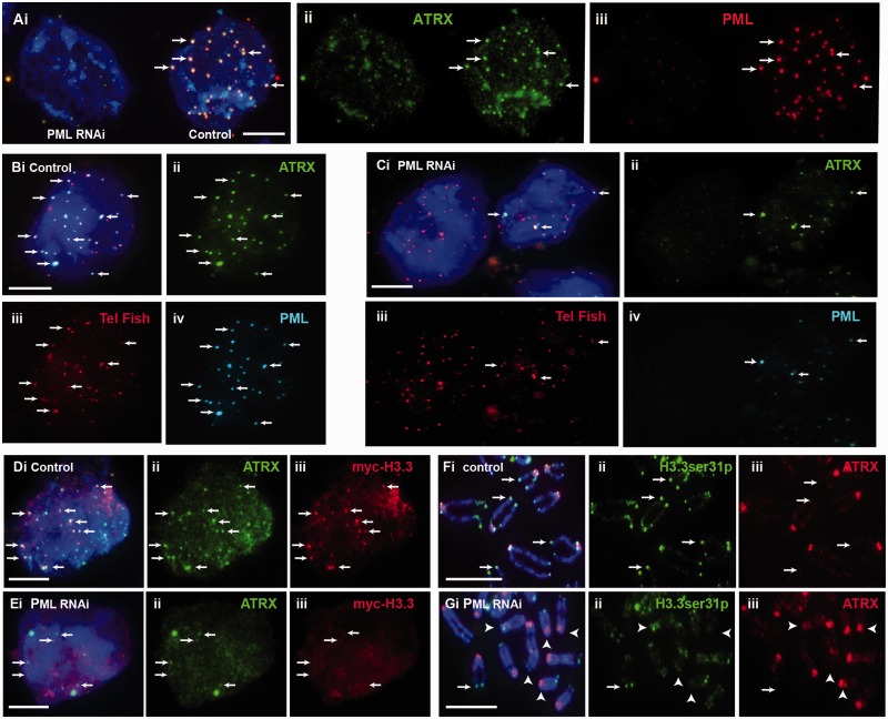
We have previously shown that α-thalassemia mental retardation X-linked (ATRX) and histone H3.3 are key regulators of telomeric chromatin in mouse embryonic stem cells. The function of ATRX and H3.3 in the maintenance of telomere chromatin integrity is further demonstrated by recent studies that show the strong association of ATRX/H3.3 mutations with alternative lengthening of telomeres in telomerase-negative human cancer cells. Here, we demonstrate that ATRX and H3.3 co-localize with the telomeric DNA and associated proteins within the promyelocytic leukemia (PML) bodies in mouse ES cells. The assembly of these telomere-associated PML bodies is most prominent at S phase. RNA interference (RNAi)-mediated knockdown of PML expression induces the disassembly of these nuclear bodies and a telomere dysfunction phenotype in mouse ES cells. Loss of function of PML bodies in mouse ES cells also disrupts binding of ATRX/H3.3 and proper establishment of histone methylation pattern at the telomere. Our study demonstrates that PML bodies act as epigenetic regulators by serving as platforms for the assembly of the telomeric chromatin to ensure a faithful inheritance of epigenetic information at the telomere.


Figures

Figure 1.
Figure 1.
ATRX, DAXX and telomeres localized at PML bodies in mouse ES cells. (A–C) In ES129.1 cells, beside ATRX (Aii) and DAXX (Bii), telomeres (Cii; TERF-1 staining) also localized at the PML bodies (A-Ciii; arrows). The co-localization is indicated by the presence of overlapped signals. (D and E) Telomeres of NIH3T3 (Dii; TERF-1 staining) and HT1080 (Eii; TERF-2 staining) cells did not localize at PML bodies (D-Eiii). Part ‘i’ is the merged figures and ‘ii and iii’ the split images of ‘i’. The arrows show some ‘examples’ of co-localized foci. (Scale bars: 5 μm).
Figure 2.
Figure 2.
ATRX co-localized with telomeres at PML bodies in mouse ES cells. (A and B) ES129.1 and W9.5 cells showed clear co-staining of ATRX (A-B ii) with telomere FISH signals (A–B iii) at the PML bodies (A and B iv). (C) The co-staining pattern of ATRX (ii) with telomeres (iii) at the PML bodies (iv) started to decrease after 6 days of differentiation. Part ‘i’ is the merged figures and ‘ii, iii and iv’ the split images of ‘i’. The arrows show some ‘examples’ of co-localized foci of ATRX/telomeres at PML bodies. (Scale bars: 5 μm).
Figure 3.
Figure 3.
Proteins that localized at telomeres-associated PML bodies. (A–C) DNA repair protein MRE11 (Aii) was present at PML bodies (Aiii) in ES129.1 cells. NBS1 (B-C iii) was also present at PML bodies (Bii) and telomeres (Cii; TERF-1 staining). (D) NBS1 (iv) co-localized with ATRX (ii) at telomeres (iii) in ES129.1 cells. Part ‘i’ is the merged figures and ‘ii, iii and iv’ the split images of ‘i’. The arrows show some ‘examples’ of co-localized foci. (Scale bars: 5 μm).
Figure 4.
Figure 4.
Effects of loss of PML bodies on telomere function. (A) ES129.1 cells were depleted of PML expression with two sets of siRNA oligonucleotides (set 3 and 4; Supplementary Table S2), followed by western blot analysis with antiserum against PML (i, short exposure; ii, long exposure of blot—the blot was overexposed to detect all PML isoforms) and β-tubulin (iii). Equal loading of protein was achieved by normalization against β-tubulin level. (iv) ‘Low bars’ were graphed according to protein bands in (i), whereas ‘High bars’ according to protein bands in (ii). In PML depleted cells, PML levels (lanes 2 and 3) were reduced by ∼90% 48 h after transfection compared with cells transfected with control siRNA oligonucleotides (lane 1). (B) Two sets of siRNA oligonucleotides (set 3 and 4) were used to deplete PML expression in ES129.1 cells. Immunofluorescence analysis was performed using antiserum against PML. Examples of ES129.1 cells showing the loss of PML bodies are shown by arrowheads. (C) TIFs were detected by co-staining of telomeres (TERF-1 staining; ii) with 53BP1 signals (iii) (see Supplementary Table S3a for scoring data). In ES129.1 cells transfected with control scramble siRNA oligonucleotides, <5 TIFs were detected (I), whereas 48 h of RNAi-mediated knockdown of PML induced the formation of TIFs (11 TIFs in II and 15 TIFs in III, respectively). Arrowheads show examples of TIFs. (D) Data were sub-grouped according to number of TIFs (<5, 5–9, 10–14 and >14) per cell. A normal cell contains ∼2–3 TIFs on average, so a threshold of >4 TIFs was used. When depleted of PML expression, percentage of cells with ≥5 TIFs (85 cells were counted) rose by 3–5-folds (from 10–14% to 38–46%).
Figure 5.
Figure 5.
Effects of loss of PML bodies on binding of ATRX and H3.3 at telomeres. ES129.1 cells transfected with control scramble siRNA oligonucleotides are indicated as ‘control’, whereas cells subjected to knockdown with PML-specific siRNA oligonucleotides are indicated as ‘PML RNAi’. (A) RNAi knockdown of PML in ES129.1 cells resulted in a loss in the formation of PML bodies (iii; cell on the left), leading to de-localization of ATRX (ii; arrows). (B and C) De-localization of ATRX from telomeres was confirmed by telomere FISH analysis. Compared with scramble control ES129.1 cells (B; control), RNAi knockdown of PML (C; PML RNAi) led to disassembly of PML bodies (Civ), resulting in ATRX de-localization (Cii) from the telomeres (Ciii; telomere FISH) (see Table 3). (D and E) RNAi knockdown of PML and loss of PML bodies (E; PML RNAi) also resulted in a reduction in the co-localization of myc-H3.3 (iii) with ATRX (ii) in ES129.1 cells. (F and G) In addition, depletion of PML expression and loss of PML bodies (G; PML RNAi) also resulted in a reduction in the level of H3.3ser31P at the telomeres on metaphase chromosomes (examples of positive H3.3ser31P staining at telomeres are indicated by arrows, whereas examples of the absence of H3.3ser31P signal at telomeres are indicated by arrowheads; see Table 4 for quantification) (Scale bars: 5 μm).
Figure 6.
Figure 6.
Effects of loss of PML bodies on establishment of chromatin states at telomeres. (A) ES129.1 cells were subjected to RNAi knockdown of PML and western blot analysis with antisera against PML (i, short exposure; ii, long exposure of blot), TERF-1 (iii), β-tubulin (iv), H4K20 monomethylation (v), H4K20 trimethylation (vi, Up, Millipore; and vii, Ab, Abcam), (viii) H3K9 dimethylation, (ix) H3K9 trimethylation and (x) histone H4. No change in expression level was seen in any of these proteins and chromatin marks, except for PML proteins. (B) ChIP analysis (Supplementary Table S4 for data) with antisera against H3K9me3, H4K20me3, ATRX and TERF-1, followed by dot blot analysis by hybridization with a telomere-specific probe. Consistent with the immunofluorescence analysis, a lower level of ATRX is detected at telomeres in PML-depleted cells, whereas, higher levels of H3K9me3 (2.4×) and H4K20me3 (3.4×) were detected at telomeres. In PML-depleted cells, a higher level of TERF-1 (2.5×) was also detected at telomeres.

References

    1. Costa A, Daidone MG, Daprai L, Villa R, Cantu S, Pilotti S, Mariani L, Gronchi A, Henson JD, Reddel RR, et al. Telomere maintenance mechanisms in liposarcomas: association with histologic subtypes and disease progression. Cancer Res. 2006;66:8918–8924. - PubMed
    1. Hakin-Smith V, Jellinek DA, Levy D, Carroll T, Teo M, Timperley WR, McKay MJ, Reddel RR, Royds JA. Alternative lengthening of telomeres and survival in patients with glioblastoma multiforme. Lancet. 2003;361:836–838. - PubMed
    1. Dunham MA, Neumann AA, Fasching CL, Reddel RR. Telomere maintenance by recombination in human cells. Nat. Genet. 2000;26:447–450. - PubMed
    1. Bryan TM, Englezou A, Dalla-Pozza L, Dunham MA, Reddel RR. Evidence for an alternative mechanism for maintaining telomere length in human tumors and tumor-derived cell lines. Nat. Med. 1997;3:1271–1274. - PubMed
    1. Bryan TM, Englezou A, Gupta J, Bacchetti S, Reddel RR. Telomere elongation in immortal human cells without detectable telomerase activity. EMBO J. 1995;14:4240–4248. - PMC - PubMed

Publication types