. 2013 Apr 25;121(17):3493-501.
doi: 10.1182/blood-2012-11-466227. Epub 2013 Feb 26.

Mi2β-mediated silencing of the fetal γ-globin gene in adult erythroid cells

Affiliations

Mi2β-mediated silencing of the fetal γ-globin gene in adult erythroid cells

Maria Amaya et al. Blood. .

Abstract

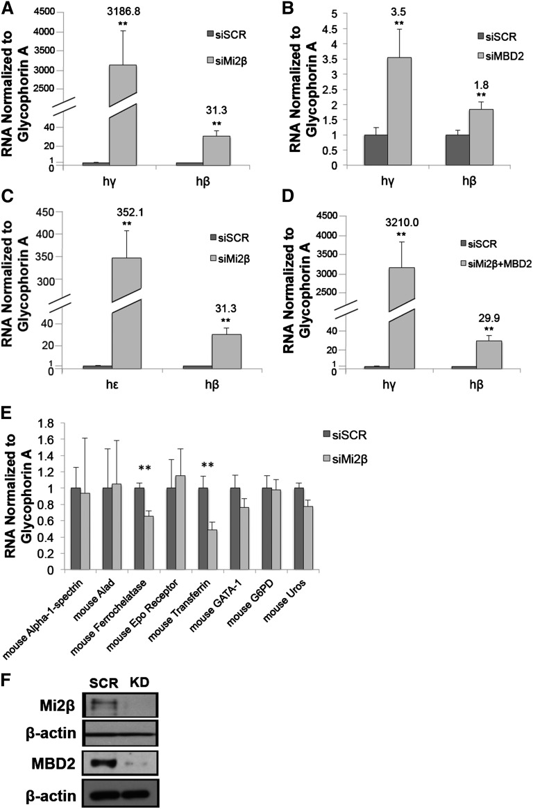
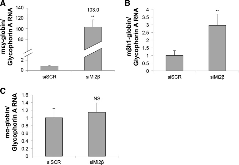
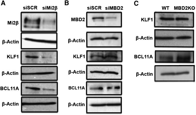
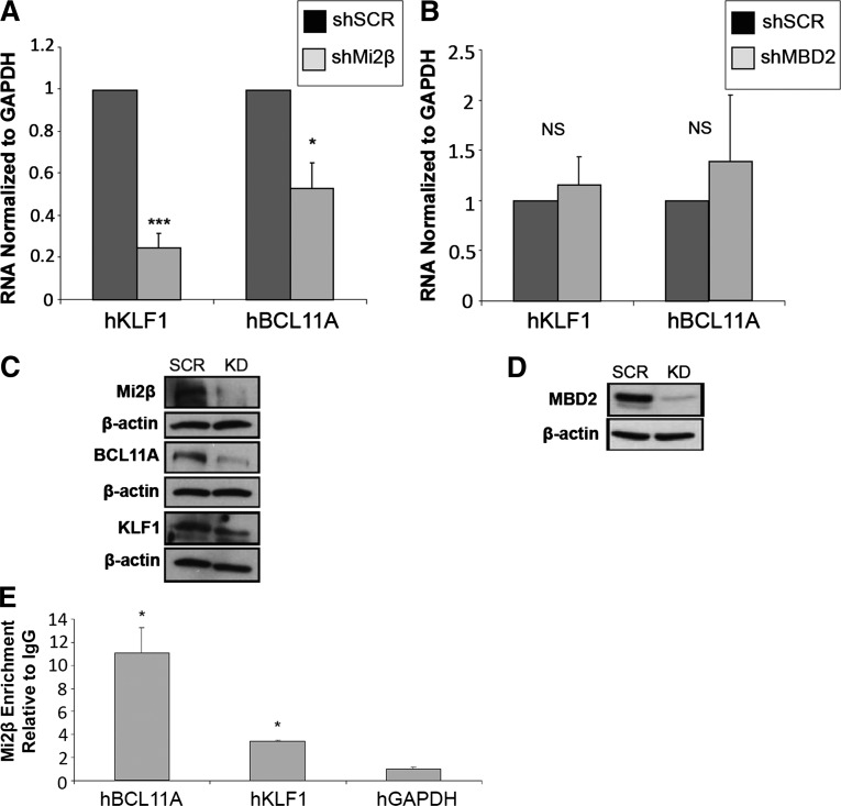
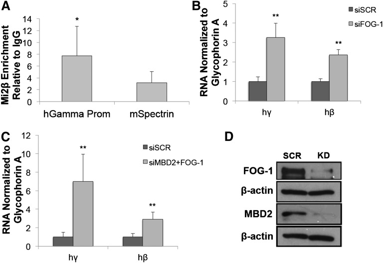
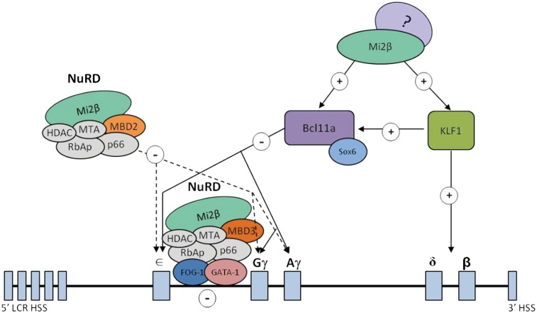
An understanding of the human fetal to adult hemoglobin switch offers the potential to ameliorate β-type globin gene disorders such as sickle cell anemia and β-thalassemia through activation of the fetal γ-globin gene. Chromatin modifying complexes, including MBD2-NuRD and GATA-1/FOG-1/NuRD, play a role in γ-globin gene silencing, and Mi2β (CHD4) is a critical component of NuRD complexes. We observed that knockdown of Mi2β relieves γ-globin gene silencing in β-YAC transgenic murine chemical inducer of dimerization hematopoietic cells and in CD34(+) progenitor-derived human primary adult erythroid cells. We show that independent of MBD2-NuRD and GATA-1/FOG-1/NuRD, Mi2β binds directly to and positively regulates both the KLF1 and BCL11A genes, which encode transcription factors critical for γ-globin gene silencing during β-type globin gene switching. Remarkably, <50% knockdown of Mi2β is sufficient to significantly induce γ-globin gene expression without disrupting erythroid differentiation of primary human CD34(+) progenitors. These results indicate that Mi2β is a potential target for therapeutic induction of fetal hemoglobin.


Figures

Figure 1
Figure 1
Mi2β preferentially regulates human β-globin locus gene expression in CID cells. CID cells were transiently transfected with siRNA for Mi2β, MBD2, or scramble control (siSCR) as indicated. (A) Transient knockdown of Mi2β by siRNA leads to a 3186.8-fold increase in the expression of γ-globin (hγ) gene expression in CID cells and a 31.3-fold increase in β-globin RNA (hβ) determined by qPCR. (B) Transient knockdown of MBD2 by siRNA leads to a 3.5-fold induction of γ-globin gene expression in CID cells and a 1.8-fold induction of β-globin RNA. (C) A 352.1-fold increase is seen in the expression of human ε-globin (hε) on knocking down Mi2β and a 31.3-fold increase in β-globin. (D) Combined knockdown of Mi2β and MBD2 leads to a 3210-fold increase in γ-globin gene expression and a 29.9-fold increase in β-globin RNA, similar to Mi2β knockdown alone. Data are expressed as human γ-, β-, or ε-globin RNA normalized to glycophorin A RNA. (E) qPCR analysis showing the expression of six murine genes (α-1-spectrin, aminolevulinate dehydratase [Alad], erythropoietin [Epo] receptor, GATA-1, glucose-6-phosphate dehydrogenase [G6PD], and uroporphynogen III synthase [Uros]) was not upregulated on Mi2β knockdown, whereas the mouse ferrochelatase and transferrin genes are slightly significantly down-regulated. (F) Western blot showing the degree of Mi2β and MBD2 protein knockdown in the CID cells used for the globin gene expression studies shown in Figures 1 and 2, respectively. Error bars represent the standard deviation of 3 independent experiments. *P < .05, and **P < .02, according to the Student t test.
Figure 2
Figure 2
Mi2β regulates the expression of endogenous mouse β-type globin genes. CID cells that were transiently transfected as described in Figure 1 were then assayed for endogenous εy-, βh1-, and α-globin RNA levels by qPCR. (A) Transient knockdown of Mi2β in CID cells leads to increased expression of the murine εy (mεy) gene by 103-fold normalized to glycophorin A. (B) Mi2β knockdown leads to a threefold increase in murine βh1 (mßh1) gene expression. (C) Murine α-globin (mα) RNA level is unchanged on Mi2β knockdown. These data are expressed as εy-, βh1-, and α-globin RNA normalized to glycophorin A, a murine erythroid-specific housekeeping gene. Error bars represent the standard deviation of 3 independent experiments. *P < .05 and **P < .02 according to the Student t test. NS = not statistically significant.
Figure 3
Figure 3
Mi2β regulates the expression of the γ-globin gene in human primary erythroid cells. CD34+ human hematopoietic progenitor cells were infected with lentivirus vectors harboring shRNA either for scramble control, 2 different Mi2β constructs, or MBD2. (A) Knockdown with shMi2β 1 leads to a 7.2-fold induction of γ-globin gene expression determined by qPCR. Knockdown with shMi2β 2 leads to a ∼20-fold increase in γ-globin expression and a slight decrease in β-globin gene expression. Shown below the graph are RNA levels in cells infected with scramble control or knockdown shRNA vectors. (B) Partial knockdown of Mi2β (construct 2) leads to a 20-fold increase in γ/γ+β-globin gene expression. (C) Knockdown of MBD2 leads to a ninefold increase in expression of γ/γ+β-globin gene expression. (D) Fluorescence activated cell sorting analysis showing erythroid differentiation of 81.1% of CD34+ progenitor cells in which Mi2β is knocked down compared with 93.8% of scramble shRNA control cells and 92.9% of cells in which MBD2 is knocked down. Values signify standard deviations for ≥3 independent experiments. (E) qPCR analysis showing a ∼40% knockdown level of Mi2β RNA in double-positive cells taken at the end of differentiation (quadrant 2). (F) qPCR analysis showing a 90% knockdown of Mi2β RNA in double-negative cells taken at the end of differentiation (quadrant 3). (G-I) Wright-Giemsa stain of scramble control, Mi2β knockdown, and MBD2 knockdown cell populations. Photomicrographs were generated using an Olympus (Center Valley, PA) BX41 compound microscope and Olympus DP71 digital camera at ×100 magnification. Images were acquired with Olympus DP Controller software. Error bars represent the standard deviation of ≥3 independent experiments. *P < .05 and **P < .02 according to the Student t test.
Figure 4
Figure 4
Mi2β positively regulates the expression of KLF1 and BCL11A in CID cells. (A) Western blot showing a decrease in murine KLF1 and murine BCL11A protein levels after Mi2β knockdown in CID cells. (B) Western blot showing no change in murine KLF1 and murine BCL11A protein levels after MBD2 knockdown in CID cells. (C) Western blot showing no change in murine KLF1 or murine BCL11A in primary adult mouse erythroblasts from MBD2 knockout mice.
Figure 5
Figure 5
Mi2β positively regulates the expression of KLF1 and BCL11A in human CD34+ hematopoietic progenitor-derived primary erythroid cells. (A) qPCR analysis showing mRNA levels of KLF1 and BCL11A following Mi2β knockdown are decreased by 70% and 40%, respectively. (B) mRNA levels of KLF1 and BCL11A following MBD2 knockdown are not affected. (C) Western blot showing a decrease in the levels of BCL11A and KLF1 protein after Mi2β knockdown in human primary erythroid cells. (D) Western blot showing the level of MBD2 protein knockdown in human primary erythroid cells. (E) ChIP assay showing significant enrichment of Mi2β at the BCL11A and KLF1 promoter regions. Glyceraldehyde-3-phosphate dehydrogenase was used as a negative control, and enrichment values are normalized to IgG controls. Error bars represent the standard deviation of ≥3 independent experiments. *P < .05, **P < .02, and ***P < .001 according to the Students t test. NS = not statistically significant.
Figure 6
Figure 6
Mi2β occupies the γ-globin gene promoter and acts in a partially independent manner from GATA-1/FOG-1/NuRD. (A) ChIP assays showing significant Mi2β enrichment at the γ-globin promoter region in CID cells. The mouse α-spectrin gene was used as a negative control. (B) qPCR results showing knockdown of FOG-1 leads to a ∼3-fold induction of the γ-globin (hγ) and an ∼2.5-fold induction of the β-globin (hβ) gene. (C) Simultaneous knockdown of MBD2 and FOG-1 leads to an approximate sevenfold induction of γ-globin and an approximate threefold induction of β-globin. (D) Western blot showing both FOG-1 and MBD2 knockdown in CID cells. Error bars represent the standard deviation of ≥3 experiments. *P < .05 and **P < .02 according to the Student t test.
Figure 7
Figure 7
Working model of Mi2β-mediated developmental globin gene silencing through multiple mechanisms. Mi2β is a critical component of the MBD2/NuRD complex that regulates developmental globin gene silencing independently of BCL11A and KLF1-EKLF in an indirect manner. Mi2β binds to the distal promoter region of the γ-globin gene as part of the MBD3/NuRD/GATA-1/FOG-1 silencing complex. Mi2β binds to and activates expression of BCL11A and KLF1/EKLF, which in turn silence γ-globin gene expression. Solid arrows represent direct interactions, and dotted arrows represent indirect interactions.

References

    1. Stamatoyannopoulos G. Control of globin gene expression during development and erythroid differentiation. Exp Hematol. 2005;33(3):259–271. - PMC - PubMed
    1. Thein SL, Menzel S. Discovering the genetics underlying foetal haemoglobin production in adults. Br J Haematol. 2009;145(4):455–467. - PubMed
    1. Sankaran VG, Xu J, Orkin SH. Advances in the understanding of haemoglobin switching. Br J Haematol. 2010;149(2):181–194. - PMC - PubMed
    1. Peschle C, Mavilio F, Carè A, et al. Haemoglobin switching in human embryos: asynchrony of zeta—alpha and epsilon—gamma-globin switches in primitive and definite erythropoietic lineage. Nature. 1985;313(5999):235–238. - PubMed
    1. Ley TJ, Maloney KA, Gordon JI, Schwartz AL. Globin gene expression in erythroid human fetal liver cells. J Clin Invest. 1989;83(3):1032–1038. - PMC - PubMed

Publication types

MeSH terms