. 2013 May 2;121(18):3594-8.
doi: 10.1182/blood-2012-12-476200. Epub 2013 Feb 26.

Erk1 and Erk2 are required for maintenance of hematopoietic stem cells and adult hematopoiesis

Affiliations

Erk1 and Erk2 are required for maintenance of hematopoietic stem cells and adult hematopoiesis

Gordon Chan et al. Blood. .

Abstract

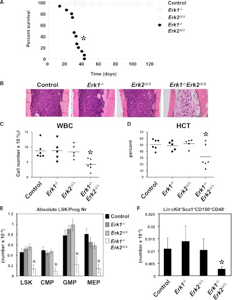
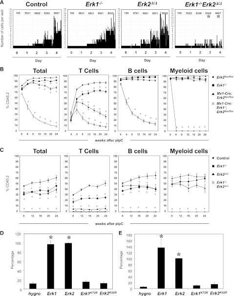
Extracellular signal-regulated kinase 1 (Erk1) and Erk2 play crucial roles in cell survival, proliferation, cell adhesion, migration, and differentiation in many tissues. Here, we report that the absence of Erk1 and Erk2 in murine hematopoietic cells leads to bone marrow aplasia, leukopenia, anemia, and early lethality. Mice doubly-deficient in Erk1 and Erk2 show rapid attrition of hematopoietic stem cells and immature progenitors in a cell-autonomous manner. Reconstitution studies show that Erk1 and Erk2 play redundant and kinase-dependent functions in hematopoietic progenitor cells. Moreover, in cells transformed by the oncogenic KRas(G12D) allele, the presence of either Erk1 or Erk2 with intact kinase activity is sufficient to promote cytokine-independent proliferation.


Figures

Figure 1
Figure 1
Deletion of Erk1 and Erk2 leads to fatal BM failure and loss of HSC. (A) Kaplan-Meier survival analysis of a cohort of Erk1−/− (n = 23), Mx1-Cre;Erk flox/flox (n = 21), Mx1-Cre Erk1−/−;Erk2 flox/flox (n = 22), and littermate control (n = 18) mice following pIpC treatment. Erk1−/−Erk2Δ/Δ mice have a median lifespan of 30 days; control mice remain healthy for >120 days. *P < .05, log-rank test. (B) Hematoxylin and eosin–stained sections from humeri of control, Erk1−/−, Erk2Δ/Δ, and Erk1−/−Erk2Δ/Δ animals 30 days after receiving their last dose of pIpC. (C-D) White blood cell count (WBC) and hematocrit (HCT) of control, Erk1−/−, Erk2Δ/Δ, and Erk1−/−Erk2Δ/Δ mice (*P < .05, ANOVA). (E) Absolute number (mean ± SEM, *P < .05, ANOVA) of LSK, CMP (LinSca1cKit+CD34+FcγRlo), GMP (LinSca1cKit+CD34+FcγRhi), MEP (LinSca1cKit+CD34FcγR−/lo) of control (n = 6), Erk1−/− (n = 5), Erk2Δ/Δ (n = 5), and Erk1−/−Erk2Δ/Δ mice (n = 6). *P < .05, ANOVA. (F) Absolute number (mean ± SEM, * P < .05, Student t test) of signaling lymphocyte activation molecule (SLAM)-HSC from control (n = 6), Erk1−/− (n = 5), Erk2Δ/Δ (n = 5), and Erk1−/−Erk2Δ/Δ mice (n = 6). *P < .05, ANOVA.
Figure 2
Figure 2
Requirement of Erk1 and Erk2 in HSCs and immature progenitors. (A) Single SLAM-HSC from of control, Erk1−/−, Erk2Δ/Δ, and Erk1−/−Erk2Δ/Δ mice were sorted into 60-well plates (1 cell per well) and cultured in media containing SCF, Tpo, Flt3l, and IL-11. Proliferation of each clone was evaluated microscopically over 4 days. Representative data from 1 of 4 experiments with similar results are shown. The average percentages (±SEM) of surviving clones from the 4 experiments are shown at the top of each panel. *P < .05, ANOVA. (B) Lethally irradiated CD45.1+ recipients (6 mice per group) were transplanted with 106 BM cells from control, Erk1−/−, Erk2Δ/Δ, and Erk1−/−Erk2Δ/Δ mice, along with 105 wild-type CD45.1+ cells. After 5 weeks of engraftment, chimeric mice were treated with 5 doses of pIpC, and the percentage of donor-derived peripheral blood cells (Total), CD3+ (T cells), B220+ (B cells), and Gr1+ (myeloid cells) were determined flow cytometry. *P < .05, ANOVA. (C) Control, Erk1−/−, Erk2−/−, and Mx1-Cre Erk1−/−Erk2flox/flox mice were treated with pIpC for 14 days before their BM cells (5 × 10⁵) were harvested and mixed with equal numbers of wild-type CD45.1+ cells and intrafemorally injected into lethally irradiated CD45.1+ recipients (5 mice per group). After 8 weeks of engraftment, the percentage of donor-derived peripheral blood cells (Total), CD3+ (T cells), B220+ (B cells), and Gr1+ (myeloid cells) were determined flow cytometry. *P < .05, ANOVA. (D) Primary lin BM cells from Erk1−/−;Erk2 flox/flox mice were transduced with the indicated retroviruses, and FACS-purified GFP+ cells were cultured in Iscove modified Dulbecco medium containing IL-3, IL-6, and SCF. MIG-Cre vectors coexpress the hygromycin resistance gene (hygro), Erk1, Erk2, Erk1KR, and Erk2KR. Relative cell proliferation in each experiment is normalized to that of Erk2. Results are shown as mean ± SEM of 4 independent experiments. *P < .05, ANOVA. (E) Lin BM cells from Erk1−/−;Erk2 flox/flox;LSL-KrasG12D mice were transduced with retroviruses as described in panel D, and GFP+ cells were cultured in IMDM medium containing no cytokines. Cells were harvested and proliferation was determined. Cell proliferation in each experiment is normalized to that of Erk2 and results are shown as mean ± SEM of 4 independent experiments. *P < .05, ANOVA.

References

    1. Roskoski R., Jr ERK1/2 MAP kinases: structure, function, and regulation. Pharmacol Res. 2012;66(2):105–143. - PubMed
    1. Geest CR, Coffer PJ. MAPK signaling pathways in the regulation of hematopoiesis. J Leukoc Biol. 2009;86(2):237–250. - PubMed
    1. Chung E, Kondo M. Role of Ras/Raf/MEK/ERK signaling in physiological hematopoiesis and leukemia development. Immunol Res. 2011;49(1-3):248–268. - PubMed
    1. Pagès G, Guérin S, Grall D, et al. Defective thymocyte maturation in p44 MAP kinase (Erk 1) knockout mice. Science. 1999;286(5443):1374–1377. - PubMed
    1. Nekrasova T, Shive C, Gao Y, et al. ERK1-deficient mice show normal T cell effector function and are highly susceptible to experimental autoimmune encephalomyelitis. J Immunol. 2005;175(4):2374–2380. - PubMed

Publication types

MeSH terms

Substances