. 2013 Feb 27:14:5.
doi: 10.1186/1471-2091-14-5.

Expression, purification and biochemical characterization of Schizosaccharomyces pombe Mcm4, 6 and 7

Affiliations

Expression, purification and biochemical characterization of Schizosaccharomyces pombe Mcm4, 6 and 7

Meng Xu et al. BMC Biochem. .

Abstract

Background: The hetero-hexamer of the eukaryotic minichromosome maintenance (MCM) proteins plays an essential role in replication of genomic DNA. The ring-shaped Mcm2-7 hexamers comprising one of each subunit show helicase activity in vitro, and form double-hexamers on DNA. The Mcm4/6/7 also forms a hexameric complex with helicase activity in vitro.

Results: We used an Escherichiai coli expression system to express various domains of Schizosaccharomyces pombe Mcm4, 6 and 7 in order to characterize their domain structure, oligomeric states, and possible inter-/intra-subunit interactions. We also successfully employed a co-expression system to express Mcm4/6/7 at the same time in Escherichiai coli, and have purified functional Mcm4/6/7 complex in a hexameric state in high yield and purity, providing a means for generating large quantity of proteins for future structural and biochemical studies.

Conclusions: Based on our results and those of others, models were proposed for the subunit arrangement and architecture of both the Mcm4/6/7 hexamer and the Mcm2-7 double-hexamer.


Figures
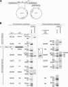
Figure 1
Figure 1
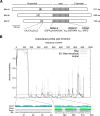
Interactions and oligomeric states of co-expressed fragments of Mcm4, 6 and 7. (A) Schematic of the polycistronic co-expression strategy that involves two compatible vectors. ORF1 and ORF2 were linked by a ribosome binding site (RBS) with a spacer. ORF3 was cloned in pXA-BN vector. Two plasmids were co-transformed into E. coli., followed by dual screening of ampicillin (50 μg/ml) and chloramphenicol (17 μg/ml). (B) Interactions of co-expressed and copurified fragments of Mcm4, 6 and 7, as identified in the two components co-expression (left side) or three components co-expression (right side) experiments. E. coli. lysates co-expressing various fragments with or without tags were passed through either glutathione or Ni-NTA resins, then the resins were washed as described under “Materials and Methods”. GST tags were cleaved by PreScission protease on the resin to release the MCM proteins. His tagged proteins were eluted by imidazole. All elutions were analyzed by SDS-PAGE. Asterisk denotes the co-lysis (instead of co-expression) of the indicated near-full-length fragments.
Figure 2
Figure 2
Designs of truncated fragments of Mcm4, 6 and 7. (A) Schematic of fission yeast Mcm4, 6 and 7. Locations of putative zinc finger (white boxes labeled with Z), the MCM core region (gray boxes) was shown. Three ATPase consensus motifs in the MCM core region were labeled with A (the Walker A motif), B (the Walker B motif) and R (the Arg-finger motif). All conversed amino acid residues that define each motif were shown. All truncation fragments reported in this paper were designed according to three domains, N-terminal, core and C-terminal domains. This figure was generated from the sequence alignment results shown in Additional File 1: Figure S1 and each Mcm protein was aligned with the MCM box region. (B) Disordered profile plot and predicted secondary structure of Mcm4. Only sampled secondary structure prediction was shown and aligned with the disordered profile. A disordered N-termini was present and aligned well with a region (1–150 aa) that lacks any defined secondary structure, while regions with very low disorder probability were predicted to show ordered secondary structures. The disordered profiles were generated by the DISOPRED server, and secondary structure prediction was generated by the PSIPRED server at University College London [15-17]. “Conf”-prediction confidence, “Pred”-predicted secondary structures, “AA”-amino acid residues. Disordered profile plots of Mcm6 and 7 were shown in Additional file 2: Figure S2.
Figure 3
Figure 3
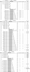
Summary of biochemical properties of fragments of Mcm4, 6 and 7. Schematic of truncated fragments of Mcm4, 6 and 7 tested in this study. The motifs are represented by: “A”-Walker A motif, “B”-Walker B motif, “R”-Arg-finger motif, “Z”-zinc finger motif. The nomenclature for the fragments is as follows, the first numbers represent the Mcm 4, 6, or 7; the letters in the middle indicate domain locations (“N”-N terminal fragments, “C”-core fragments, “F”-near-full-length fragments); the last numbers denotes construct number. a, decreased expression level or plasmid instability; b, oligomeric states depended on protein concentration; c, little equilibrium between monomeric and dimeric states and proteins in the two states could be separated by ion-exchange chromatography; d, a stable large complex identified with a molecular weight equal to a double-hexamer; n/a, not available, due to lack of enough samples.
Figure 4
Figure 4
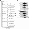
Oligomeric states and interactions of N-terminal fragments of Mcm4, 6 and 7. (A) Gel filtration chromatography profiles of N-terminal fragments of Mcm4, 6 and 7. Schematic of each fragment was shown in accordance with its gel filtration profile. N-terminal fragments of Mcm6 were aligned with the zinc finger motif and a 62 amino acid residues protruding N-termini was shown. 7N1a, separated monomeric 7 N1 fragment; 7N1b, separated dimeric 7 N1 fragment. Gel filtration analysis was carried out a described under “Materials and Methods”. (B) In vitro incubation of purified N-terminal fragments of Mcm4, 6 and 7. Interactions among the N-terminal fragments of Mcm4, 6 and 7 were characterized by gel filtration analysis. Samples from peak fractions (pointed by arrows) were quantitated by SDS-PAGE and mixed together in approximate equal molar ratio. The mixture was buffer-exchanged to 50 mM NaCl, 50 mM Tris pH8 and 1 mM DTT and then incubated on ice for 30 minutes. For 7 N1 and 7 N2, only samples from peak fraction of monomeric states were used. The incubation mixtures were subjected to gel filtration analysis and no large complex was detected. Two groups of N-terminal fragments of Mcm4, 6 and 7 were used, as shown in top and bottom panels.
Figure 5
Figure 5
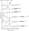
Gel filtration chromatography profiles of core and near-full-length fragments of Mcm4, 6 and 7. Schematic of each fragment was shown in accordance with its gel filtration profile. Gel filtration analysis was carried out a described under “Methods”. (a) Gel filtration profile of 7 N1 was chosen as a reference, and its dimer peak was used to align with monomer peaks of 7 F4. The other molecular weight shown was determined by Bio-Rad Gel Filtration Standard (data not shown). (b-c) Concentration dependent oligomerization of a core fragment of Mcm4, 4C1. (d) Large and heterogeneous aggregates composed of a nFL fragment of Mcm6, 6 F9. (e-f) Two oligomeric states of a nFL fragment of Mcm7, 7 F4. Peaks on the gel filtration profile correspond to the monomer and the double-hexamer.
Figure 6
Figure 6
Identification of stable Mcm complexes and helicase activity of Mcm4/6/7 hexamers. (A) Gel filtration chromatography profiles of purified complexes of Mcm4, 6 and 7. Gel filtration analysis was carried out a described under “Materials and Methods”. Asterisk: Gel filtration profile of Mcm4/6/7 hexamers expressed and purified from insect cells in our laboratory. (B) SDS-PAGE analysis of purified complexes of Mcm4, 6 and 7 from the gel filtration fractions shown in Panel C. (C) Helicase assay results of the Mcm4/6/7 hexamers. No protein added in lane1 and 2. B, boiled substrate; UB, un-boiled substrate; lane 3 and 5, 100 ng protein added; lane 4 and 6, 200 ng protein added; Asterisk, Mcm4/6/7 hexamers expressed and purified from insect cells.
Figure 7
Figure 7
Schematics of proposed models of the Mcm4/6/7 hexamer and the Mcm2-7 double-hexamer. (A) Summary of interactions identified in this report. Double arrows: reciprocal interactions. Single arrows: unidirectional interactions. Hallow arrows: weak interactions. Solid lines: stable homogeneous oligomeric states, such as dimers. Dashed lines: heterogeneous oligomeric states: such as aggregates. (B) Model of the Mcm4/6/7 hexamer. This model is based on the interactions identified in Panel A, which is consistent with the model proposed previouslyfor S. cerevisiae MCM [26], and human MCM [23,24,42]. (C-D) Model of hexamer-hexamer interactions for the Mcm2-7 double-hexamer. This model is based on Figure 1A of [6], showing a proposed Mcm7/7 interaction that locks the orientation of two hexamers. The convex and the concave on each subunit in this figure represent the P-loop of the Walker A motif and the Arg-finger motif, respectively.

Similar articles

Cited by

References

    1. Tye BK. The MCM2-3-5 proteins: are they replication licensing factors? Trends Cell Biol. 1994;4:160–166. doi: 10.1016/0962-8924(94)90200-3. - DOI - PubMed
    1. Chong JP, Mahbubani HM, Khoo CY, Blow JJ. Purification of an MCM-containing complex as a component of the DNA replication licensing system. Nature. 1995;375:418–421. doi: 10.1038/375418a0. - DOI - PubMed
    1. Todorov IT, Attaran A, Kearsey SE. BM28, a human member of the MCM2-3-5 family, is displaced from chromatin during DNA replication. J Cell Biol. 1995;129:1433–1445. doi: 10.1083/jcb.129.6.1433. - DOI - PMC - PubMed
    1. Kearsey SE, Labib K. MCM proteins: evolution, properties, and role in DNA replication. Biochim Biophys Acta. 1998;1398:113–136. doi: 10.1016/S0167-4781(98)00033-5. - DOI - PubMed
    1. Evrin C, Clarke P, Zech J, Lurz R, Sun J, Uhle S, Li H, Stillman B, Speck C. A double-hexameric MCM2-7 complex is loaded onto origin DNA during licensing of eukaryotic DNA replication. Proc Natl Acad Sci U S A. 2009;106:20240–20245. doi: 10.1073/pnas.0911500106. - DOI - PMC - PubMed

Publication types

MeSH terms

Substances

LinkOut - more resources