. 2013 Apr;17(4):482-96.
doi: 10.1111/jcmm.12028. Epub 2013 Feb 27.

Evidence for progressive reduction and loss of telocytes in the dermal cellular network of systemic sclerosis

Affiliations

Evidence for progressive reduction and loss of telocytes in the dermal cellular network of systemic sclerosis

Mirko Manetti et al. J Cell Mol Med. 2013 Apr.

Abstract

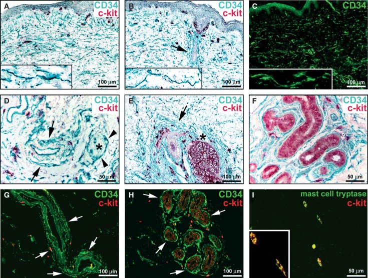
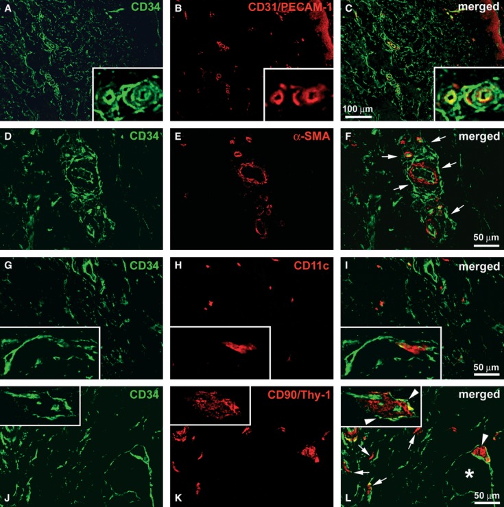
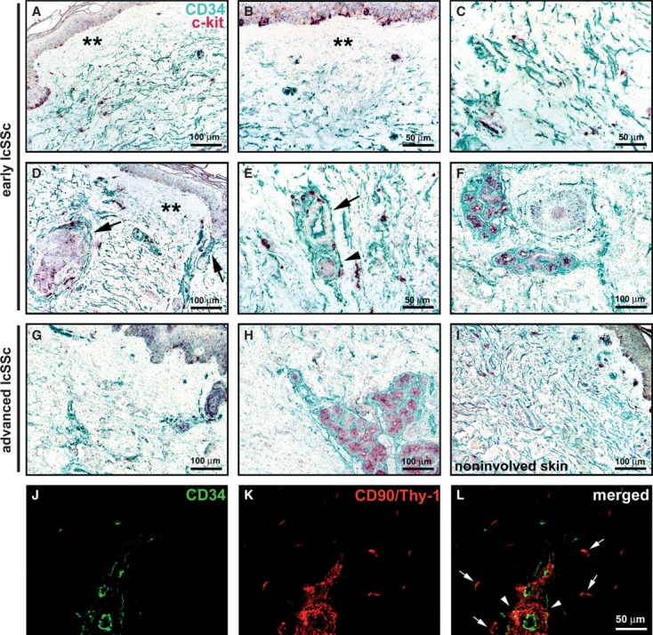
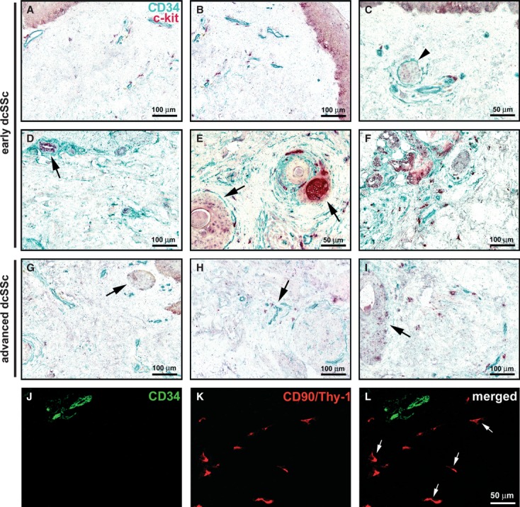
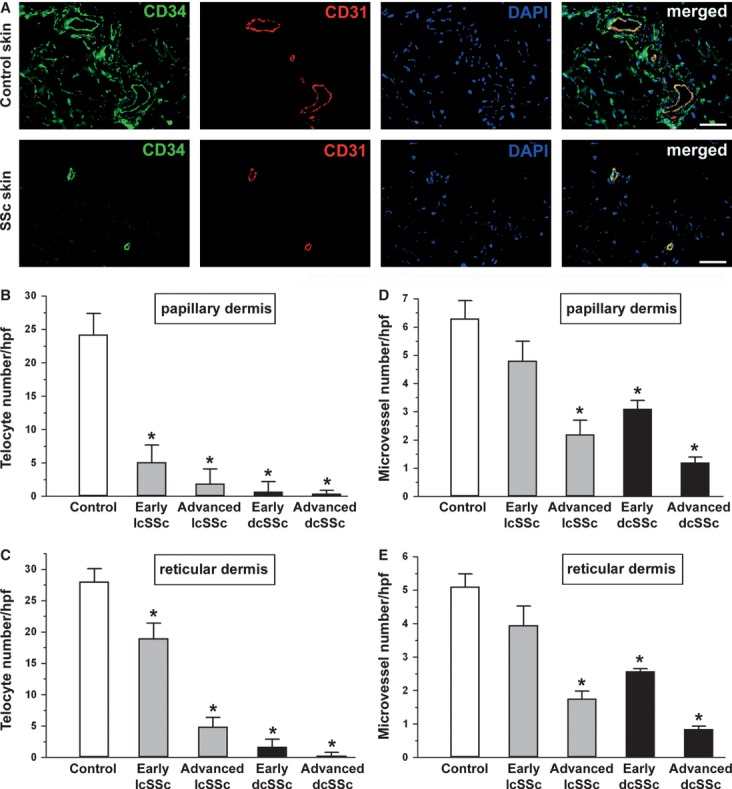
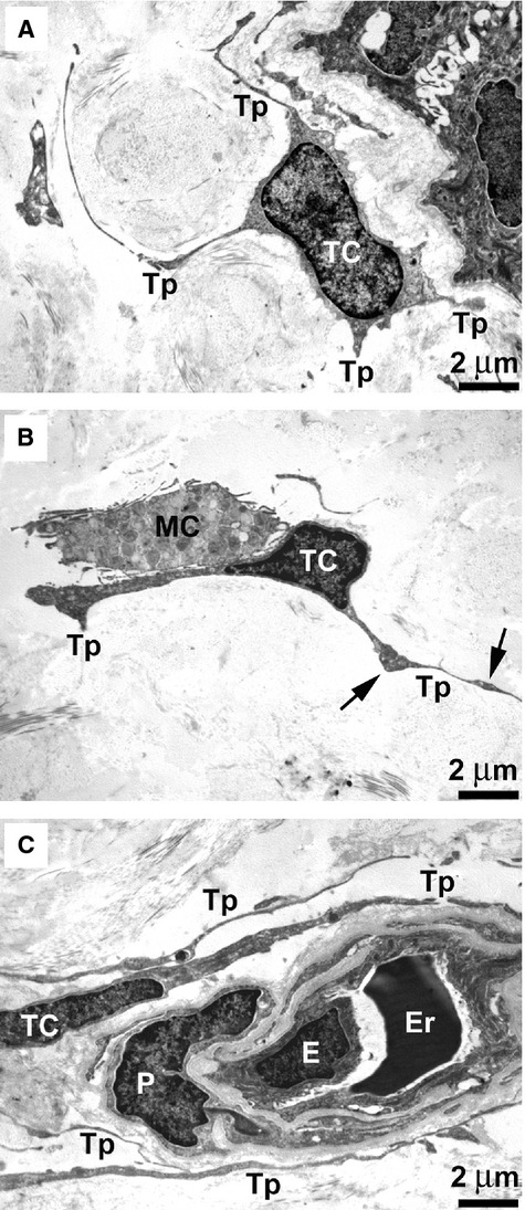
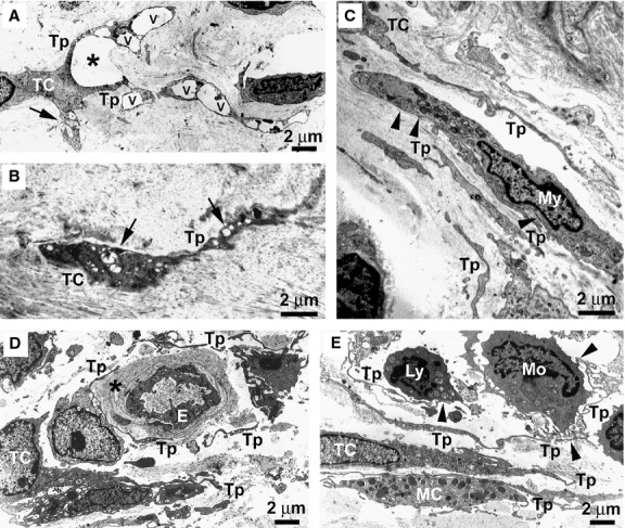
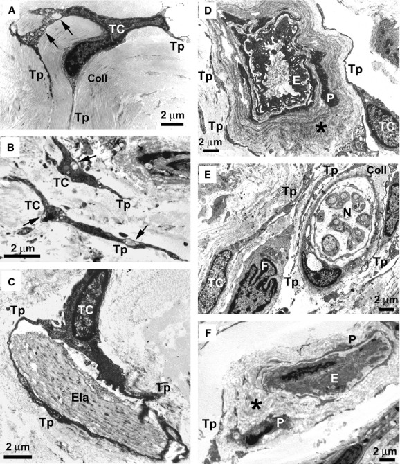
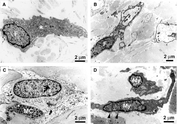
Telocytes, a peculiar type of stromal cells, have been recently identified in a variety of tissues and organs, including human skin. Systemic sclerosis (SSc, scleroderma) is a complex connective tissue disease characterized by fibrosis of the skin and internal organs. We presently investigated telocyte distribution and features in the skin of SSc patients compared with normal skin. By an integrated immunohistochemical and transmission electron microscopy approach, we confirmed that telocytes were present in human dermis, where they were mainly recognizable by their typical ultrastructural features and were immunophenotypically characterized by CD34 expression. Our findings also showed that dermal telocytes were immunophenotypically negative for CD31/PECAM-1 (endothelial cells), α-SMA (myofibroblasts, pericytes, vascular smooth muscle cells), CD11c (dendritic cells, macrophages), CD90/Thy-1 (fibroblasts) and c-kit/CD117 (mast cells). In normal skin, telocytes were organized to form three-dimensional networks distributed among collagen bundles and elastic fibres, and surrounded microvessels, nerves and skin adnexa (hair follicles, sebaceous and sweat glands). Telocytes displayed severe ultrastructural damages (swollen mitochondria, cytoplasmic vacuolization, lipofuscinic bodies) suggestive of ischaemia-induced cell degeneration and were progressively lost from the clinically affected skin of SSc patients. Telocyte damage and loss evolved differently according to SSc subsets and stages, being more rapid and severe in diffuse SSc. Briefly, in human skin telocytes are a distinct stromal cell population. In SSc skin, the progressive loss of telocytes might (i) contribute to the altered three-dimensional organization of the extracellular matrix, (ii) reduce the control of fibroblast, myofibroblast and mast cell activity, and (iii) impair skin regeneration and/or repair.


Figures

Fig. 1
Fig. 1
Normal skin, immunohistochemistry. (A, B, DF) Double immunoenzymatic labelling for CD34 and c-kit with horseradish peroxidase (green) and alkaline phosphatase (red) detection systems respectively. (C) CD34 immunofluorescence labelling. (G and H) Double immunofluorescence labelling for CD34 (green) and c-kit (red). (I) Double immunofluorescence labelling for mast cell tryptase (green) and c-kit (red). (A–H) Dermal interstitial cells, presumably telocytes and endothelial cells display CD34-immunoreactivity. The CD34-positive cells do not display c-kit-immunoreactivity. In the papillary and reticular dermis, telocytes form an interstitial network (A–C). Telocytes have an elongated shape with an oval or triangular body and two or three long and thin cell processes with knobs/dilations along their length (A–C, higher magnification view in insets). Telocytes form a thin and almost continuous layer encircling arterioles (D and G, arrows). A nerve [denoted by an asterisk in (D)] is surrounded by telocytes (arrowheads). Telocytes make an almost continuous multilayered sheath around hair follicles (B and E, arrows), sebaceous glands (E, asterisk) and the secretory and excretory portions of eccrine sweat glands (F and H, arrows). Many c-kit-positive cells, round or oval in shape, are observed in the dermis, often in close contact with telocytes (A, B, D-H). Melanocytes (B) and epithelial cells of sebaceous (E) and sweat (F, H) glands are c-kit-immunoreactive. (I) The c-kit-positive cells show mast cell tryptase immunoreactivity. A higher magnification view of two double-positive mast cells is shown in the inset. Scale bars are indicated in each panel.
Fig. 2
Fig. 2
Normal skin, immunohistochemistry. Double immunofluorescence labelling for CD34 (green) and (AC) CD31/platelet-endothelial cell adhesion molecule-1 (PECAM-1, red), (DF) α-smooth muscle actin (α-SMA, red), (GI) CD11c (red), (JL) CD90/Thy-1 (red). (A–C) Endothelial cells are CD34/CD31-double-positive, while telocytes are CD34-positive/CD31-negative. The inset is a higher magnification view showing microvessels surrounded by telocytes. (D–F) Telocytes do not display α-SMA-immunoreactivity. α-SMA-positive pericytes and vascular smooth muscle cells are surrounded by telocytes (F, arrows). (G–I) Telocytes are CD11c-negative, while dendritic cells and macrophages show CD11c-immunoreactivity. A dendritic cell is embraced by the long processes of a telocyte (I, inset). (J–L) Telocytes do not display immunoreactivity for CD90/Thy-1, while fibroblasts are CD90-positive/CD34-negative (L, arrows). Telocyte prolongations surround a CD90-positive stem cell niche (L, arrowhead) near a sebaceous gland (L, asterisk) and a vascular wall-resident stem cell niche (L, inset, arrowheads). Scale bars are indicated in each panel.
Fig. 3
Fig. 3
Limited cutaneous systemic sclerosis (lcSSc) skin, immunohistochemistry. (A–I) Double immunoenzymatic labelling for CD34 and c-kit with horseradish peroxidase (green) and alkaline phosphatase (red) detection systems respectively. Everywhere in the dermis, telocytes and endothelial cells are CD34-positive, mast cells are c-kit-positive. Melanocytes (A, B, D, G, I) and epithelial cells of sebaceous (D and G) and sweat (F and H) glands are c-kit-immunoreactive. (A–F) Early lcSSc skin. Telocytes are absent from the papillary dermis (A, B, D, double asterisks) and patchily reduced in the reticular dermis. In the reticular dermis, most telocytes are enlarged in shape (C). Telocytes are present around arterioles (E, arrow), nerves (E, arrowhead), hair follicles and sebaceous glands (D, arrows), and eccrine sweat glands (F). (G–I) Advanced lcSSc skin. Telocytes are absent from the papillary dermis and severely reduced in the reticular dermis and around most of the adnexal structures (G). In the deep reticular dermis, telocytes are preserved around sweat glands (H). A normal distribution of telocytes is observed in clinically non-involved lcSSc skin (I). (J–L) Advanced lcSSc skin, double immunofluorescence labelling for CD34 (green) and CD90/Thy-1 (red). Very few telocytes are observed (J and L). Fibroblasts are CD90-positive/CD34-negative (K, L, arrows). A CD90-positive vascular wall-resident stem cell niche is scarcely surrounded by telocytes (L, arrowheads). Scale bars are indicated in each panel.
Fig. 4
Fig. 4
Diffuse cutaneous systemic sclerosis (dcSSc) skin, immunohistochemistry. (AI) Double immunoenzymatic labelling for CD34 and c-kit with horseradish peroxidase (green) and alkaline phosphatase (red) detection systems respectively. Everywhere in the dermis, telocytes and endothelial cells are CD34-positive, mast cells are c-kit-positive. Melanocytes (A–C) and epithelial cells of sebaceous (E and G) and sweat (D and F) glands display c-kit-immunoreactivity. (A–F) Early dcSSc skin. Telocytes are very few or absent in the papillary and reticular dermis (A and B). (C) Some telocytes are observed around a nerve (arrowhead) and microvessels. Telocytes form a single discontinuous layer surrounding hair follicles and sebaceous glands (E, arrows), and eccrine sweat glands (D, arrow and F). (G–I) Advanced dcSSc skin. Telocytes are almost completely absent in the papillary and reticular dermis (G–I), in the connective tissue surrounding skin adnexa (G and I, arrows) and around occluded microvessels (H, arrow). (JL) Advanced dcSSc skin, double immunofluorescence labelling for CD34 (green) and CD90/Thy-1 (red). Two microvessels display CD34-positive endothelial cells (J and L). Telocytes are not identifiable, while several fibroblasts are present (K and L, arrows). Scale bars are indicated in each panel.
Fig. 5
Fig. 5
Quantitative analysis of telocytes and microvessels in skin sections from controls and systemic sclerosis (SSc) patients double-immunolabelled for CD34 (green) and CD31 (red) and counterstained with DAPI (blue) for nuclei. (A) Representative photomicrographs from control and clinically affected SSc skin samples are shown. Scale bar = 50 μm. CD34-positive/CD31-negative spindle-shaped cells (telocytes) and CD34/CD31-double-positive microvessels were counted in 10 randomly chosen microscopic high-power fields (hpf; 40× original magnification) of the papillary dermis (B and D) and 10 hpf of the reticular dermis (C and E) per sample. Only the cells with well defined nuclei were counted. Data are represented as mean ± SD. *P < 0.05 vs control (by Student's t-test). Limited cutaneous SSc: lcSSc; diffuse cutaneous SSc: dcSSc.
Fig. 6
Fig. 6
Normal skin, transmission electron microscopy. (AC) In both the papillary dermis (A) and reticular dermis (B and C), telocytes have a small cell body and very long and thin processes (telopodes) that are collagen-embedded or lining elastic fibres. Telocytes lack a basal lamina and have a scarce cytoplasm surrounding the nucleus with few mitochondria and cisternae of endoplasmic reticulum, and a small Golgi apparatus. Telopodes display a moniliform aspect due to the alternation of thin segments (podomers) and dilated segments (podoms) oval or triangular in shape (B, arrows). (B) A telocyte is in contact with a mast cell. (C) Telocytes are closely associated with each other. The telopodes of perivascular telocytes encircle the basal lamina of a blood microvessel, pericytes are embedded in the vessel basal lamina (C). TC: telocyte; Tp: telopode; MC: mast cell; E: endothelial cell; Er: erythrocyte; P: pericyte. Scale bars are indicated in each panel.
Fig. 7
Fig. 7
Limited cutaneous systemic sclerosis (lcSSc) skin, transmission electron microscopy. (A) Early lcSSc. A telocyte displaying an enlarged shape, due to the presence of large vacuoles (v) in its telopodes, surrounds an area of dermal oedema (asterisk). Both normal mitochondria and swollen mitochondria with a clear matrix and few cristae (arrow) are identifiable in the cytoplasm. (B) Advanced lcSSc. A degenerating telocyte entrapped in the fibrotic extracellular matrix shows numerous swollen mitochondria (arrows). The cytoplasm is dark and contains vacuoles and lipofuscinic bodies. (C) Early lcSSc. Telocytes and telopodes displaying a normal morphology are present in the close vicinity of or even in contact with a myofibroblast which shows a large body rich in rough endoplasmic reticulum, mitochondria and myofilaments. Subplasmalemmal focal densities are evident (arrowheads). (D) Early lcSSc. Some telocytes and telopodes with a normal morphology are present around a blood vessel displaying a patent lumen. The vessel basal lamina is markedly thickened (asterisk). (E) Early lcSSc. Normal telocytes with very long and convoluted telopodes surround a perivascular inflammatory infiltrate composed of monocytes and lymphocytes. Telopodes establish cell-to-cell contacts with inflammatory cells (arrowheads). A mast cell is also in contact with telopodes. TC: telocyte; Tp: telopode; My: myofibroblast; E: endothelial cell; Ly: lymphocyte; Mo: monocyte; MC: mast cell. Scale bars are indicated in each panel.
Fig. 8
Fig. 8
Diffuse cutaneous systemic sclerosis (dcSSc) skin, transmission electron microscopy. (A) Early dcSSc. A telocyte with a small perinuclear cytoplasm and slender telopodes is embedded in a matrix composed of closely packed collagen bundles. Swollen mitochondria and vacuoles (arrows) are present in the cytoplasm. (B) Advanced dcSSc. Telocytes and telopodes embedded in the fibrotic extracellular matrix show features of degenerating cells. The cytoplasm is dark and contains swollen mitochondria (arrows), vacuoles and lipofuscinic bodies. Many cell debris are evident. (C) Early dcSSc. A telocyte displaying a normal morphology embraces with telopodes a large and abnormal elastin fibre. (D) Early dcSSc. Normal telocytes surround the thickened basal lamina (asterisk) of a blood vessel displaying a patent lumen. (E) Early dcSSc. Telocytes with a normal morphology are evident around nerve bundles. Abundant collagen fibres separate telopodes from the nerve bundle. A fibroblast is in the close vicinity of a telocyte and is surrounded by telopodes. (F) Advanced dcSSc. Telocytes are not identifiable around an occluded microvessel. Only a few cell debris are observed. The vessel basal lamina is markedly thickened (asterisk). TC: telocyte; Tp: telopode; Coll: collagen; Ela: elastin; E: endothelial cell; P: pericyte; N: nerve; F: fibroblast. Scale bars are indicated in each panel.
Fig. 9
Fig. 9
Diffuse cutaneous systemic sclerosis (dcSSc) skin, transmission electron microscopy. (A) Early dcSSc. An activated fibroblast displays a particularly extended cytoplasm containing an abundant rough endoplasmic reticulum and a large Golgi apparatus (asterisk). (B and C) Advanced dcSSc. Some fibroblasts embedded in the fibrotic extracellular matrix show ultrastructural features suggestive of a functional exhaustion, such as extremely dilated endoplasmic reticulum cisternae (B) or a clear and empty cytoplasm (C). Note a mast cell in close contact with the fibroblast (C). (D) Advanced dcSSc. Myofibroblasts show a large body with short and thick processes and are rich in cisternae of rough endoplasmic reticulum, mitochondria and myofilaments. Subplasmalemmal focal densities are evident (arrowheads). F: fibroblast; MC: mast cell; My: myofibroblast. Scale bars are indicated in each panel.

Similar articles

Cited by

References

    1. Varga J, Abraham D. Systemic sclerosis: a prototypic multisystem fibrotic disorder. J Clin Invest. 2007;117:557–67. - PMC - PubMed
    1. Gabrielli A, Avvedimento EV, Krieg T. Scleroderma. N Engl J Med. 2009;360:1989–2003. - PubMed
    1. Jain S, Shahane A, Derk CT. Interstitial lung disease in systemic sclerosis: pathophysiology, current and new advances in therapy. Inflamm Allergy Drug Targets. 2012;11:266–77. - PubMed
    1. Manetti M, Neumann E, Milia AF, et al. Severe fibrosis and increased expression of fibrogenic cytokines in the gastric wall of systemic sclerosis patients. Arthritis Rheum. 2007;56:3442–7. - PubMed
    1. Bhattacharyya S, Wei J, Varga J. Understanding fibrosis in systemic sclerosis: shifting paradigms, emerging opportunities. Nat Rev Rheumatol. 2011;8:42–54. - PMC - PubMed

Publication types

Substances