

doi: 10.1021/bm3019906.
Epub 2013 Mar 11.
Synergistic antitumor activity from two-stage delivery of targeted toxins and endosome-disrupting nanoparticles
Affiliations
- PMID: 23444913
- PMCID: PMC3646422
- DOI: 10.1021/bm3019906
Item in Clipboard
Synergistic antitumor activity from two-stage delivery of targeted toxins and endosome-disrupting nanoparticles
Biomacromolecules.
.
Abstract
Plant-derived Type I toxins are candidate anticancer therapeutics requiring cytosolic delivery into tumor cells. We tested a concept for two-stage delivery, whereby tumor cells precoated with an antibody-targeted gelonin toxin were killed by exposure to endosome-disrupting polymer nanoparticles. Co-internalization of particles and tumor cell-bound gelonin led to cytosolic delivery and >50-fold enhancement of toxin efficacy. This approach allows the extreme potency of gelonin to be focused on tumors with significantly reduced potential for off-target toxicity.
Figures
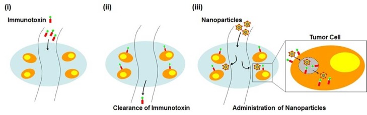
Schematic of temporally
staggered, staged delivery of a tumor-targeted
toxin and a cytosolic delivery chaperone.

Structure and composition of PEGylated
lipid-coated PBAE particles.
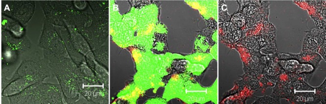
pH-responsive lipid-enveloped PBAE particles disrupt endosomes
and deliver coendocytosed calcein into the cytosol and nucleus of
tumor cells. B16F10 cells were incubated for 1 h at 37 °C with
calcein alone or calcein and rhodamine-labeled lipid-coated PBAE particles,
washed to remove unbound particles, then imaged live by confocal microscopy.
Representative confocal images of B16F10 melanoma cells incubated
with calcein (green) alone (A) or coincubated with calcein and lipid-coated
PBAE particles (red, B = brightfield-calcein-particle fluorescence
overlay, C = brightfield-particle fluorescence overlay). Scale bars
20 μm.
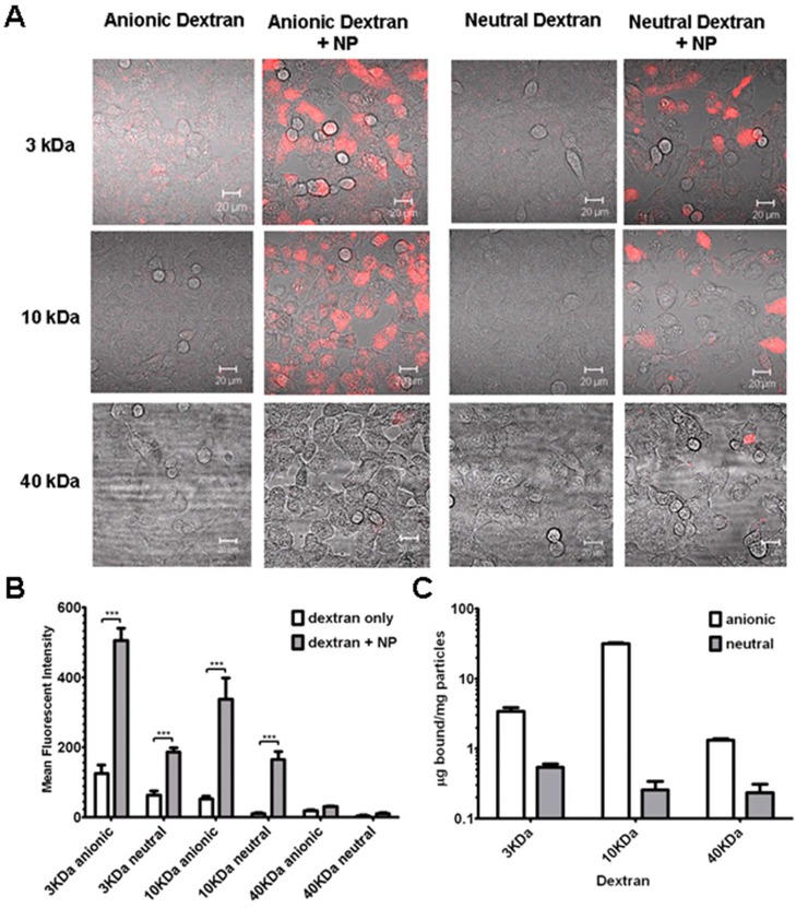
Effect of molecular weight
and charge of polar cargo macromolecules
on cytosolic delivery through coendocytosis with lipid-coated PBAE
particles. (A) B16F10 cells were incubated with anionic or neutral
fluorescent dextran conjugates (red, 150 μg/mL) of various molecular
weights, with or without lipid-coated PBAE nanoparticles (unlabeled,
75 μg/mL) for 1 h at 37 °C. Cells were washed and imaged
live via confocal microscopy. Scale bars 20 μm. (B) Plot of
mean fluorescence intensity detected in cells computed from replicate
fields of view for each dextran relative to cotreatment with particles
(***, p < 0.0001). (C) Binding of various dextrans
to nanoparticles coincubated in DMEM containing 10% serum for 18 h
at 37 °C at concentrations identical to the conditions of A and
B.
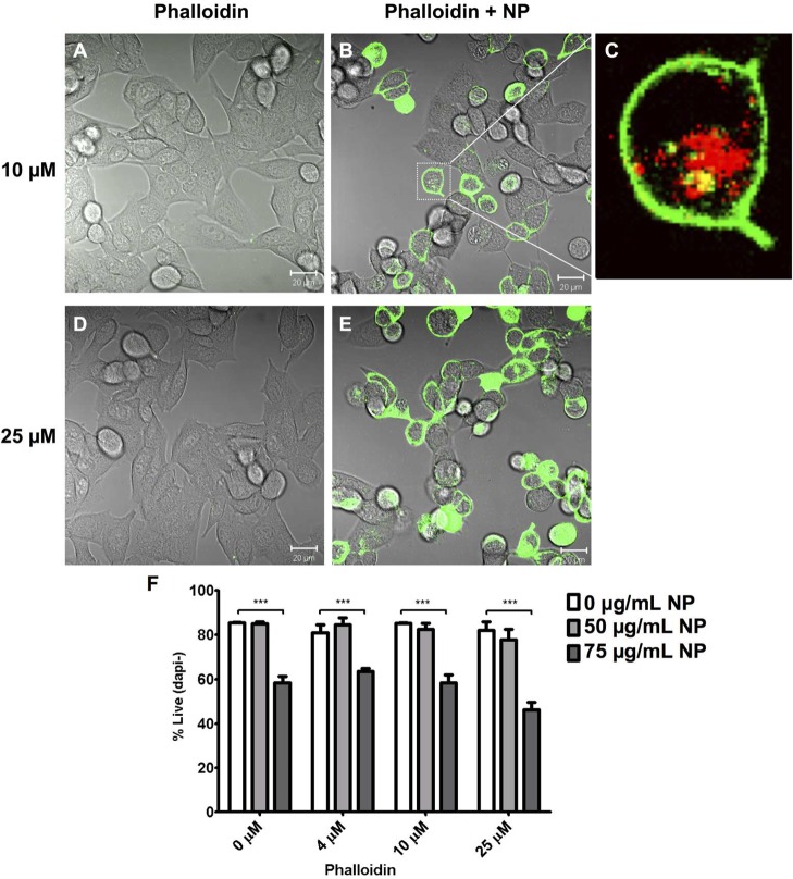
Cytosolic delivery of soluble phalloidin by lipid-coated
PBAE particles.
Confocal images of B16F10 cells incubated with (A) 10 μM or
(D) 25 μM phalloidin alone or coincubated with phalloidin and
75 μg/mL rhodamine-labeled lipid-coated PBAE particles (B–C,E).
(A,B,D,E = brightfield-phalloidin fluorescence overlays; C = magnified,
phalloidin-particle fluorescence image of boxed cell in B; red, nanoparticles;
green, phalloidin-alexa 488 conjugate). (F) Cytotoxicity of B16F10
cells treated with various concentrations of phalloidin alone or combined
with 50 or 75 μg/mL particles for 24 h. (***, p < 0.0001).
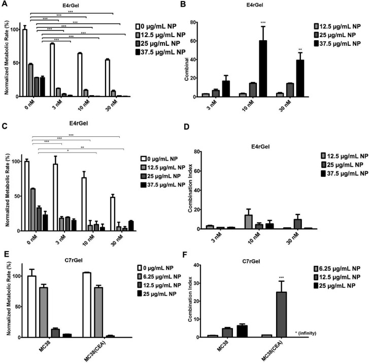
Killing of tumor cells by particle-chaperoned immunotoxins. (A–D)
Tumor cell lines A431 (EGFR-expressing, A,B) or B16F10 (EGFR-negative,
C,D) were incubated with E4rGel immunotoxin and/or lipid-coated PBAE
particles at the indicated concentrations for 24 h and then washed
into fresh medium. Viability was measured at 72 h via the WST-1 metabolic
assay (A,C) and used to compute the combination index where a value
>1 indicates synergistic tumor cell killing in the combination
treatment
(particles + E4rGel) compared to immunotoxin alone (B,D). (E–F)
The normalized metabolic rate (E) and combination index (F) for MC38
tumor cells or MC38 cells expressing CEA following incubation with
1 nM of the CEA-targeted C7rGel immunotoxin and lipid-coated PBAE
particles at the indicated concentrations. (A: ***, p < 0.0001; B,D: ***, p < 0.0001, **, p < 0.01 relative to combination index = 1).
Similar articles
-
Fusion of gelonin and anti-insulin-like growth factor-1 receptor (IGF-1R) affibody for enhanced brain cancer therapy.Arch Pharm Res. 2017 Sep;40(9):1094-1104. doi: 10.1007/s12272-017-0953-7. Epub 2017 Sep 12. Arch Pharm Res. 2017. PMID: 28900896
-
Preparation and Characterization of Gelonin-Melittin Fusion Biotoxin for Synergistically Enhanced Anti-Tumor Activity.Pharm Res. 2016 Sep;33(9):2218-2228. doi: 10.1007/s11095-016-1959-4. Epub 2016 Jun 1. Pharm Res. 2016. PMID: 27251414 Free PMC article.
-
Combination of antibody targeting and PTD-mediated intracellular toxin delivery for colorectal cancer therapy.J Control Release. 2014 Nov 28;194:197-210. doi: 10.1016/j.jconrel.2014.08.030. Epub 2014 Sep 7. J Control Release. 2014. PMID: 25204286 Free PMC article.
-
15 years of ATTEMPTS: a macromolecular drug delivery system based on the CPP-mediated intracellular drug delivery and antibody targeting.J Control Release. 2015 May 10;205:58-69. doi: 10.1016/j.jconrel.2014.12.002. Epub 2014 Dec 4. J Control Release. 2015. PMID: 25483423 Review.
-
Advanced targeted therapies in cancer: Drug nanocarriers, the future of chemotherapy.Eur J Pharm Biopharm. 2015 Jun;93:52-79. doi: 10.1016/j.ejpb.2015.03.018. Epub 2015 Mar 23. Eur J Pharm Biopharm. 2015. PMID: 25813885 Review.
Cited by
-
Engineered Polymeric Materials for Biological Applications: Overcoming Challenges of the Bio-Nano Interface.Polymers (Basel). 2019 Sep 2;11(9):1441. doi: 10.3390/polym11091441. Polymers (Basel). 2019. PMID: 31480780 Free PMC article. Review.
-
Plant Ribosome-Inactivating Proteins: Progesses, Challenges and Biotechnological Applications (and a Few Digressions).Toxins (Basel). 2017 Oct 12;9(10):314. doi: 10.3390/toxins9100314. Toxins (Basel). 2017. PMID: 29023422 Free PMC article. Review.
-
Cetuximab Immunoliposomes Enhance Delivery of 5-FU to Skin Squamous Carcinoma Cells.Anticancer Agents Med Chem. 2017;17(2):301-308. doi: 10.2174/1871520616666160526110913. Anticancer Agents Med Chem. 2017. PMID: 27225449 Free PMC article.
-
Immunotoxins constructed with ribosome-inactivating proteins and their enhancers: a lethal cocktail with tumor specific efficacy.Curr Pharm Des. 2014;20(42):6584-643. doi: 10.2174/1381612820666140826153913. Curr Pharm Des. 2014. PMID: 25341935 Free PMC article. Review.
-
Two-Step Targeted Drug Delivery via Proteinaceous Barnase-Barstar Interface and Doxorubicin-Loaded Nano-PLGA Outperforms One-Step Strategy for Targeted Delivery to HER2-Overexpressing Cells.Pharmaceutics. 2022 Dec 24;15(1):52. doi: 10.3390/pharmaceutics15010052. Pharmaceutics. 2022. PMID: 36678681 Free PMC article.
References
-
- Atkinson S. F.; Bettinger T.; Seymour L. W.; Behr J. P.; Ward C. M. J. Biol. Chem. 2001, 276, 27930–27935. - PubMed
-
- Cao Y.; Marks J. D.; Huang Q.; Rudnick S. I.; Xiong C.; Hittelman W. N.; Wen X.; Marks J. W.; Cheung L. H.; Boland K.; Li C.; Adams G. P.; Rosenblum M. G. Mol. Cancer Ther. 2012, 11, 143–153. - PubMed
-
- Leader B.; Baca Q. J.; Golan D. E. Nat. Rev. Drug Discovery 2008, 7, 21–39. - PubMed
-
- Kreitman R. J.; Pastan I. Adv. Drug Delivery Rev. 1998, 31, 53–88. - PubMed
Publication types
MeSH terms
Substances
Grants and funding
LinkOut - more resources
Full Text Sources
Other Literature Sources
