Review
. 2013 Feb;135(2):020301.
doi: 10.1115/1.4023479.

Evolving strategies in mechanobiology to more effectively treat damaged musculoskeletal tissues

Affiliations
Review

Evolving strategies in mechanobiology to more effectively treat damaged musculoskeletal tissues

David L Butler et al. J Biomech Eng. 2013 Feb.

Abstract

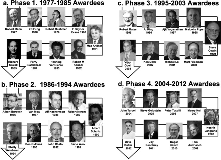
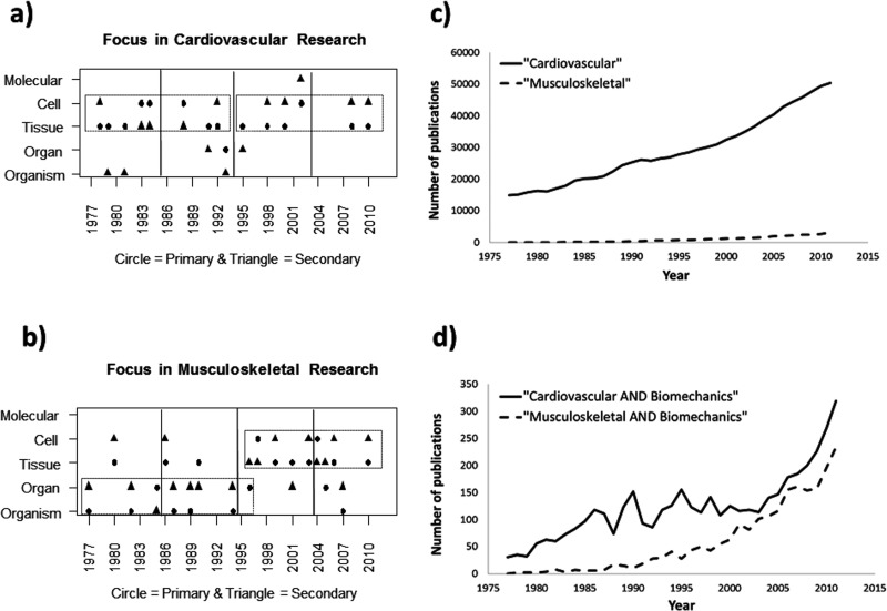
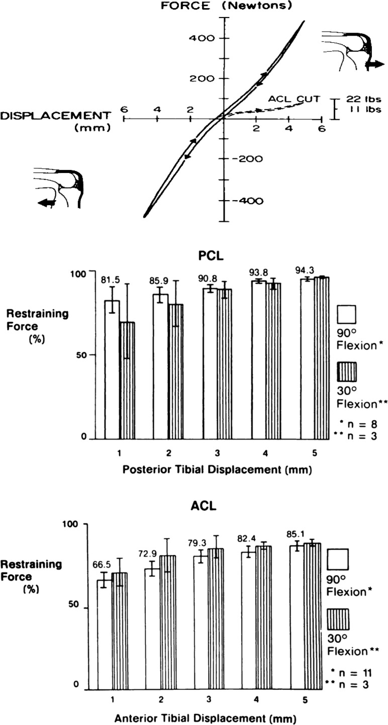
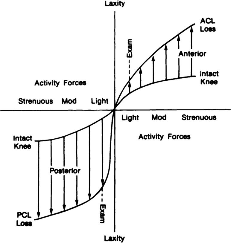
In this paper, we had four primary objectives. (1) We reviewed a brief history of the Lissner award and the individual for whom it is named, H.R. Lissner. We examined the type (musculoskeletal, cardiovascular, and other) and scale (organism to molecular) of research performed by prior Lissner awardees using a hierarchical paradigm adopted at the 2007 Biomechanics Summit of the US National Committee on Biomechanics. (2) We compared the research conducted by the Lissner award winners working in the musculoskeletal (MS) field with the evolution of our MS research and showed similar trends in scale over the past 35 years. (3) We discussed our evolving mechanobiology strategies for treating musculoskeletal injuries by accounting for clinical, biomechanical, and biological considerations. These strategies included studies to determine the function of the anterior cruciate ligament and its graft replacements as well as novel methods to enhance soft tissue healing using tissue engineering, functional tissue engineering, and, more recently, fundamental tissue engineering approaches. (4) We concluded with thoughts about future directions, suggesting grand challenges still facing bioengineers as well as the immense opportunities for young investigators working in musculoskeletal research. Hopefully, these retrospective and prospective analyses will be useful as the ASME Bioengineering Division charts future research directions.


Figures

Fig. 1
Fig. 1
(a)–(d) Prior Lissner Award winners assigned to four 9-year phases between 1977 and 2012
Fig. 2
Fig. 2
An analysis of cardiovascular versus musculoskeletal research between 1977 and 2011. (a) Primary and secondary research areas for Lissner awardees working in the cardiovascular area mostly remained at the tissue and cell level during the four 9-year phases. (b) By contrast, primary and secondary areas for Lissner winners working in the musculoskeletal area moved from the organism/organ level to the tissue/cell level around 1998 (p = 0.05). Also interesting to note from PubMed is that, during this same period, (c) the number of cardiovascular publications far exceeded those in the musculoskeletal field (e.g., 50,355 versus 3152 in 2011). (d) Despite this discrepancy, when this data was restricted to biomechanics publications, the two groups were much more similar and began to converge in the late 1990 s.
Fig. 3
Fig. 3
The ACL (bottom graph) is the primary ligamentous restraint up to 5 mm of anterior tibial translation (85% of total anterior restraining force). The PCL (middle graph) is the primary restraint up to 5 mm of posterior tibial translation (94%–96% of total restraining force). Adapted with permission from Ref. [9].
Fig. 4
Fig. 4
Anterior knee laxity versus activity forces in intact cadaveric knee and after individual sectioning of the ACL and PCL. The surgeon may not detect a small increase in anterior laxity in the ACL-deficient knee under “light” forces of the clinical exam, but the patient definitely experiences the greater increases in laxity under more strenuous forces. The increases in posterior laxity after loss of the PCL are more pronounced at both load levels. Adapted with permission from Ref. [9].
Fig. 5
Fig. 5
Maximum forces generated by graft tissues compared to the young adult anterior cruciate ligament-bone unit. Central- and medial bone-patellar tendon-bone units were the strongest tissues (159%–168% of ACL failure force). The semitendinosis (70%) and gracilis (49%) tendons were somewhat weaker than the ACL. All other structures were still weaker, with the retinacular tissues transmitting only 14%–21% of ACL maximum force. Adapted with permission from Ref. [12].
Fig. 6
Fig. 6
Designing a graft to withstand normal ligament failure forces is ideal. However, designing grafts within “safety zones” for normal and strenuous ADLs might matter more. Unfortunately, researchers in the mid-1980s could only estimate these force limits. Adapted with permission from Ref. [12].
Fig. 7
Fig. 7
Unloading the rabbit PT with K-wire and sutures produced 70%–80% reductions in tissue material properties by 6 weeks postsurgery. Adapted with permission from Ref. [94].
Fig. 8
Fig. 8
Peak in vivo forces in the patellar tendon are larger than those in the anterior cruciate ligament. (a) Note that peak IVFs in the goat ACL are negligible during the swing phase of gait, increasing rapidly during stance but never exceeding 7%–10% of the tissue's failure force. (b) Peak PT force is 8% of failure force during stance phase, increasing rapidly during gait to 32%–40% of failure force at 2.0–2.5 m/s. Adapted with permission from Refs. [97] and [33].
Fig. 9
Fig. 9
Functional design limits for the goat anterior cruciate ligament were found to be less than those for the goat patellar tendon. Adapted with permission from Ref. [12].
Fig. 10
Fig. 10
Functional tissue engineering roadmap. Shown are the in vitro, tissue engineering phase required to create a tissue engineering substitute or construct as well as the important surgery and evaluation phase to determine if the repair regenerates the tissue to exceed in vivo forces or at least repairs to achieve functional efficacy. Adapted from Ref. [47].
Fig. 11
Fig. 11
Continuous improvement in traditional biomechanical properties, including maximum force, stiffness, maximum stress, and linear modulus. These improvements involved changes in cell density, collagen scaffold stiffness, and the use of mechanical preconditioning of the TEC before surgery. Adapted from Ref. [69].
Fig. 12
Fig. 12
Tissue-engineered constructs containing MSCs in a collagen scaffold improve central rabbit PT repair. (a) Constructs containing a high cell density (1 × 106 cells/ml) produce a small but significant improvement in the force-displacement repair curve compared to natural healing. Not only does the failure curve for the TEC repair not match that for the normal unoperated PT, the 12-week repair also does not reach the peak in vivo forces (IVFs) acting on the normal central PT or match normal tangent stiffness. (b) Lowering the cell density, stiffening the collagen scaffold, and mechanically preconditioning the constructs before surgery resulted in improvements in failure properties as well as functional parameters (exceeding peak IVFs and matching normal PT tangent stiffness with a 50% safety factor). Adapted from Ref. [56].
Fig. 13
Fig. 13
The murine patellar tendon rapidly changes its structure and cellularity from late fetal life to 2 weeks after birth. The tendon midsubstance and insertion are cellular and their extracellular matrices are rather poorly aligned at E17.5. Postnatally, the tissue midsubstance shows decreasing cellularity and increasing collagen alignment from P1 to P14. The insertion is also maturing into fibrocartilage and bone. Adapted with permission from Ref. [80].
Fig. 14
Fig. 14
Natural healing of murine central patellar tendon defect injury. (a) Healing occurs slowly between 2 and 8 weeks postinjury when compared to the normal tendon failure curve. Estimated upper and lower peak in vivo force bounds are shown (using rabbit results from Ref. [37] and goat results from Ref. [33]) (from Ref. [81]). (b) Panels of genes for normal, sham, and defect healing groups at 1, 2, and 3 weeks postsurgery analyzed using principal component analysis.
Fig. 15
Fig. 15
A fundamental tissue engineering strategy that seeks to more rapidly design, evaluate, and optimize tissue-engineered constructs using normal tissue development, natural healing, and TEC manipulation across species. Adapted from Ref. [82].
Fig. 16
Fig. 16
Tendon healing shares similar characteristics with bone healing. Central PT healing in the mouse (upper panel, cross-sectional view) results in paratenon progenitor cells proliferating and migrating to form a bridge over the anterior surface of the defect space. This response is similar to fracture callus formation in tibial fractures (lower panel). Scleraxis (Scx) GFP reporter expression and smooth muscle actin α (SMAA) immunostaining (red) label potential early progenitor cells in these healing scenarios. White arrows indicate coexpressing cells.

References

    1. Bao, G. , Kamm, R. D. , Thomas, W. , Hwang, W. , Fletcher, D. A. , Grodzinsky, A. J. , Zhu, C. , and Mofrad, M. R. , 2010, “Molecular Biomechanics: The Molecular Basis of How Forces Regulate Cellular Function,” Mol. Cell Biomech., 3(2), pp. 91–105.10.1007/s12195-010-0109-z - DOI - PMC - PubMed
    1. Discher, D. , Dong, C. , Fredberg, J. J. , Guilak, F. , Ingber, D. , Janmey, P. , Kamm, R. D. , Schmid-Schonbein, G. W. , and Weinbaum, S. , 2009, “Biomechanics: Cell Research and Applications for the Next Decade,” Ann. Biomed. Eng., 37(5), pp. 847–859.10.1007/s10439-009-9661-x - DOI - PMC - PubMed
    1. Butler, D. L. , Goldstein, S. A. , Guldberg, R. E. , Guo, X. E. , Kamm, R. , Laurencin, C. T. , McIntire, L. V. , Mow, V. C. , Nerem, R. M. , Sah, R. L. , Soslowsky, L. J. , Spilker, R. L. , and Tranquillo, R. T. , 2009, “The Impact of Biomechanics in Tissue Engineering and Regenerative Medicine,” Tissue Eng. Part B Rev., 15(4), pp. 477–484.10.1089/ten.teb.2009.0340 - DOI - PMC - PubMed
    1. Ateshian, G. A. , and Friedman, M. H. , 2009, “Integrative Biomechanics: A Paradigm for Clinical Applications of Fundamental Mechanics,” J. Biomech., 42(10), pp. 1444–1451.10.1016/j.jbiomech.2009.04.001 - DOI - PubMed
    1. Dalgaard, P. , 2008, Introductory Statistics With R, Springer, New York, p. 364.

Publication types