. 2013 Mar 28;495(7442):481-6.
doi: 10.1038/nature11991. Epub 2013 Feb 27.

Structural visualization of key steps in human transcription initiation

Affiliations

Structural visualization of key steps in human transcription initiation

Yuan He et al. Nature. .

Abstract

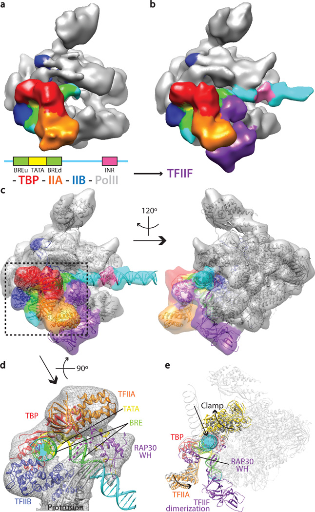
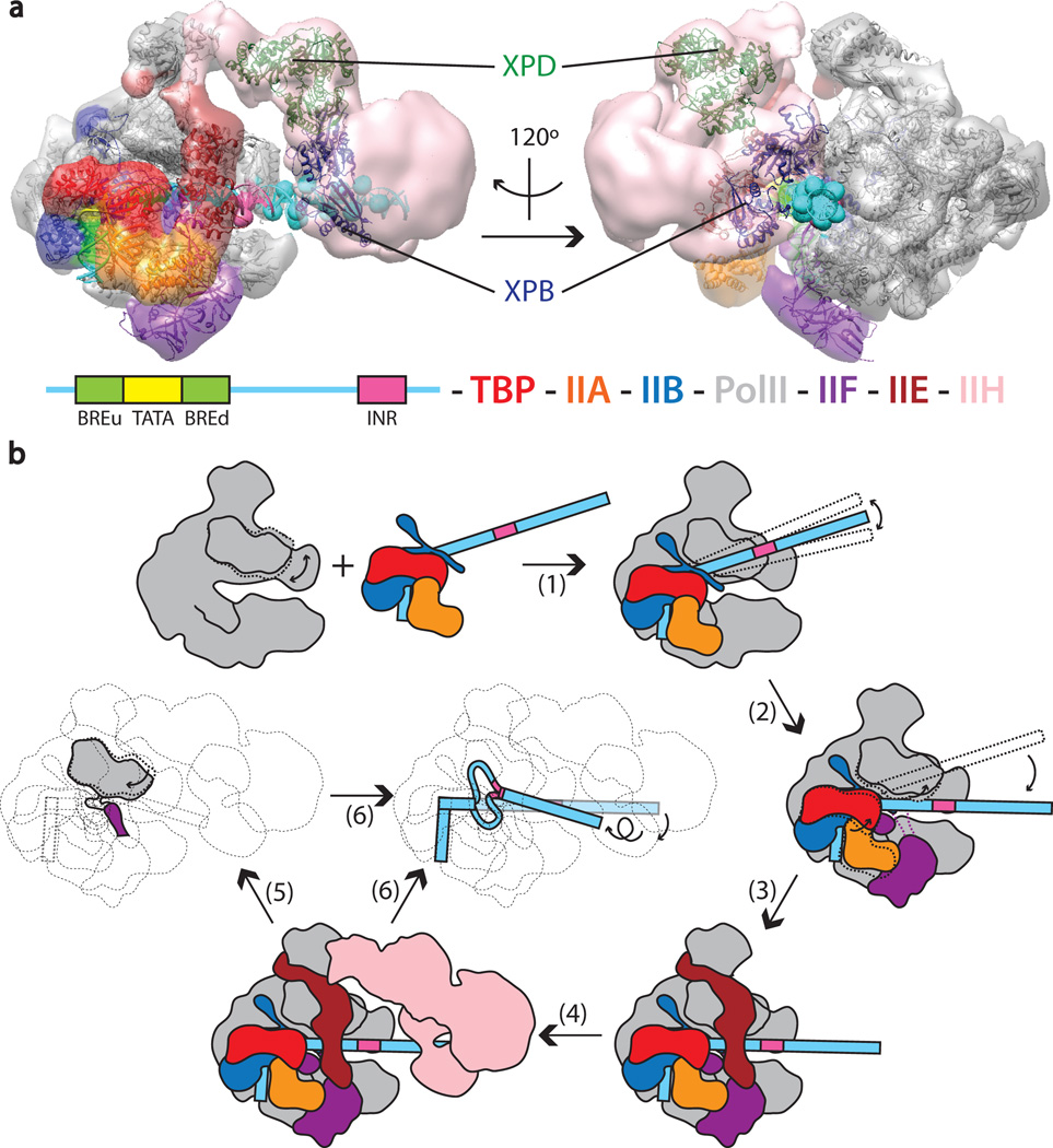
Eukaryotic transcription initiation requires the assembly of general transcription factors into a pre-initiation complex that ensures the accurate loading of RNA polymerase II (Pol II) at the transcription start site. The molecular mechanism and function of this assembly have remained elusive due to lack of structural information. Here we have used an in vitro reconstituted system to study the stepwise assembly of human TBP, TFIIA, TFIIB, Pol II, TFIIF, TFIIE and TFIIH onto promoter DNA using cryo-electron microscopy. Our structural analyses provide pseudo-atomic models at various stages of transcription initiation that illuminate critical molecular interactions, including how TFIIF engages Pol II and promoter DNA to stabilize both the closed pre-initiation complex and the open-promoter complex, and to regulate start--initiation complexes, combined with the localization of the TFIIH helicases XPD and XPB, support a DNA translocation model of XPB and explain its essential role in promoter opening.


Figures

Figure 1
Figure 1. Stepwise assembly of the human PIC
(a) Reconstitution strategy for human PIC by sequential assembly. Schematic of the DNA highlighting the relative positions of the core promoter elements used and SalI restriction site (top). Color scheme for the components of the PIC (bottom). Negative stain reconstructions of PIC assembly intermediates for TBP-TFIIA-TFIIB-DNA-Pol II (b), plus TFIIF (c), plus TFIIE (d), and plus TFIIH (e).
Figure 2
Figure 2. TFIIF engagement triggers a concerted conformational change in the PIC
The positions of TBP, TFIIA, TFIIB and DNA promoter elements relative to Pol II are indicated in the cryo-EM reconstruction in the absence (a) or presence of TFIIF (b). TFIIF stabilizes promoter DNA (cyan). (c) Crystal structures for TBP-TFIIA-DNA (PDB ID: 1NVP), TBP-TFIIB-DNA (PDB ID: 1C9B), Pol II-TFIIB (PDB ID: 4BBR), RAP30/74 dimerization domain (PDB ID: 1F3U), RAP30 WH domain (PDB ID: 1BBY), and modeled B-form DNA (globally bent between −23 and +7 by 18°) are shown docked into transparent EM densities. The mobile clamp of Pol II is docked as a separate domain of Pol II. (d) Bottom view showing the presence of a nucleoprotein complex by the upstream core promoter elements. The DNA densities have been segmented out for clarity. Perturbed residues within the RAP30 WH domain during DNA titrations are colored in gold. The possible path for RAP30 N-terminus is highlighted with dotted purple lines. DNA is shown in ribbon representation. (e) Position of moving structural elements before (grey) and after (colored) TFIIF binding. The rest of the PIC components are shown in transparency and major structural rearrangements are depicted by arrows.
Figure 3
Figure 3. Stabilization of the PIC in the closed conformation by TFIIE
(a) Segmentation of the cryo-EM reconstruction of human PIC containing TFIIE (left), and docking of existing crystal structures (right). (b) Same view as the right panel in (a) depicting the cryo-EM density corresponding to TFIIE and the RAP30 WH domain. A chain of four WH domains formed by the C-terminus of RAP30 and both subunits of TFIIE (modeled based on cross-linking data) can be roughly fitted into the cryo-EM density. (c) Regions of contact between TFIIE and Pol II. The clamp and stalk domain of Pol II are shown in grey, TFIIE in maroon. The rest of the PIC components are shown in transparency. The clamp coiled-coil domain is shown in gold and the residues that crosslinks to TFIIE are colored in navy blue. (d) EM density corresponding to promoter DNA is shown together with the PIC ribbon model. Structural elements making direct contacts with the promoter DNA are depicted.
Figure 4
Figure 4. Conformational rearrangements of the PIC upon promoter opening
a) Nucleic acid scaffold used to generate a mimic of the OC. Filled and open circles correspond, respectively, to the core promoter used in Fig. 1a, and to a replacement sequence containing a 3’-tailed sequence previously designed to generate an arrested Pol II. The schematic indicates in pink the position that would correspond to the INR. (b) Segmentation of the cryo-EM reconstruction of the human PIC in the open state (left), and docking of existing crystal structures, together with a modeled DNA bubble (right). GTFs adopt the same architecture as in the closed PIC. (c) EM densities of promoter DNA in the closed PIC and OC were segmented and superimposed with respect to Pol II. Movements of the DNA between the two states are shown by arrows. The rotation accompanying translocation of the downstream DNA occurs within the plane of the view shown on the left panel (and thus perpendicular to view shown on the left). (d) Structures, before (grey) and after (colored) promoter opening, are shown using fitted crystal structures. The Pol II clamp comes down over the open bubble, in a conformation similar to that seen for the elongation state. (e) Segmented cryo-EM density showing the now visible arm domain of TFIIF connecting the rest of TFIIF with the rudder of RPB1. Crystal structures of TFIIB and TFIIF were docked as rigid bodies into the cryo-EM density.
Figure 5
Figure 5. Positioning of TFIIH helicases and model of PIC assembly and promoter opening
(a) Negative-stain structure of the full human PIC in the closed conformation (as in Fig. 1e). EM density corresponding to the TFIIH core complex, lacking any visible CAK sub-complex, is colored in pink. A homology model for XPB (navy blue) and the crystal structure of XPD (PDB ID: 3CRV, dark green) are shown docked into the core TFIIH density. The docking suggests different roles for the XPB and XPD helicases in promoter opening. DNA phosphates crosslinked to XPB are indicated by pink or cyan spheres. (b) Schematic of PIC assembly and promoter melting. Pol II is recruited through interaction with TFIIB to the promoter, which is engaged by TBP–TFIIA–TFIIB (1). TFIIF stabilizes the TBP-TFIIA-IIB-Pol II protrusion interaction hub and also positions the downstream DNA onto the cleft, forcing the clamp to swing into a slightly open state (2). TFIIE binding further stabilizes the PIC by interacting with the Pol II stalk, the clamp, and with TFIIF on the other side of the Pol II cleft (3). The TFIIE-containing PIC serves as the platform for TFIIH binding and correctly positions XPB downstream of the INR element (4). During strand separation, the clamp domain starts to swing down. The arm domain of TFIIF comes close to the Pol II rudder and the TFIIB B-linker. Stabilization of these interactions forms a physical barrier for DNA re-annealing (5). During promoter opening, the translocase activity of XPB would “screw in” the DNA toward the Pol II active site, leading to a Pol II open-promoter state ready for RNA synthesis.

References

    1. Matsui T, Segall J, Weil PA, Roeder RG. Multiple factors required for accurate initiation of transcription by purified RNA polymerase II. J Biol Chem. 1980;255:11992–11996. - PubMed
    1. Roeder RG. The role of general initiation factors in transcription by RNA polymerase II. Trends Biochem Sci. 1996;21:327–335. - PubMed
    1. Goodrich JA, Cutler G, Tjian R. Contacts in context: promoter specificity and macromolecular interactions in transcription. Cell. 1996;84:825–830. - PubMed
    1. Kornberg RD. The molecular basis of eukaryotic transcription. Proc Natl Acad Sci U S A. 2007;104:12955–12961. - PMC - PubMed
    1. Cramer P, et al. Structure of eukaryotic RNA polymerases. Annu Rev Biophys. 2008;37:337–352. - PubMed

Publication types

MeSH terms