

Increased skin inflammation and blood vessel density in human and experimental diabetes
- PMID: 23446362
- PMCID: PMC3688045
- DOI: 10.1177/1534734612474303
Increased skin inflammation and blood vessel density in human and experimental diabetes
Abstract
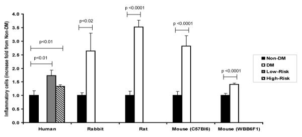
Systemic inflammation is associated with impaired wound healing in diabetes mellitus (DM) patients. Using immunohistochemistry techniques, the authors investigated changes in skin inflammation and skin blood vessels in human and experimental diabetes. Comparing to the non-DM human subjects, the total number of inflammatory cells per biopsy and the number of inflammatory cells around blood vessels, a strong indication of inflammation, were higher in DM subjects irrespective of their risk for developing diabetic foot ulcer. Inflammatory cell infiltration was robustly increased in all DM animal models compared with their non-DM controls. The number and density of blood vessels and CD31 positive proliferating endothelial cells around preexisting skin vessels was also higher in the DM patients. However, there were no differences in the skin blood flow between the non-DM and DM subjects. The number of skin blood vessels was also increased in the DM animals; however, these differences were less obvious than the ones observed for inflammatory cells. We conclude that skin inflammation and skin blood vessel density is increased in diabetic human subjects and in rodent and rabbit models of diabetes.
Figures

References
-
- Vital Statistics. Alexandria, VA: 1996.
-
- Ramsey SD, Newton K, Blough D, et al. Incidence, outcomes, and cost of foot ulcers in patients with diabetes. Diabetes Care. 1999 Mar;22(3):382–387. - PubMed
-
- Veves A, Akbari CM, Primavera J, et al. Endothelial dysfunction and the expression of endothelial nitric oxide synthetase in diabetic neuropathy, vascular disease, and foot ulceration. Diabetes. 1998 Mar;47(3):457–463. - PubMed
-
- Wagner N, Morrison H, Pagnotta S, et al. The podocyte protein nephrin is required for cardiac vessel formation. Hum Mol Genet. 2011 Jun 1;20(11):2182–2194. - PubMed
Publication types
MeSH terms
Grants and funding
LinkOut - more resources
Full Text Sources
Other Literature Sources
