. 2013 Apr 11;88(4):90.
doi: 10.1095/biolreprod.112.107128. Print 2013 Apr.

Disruption of a spermatogenic cell-specific mouse enolase 4 (eno4) gene causes sperm structural defects and male infertility

Affiliations

Disruption of a spermatogenic cell-specific mouse enolase 4 (eno4) gene causes sperm structural defects and male infertility

Noriko Nakamura et al. Biol Reprod. .

Abstract

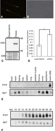
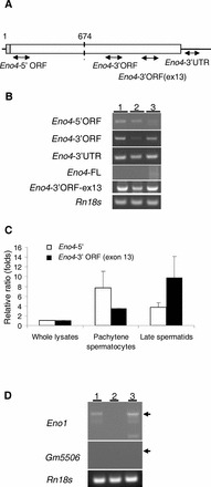
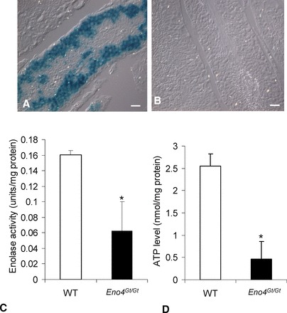
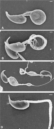
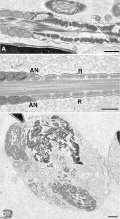
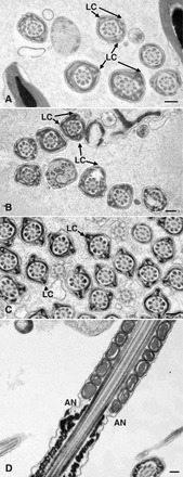
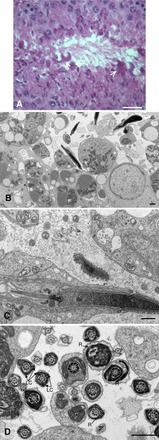
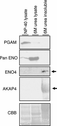
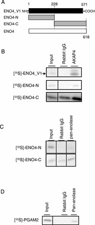
Sperm utilize glycolysis to generate ATP required for motility, and several spermatogenic cell-specific glycolytic isozymes are associated with the fibrous sheath (FS) in the principal piece of the sperm flagellum. We used proteomics and molecular biology approaches to confirm earlier reports that a novel enolase is present in mouse sperm. We then found that a pan-enolase antibody, but not antibodies to ENO2 and ENO3, recognized a protein in the principal piece of the mouse sperm flagellum. Database analyses identified two previously uncharacterized enolase family-like candidate genes, 64306537H0Rik and Gm5506. Northern analysis indicated that 64306537H0Rik (renamed Eno4) was transcribed in testes of mice by Postnatal Day 12. To determine the role of ENO4, we generated mice using embryonic stem cells in which an Eno4 allele was disrupted by a gene trap containing a beta galactosidase (beta-gal) reporter (Eno4(+/Gt)). Expression of beta-gal occurred in the testis, and male mice homozygous for the gene trap allele (Eno4(Gt/Gt)) were infertile. Epididymal sperm numbers were 2-fold lower and sperm motility was reduced substantially in Eno4(Gt/Gt) mice compared to wild-type mice. Sperm from Eno4(Gt/Gt) mice had a coiled flagellum and a disorganized FS. The Gm5506 gene encodes a protein identical to ENO1 and also is transcribed at a low level in testis. We conclude that ENO4 is required for normal assembly of the FS and provides most of the enolase activity in sperm and that Eno1 and/or Gm5506 may encode a minor portion of the enolase activity in sperm.


Figures

FIG. 1
FIG. 1
AC) Enolase in sperm. A) Immunofluorescence localization of enolase by pan-enolase antibody in the PP of the sperm flagellum. B) Phase contrast of same sperm (bar = 8 μm). C) Enolase (ENO) was detected in pellets but not in supernatants of sperm lysates. The same gel stained with Coomassie brilliant blue (CBB) before transfer is shown below to demonstrate protein loading. D) Enolase activity was detected in pellets and at lower levels in supernatants of sperm lysates. Enolase activity in the pellets was significantly higher than in supernatants (*P < 0.05). Data are expressed as means ± SD of three experiments. E and F) Northern blot analysis of Eno4 expression. Northern blot analyses using a probe from the translated region (715-1869 nt) of Eno4 transcript and RNA from various tissues of adult mice (E) or whole testes of Day 10–35 juvenile mice (F). The Rpl7 transcript was used as an internal control.
FIG. 2
FIG. 2
AC) Expression of Eno1 transcripts and variants. A) The location of primers specific for each of the Eno4 transcripts used in the PCR assays. B) Conventional RT-PCR results with specific primer pairs detecting Eno4 transcript and variants in testes and isolated germ cells. Lanes contained total RNA from testes (lane 1), StaPut-isolated pachytene spermatocytes (lane 2), and StaPut-isolated round spermatids (lane 3). Thirty-five amplification cycles were used for the Eno4 transcript and variants, and the annealing temperature used was 63°C. C) Expression of Eno4 transcripts in pachytene spermatocytes and late spermatids isolated by LCM. Primers specific for each of the Eno4 transcripts was used to assay by qPCR their expression in whole lysates, pachytene spermatocyte, and late spermatids; Eno4-5′, white bars; Eno4-3′ORF (exon 13), black bars. Expression levels were determined as described in Materials and Methods and shown here as ratios (folds) relative to the level on whole lysate, with that level set at one. Data are expressed as means ± SEM. D) Expression of Gm5506 and Eno1 transcripts in mouse testes. Gene specific primers were used to detect Eno1 and Gm5506 using cDNA from testes and isolated germ cells by conventional RT-PCR. The amplification cycles of PCR for Eno1 and Gm5506 were 40 or 50, respectively. The annealing temperature used for Eno1 or Gm5506 was 60°C. Lanes contained total RNA from testes (lane 1), StaPut-isolated pachytene spermatocytes (lane 2), and Sta-Put-isolated round spermatids (lane 3). 18S ribosomal RNA (Rn18s) was used as a control.
FIG. 3
FIG. 3
β-galactosidase activity and ATP levels in Eno4Gt/Gt and WT testes. A) β-galactosidase activity was detected in germ cells in testis from Eno4Gt/Gt mice by X-gal staining. B) No β-galactosidase activity was detected in testis from WT mice by X-gal staining. Bars = 25 μm. C) Enolase activity. Sperm were suspended in an imidazole buffer containing 2 mM MgSO4 and 50 mM imidazol-HCl (pH 6.8) and sonicated, and total enolase activity was assayed. Enolase activity in sperm from Eno4Gt/Gt mice was significantly lower than in WT mice (n = 4; *P < 0.05). D) ATP levels. The ATP levels in sperm from Eno4Gt/Gt mice were significantly lower than in WT mice (n = 3; *P < 0.05).
FIG. 4
FIG. 4
SEM of sperm from Eno4Gt/Gt and WT mice. A and D) Sperm collected from the cauda epididymis and fixed with glutaraldehyde. B and C) Sperm collected from the cauda epididymis extracted with 0.1% Triton X-100 for 5 min before fixation in glutaraldehyde. AC) Sperm from Eno4Gt/Gt mouse. D) Sperm from WT mouse. Bars = 1 μm.
FIG. 5
FIG. 5
TEM of sperm from Eno4Gt/Gt and WT mice. Sperm collected from cauda epididymis were fixed, sectioned, and examined by TEM. A and C) Sperm from Eno4Gt/Gt mouse. B) Sperm from WT mouse. R, ribs of the FS; AN, annulus. Bars = 0.5 μm.
FIG. 6
FIG. 6
TEM of sperm from Eno4Gt/Gt and WT mice. Sperm collected from cauda epididymis were fixed, sectioned, and examined by TEM. Cross sections (AC) and sagittal section (D) of sperm flagellum are shown. A, B, and D) Sperm from Eno4Gt/Gt mouse. C) Sperm from WT mouse. LC, longitudinal column; AN, annulus. Bars = 0.2 μm.
FIG. 7
FIG. 7
Testis morphology in Eno4Gt/Gt mice. A) Hematoxylin and eosin staining in testis of Eno4Gt/Gt mice. Lobular structures (arrows) were observed along lumen of testis of Eno4Gt/Gt mice. Bar = 25 μm. B, C, and D) Testes were fixed, sectioned, and examined by TEM. Sagittal section (C) and cross section (D) of sperm flagellum are shown. LC, longitudinal column; R, ribs of FS. Bars = 1 μm.
FIG. 8
FIG. 8
Differences in solubility of PGAM2, enolase, ENO4, and AKAP4. Most of the PGAM2 (detected with PGAM antibody) and some of the enolase (detected with pan-enolase antibody) was detected in the 1% NP-40 sperm lysate. The majority of the enolase was solubilized by 6 M urea. ENO4 and AKAP4 were present in the 6 M urea insoluble sperm fraction.
FIG. 9
FIG. 9
Coimmunoprecipitation assay with ENO4, AKAP4, ENO, and PGAM2. A) Regions of ENO4_V1 (aa 1–571), truncated N-terminal ENO4-N (aa 1–228), and ENO4-C (aa 228–618) used for in vitro synthesized [35S]-labeled probes. B) [35S]-ENO4_V1 and [35S]-ENO4-C were coimmunoprecipitated from sperm lysates with AKAP4 by the antiserum to AKAP4 but not by rabbit IgG. C) [35S]-ENO4-C was coimmunoprecipitated from sperm lysates with ENO1 or GM5506 by the antibody to pan-enolase but not by rabbit IgG. D) [35S]-PGAM2 was coimmunoprecipitated from sperm lysates with ENO1 or GM5506 by the antibody to pan-enolase but not by rabbit IgG.

References

    1. Mori C, Nakamura N, Welch JE, Gotoh H, Goulding EH, Fujioka M, Eddy EM. Mouse spermatogenic cell-specific type 1 hexokinase (mHk1-s) transcripts are expressed by alternative splicing from the mHk1 gene and the HK1-S protein is localized mainly in the sperm tail. Mol Reprod Dev 1998; 49: 374–385. - PubMed
    1. Nakamura N, Shibata H, O'Brien DA, Mori C, Eddy EM. Spermatogenic cell-specific type 1 hexokinase is the predominant hexokinase in sperm. Mol Reprod Dev 2008; 75: 632–640. - PMC - PubMed
    1. Nakamura N, Mori C, Eddy EM. Molecular complex of three testis-specific isozymes associated with the mouse sperm fibrous sheath: hexokinase 1, phosphofructokinase M, and glutathione S-transferase mu class 5. Biol Reprod 2010; 82: 504–515. - PMC - PubMed
    1. Vemuganti SA, Bell TA, Scarlett DO, Parker CE, Pardo-Manuel de Villena F, O'Brien DA. Three male germline-specific aldolase A isozymes are generated by alternative splicing and retrotransposition. Dev Biol 2007; 309: 18–31. - PubMed
    1. McCarrey JR, Thomas K. Human testis-specific PGK gene lacks introns and possesses characteristics of a processed gene. Nature 1987; 326: 501–505. - PubMed

Publication types

Substances