. 2013 Feb 27;280(1757):20130071.
doi: 10.1098/rspb.2013.0071. Print 2013 Apr 22.

Embryos, polyps and medusae of the Early Cambrian scyphozoan Olivooides

Affiliations

Embryos, polyps and medusae of the Early Cambrian scyphozoan Olivooides

Xi-Ping Dong et al. Proc Biol Sci. .

Abstract

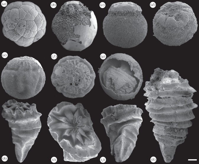
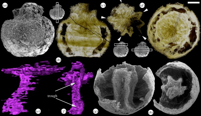
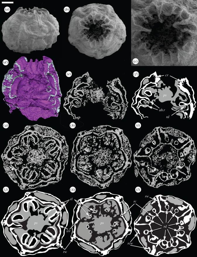
The Early Cambrian organism Olivooides is known from both embryonic and post-embryonic stages and, consequently, it has the potential to yield vital insights into developmental evolution at the time that animal body plans were established. However, this potential can only be realized if the phylogenetic relationships of Olivooides can be constrained. The affinities of Olivooides have proved controversial because of the lack of knowledge of the internal anatomy and the limited range of developmental stages known. Here, we describe rare embryonic specimens in which internal anatomical features are preserved. We also present a fuller sequence of fossilized developmental stages of Olivooides, including associated specimens that we interpret as budding ephyrae (juvenile medusae), all of which display a clear pentaradial symmetry. Within the framework of a cnidarian interpretation, the new data serve to pinpoint the phylogenetic position of Olivooides to the scyphozoan stem group. Hypotheses about scalidophoran or echinoderm affinities of Olivooides can be rejected.


Figures

Figure 1.
Figure 1.
Embryos and polyps of Olivooides. (a) Cleavage-stage embryo (GMPKU3080). (b) Stellate embryo with raised apertural rays (GMPKU3081). (c) Stellate embryo with a raised apertural region (GMPKU3082). (d) Stellate embryo with an infolded apertural region (GMPKU3084). (ef) Stellate embryo showing a characteristic pattern of bulges in (e) lateral, and (f) apertural aspect (GMPKU3083). (g) Embryo within fertilization envelope that preserves the characteristic pattern of bulges, but not the stellate tissue (GMPKU3085). (h–j) Early post-embryonic polyp stage exhibiting characteristic stellate integument of embryonic stage, but exhibiting the annulated striated adapertural tissue, and the pentamerally enfolded aperture, in (h) lateral, (i) apertural and (j) oblique aspects (GMPKU2317). (k) Later polyp-stage specimen in lateral aspect (GMPKU3086). Scale bars: (a) 69 µm, (b) 116 µm, (c) 139 µm, (d) 125 µm, (e,f) 132 µm, (g) 164 µm, (h) 275 µm, (i) 118 µm, (j) 250 µm, (k) 262 µm.
Figure 2.
Figure 2.
Embryonic Olivooides specimens with internal preservation. (a–f) Specimen with five-rayed internal structures (GMPKU3087). (a) SEM image of whole specimen in lateral view. (b–d) SRXTM-based virtual thin sections with insets showing the positions of the sections within the specimen. (b) Longitudial section showing biological preservation in the upper third. (c) Transverse section showing apertural rays, arrows indicate primary rays which are intercalated by interrays. (d) Transverse section showing the rays at depth. (e,f) SRXTM renderings of a single ray in lateral (e) and adaxial (f) aspects; arrows indicate the trough formed at depth within the ray. (g,h) Specimen preserving a pentagonal axial mould (GMPKU3088) imaged using SEM. (g) Lateral view. (h) Apertural view. Scale bars: (a,b,d) 155 µm, (c) 132 µm, (e,f) 46 µm, (g) 150 µm, (h) 170 µm. (Online version in colour.)
Figure 3.
Figure 3.
Embryonic Olivooides specimen with internal preservation (GMPKU3089) imaged using SEM (a–c) and SRXTM (d–l). (a) Whole specimen in lateral view. (b) Whole specimen in apertural view. (c) Detail of the aperture. (d) Surface rendering showing the internal anatomy. (e,f) Longitudinal section and interpretative drawing. (g,j) Transverse section and interpretative drawing showing paired projections from the inner wall and radial structures that connect the inner and outer walls. (h,k) Transverse section and interpretative drawing in particular showing interadial canals. (i,l) Transverse section and interpretative drawing showing the inner wall extending into the lumen to form the upper abradial ridge; depressions in the lower surface of the ridge appear as paired circular structures in section. Within the interpretative drawings, white represent a dense highly X-ray attenuating mineral phase, while grey reflects a more diffuse lowly attenuating mineral phase; both are interpreted to reflect original biological structure. ab.r, abradial ridges; cr, circumferential ridges; irr, interradial ridge; ps, polygonal axial structure; rc, radial canal; rcw, recurved wall; rd, radial depression; rl, radial lobe; rw, radial wall. Scale bars: (a,b) 153 µm, (c) 73 µm, (d) 78 µm, (e,f) 124 µm, (g,j) 119 µm, (h,k) 136 µm, (i,l) 143 µm. (Online version in colour.)
Figure 4.
Figure 4.
Two adpressed ephyra-stage Olivooides specimens (GMPKU3090). (a) SEM image showing the whole specimen in aboral view. (b) SEM image showing a detail of the aboral surface; note the alternately overlying and underlying arms. (c) SRXTM surface rendering showing a detail of the adoral surface including the presumed mouth. Scale bars: (a) 49 µm, (b) 27 µm, (c) 28 µm. (Online version in colour.)

Similar articles

Cited by

References

    1. Bengtson S, Yue Z. 1997. Fossilized metazoan embryos from the earliest Cambrian. Science 277, 1645–164810.1126/science.277.5332.1645 (doi:10.1126/science.277.5332.1645) - DOI - DOI
    1. Xiao S, Zhang Y, Knoll AH. 1998. Three-dimensional preservation of algae and animal embryos in a Neoproterozoic phosphate. Nature 391, 553–55810.1038/35318 (doi:10.1038/35318) - DOI - DOI
    1. Dong X-P, Donoghue PCJ, Cheng H, Liu J. 2004. Fossil embryos from the Middle and Late Cambrian period of Hunan, south China. Nature 427, 237–24010.1038/nature02215 (doi:10.1038/nature02215) - DOI - DOI - PubMed
    1. Donoghue PCJ, Dong X-P. 2005. Embryos and ancestors. In Evolving form and function: fossils and development (ed. Briggs DEG.), pp. 81–99 New Haven, CT: Yale Peabody Museum of Natural History, Yale University
    1. Yue Z, Bengtson S. 1999. Embryonic and post-embryonic development of the Early Cambrian cnidarian Olivooides. Lethaia 32, 181–19510.1111/j.1502-3931.1999.tb00538.x (doi:10.1111/j.1502-3931.1999.tb00538.x) - DOI - DOI

Publication types

LinkOut - more resources