. 2013 Apr 12;288(15):10588-98.
doi: 10.1074/jbc.M112.449033. Epub 2013 Feb 27.

Mutational tail loss is an evolutionary mechanism for liberating marapsins and other type I serine proteases from transmembrane anchors

Affiliations

Mutational tail loss is an evolutionary mechanism for liberating marapsins and other type I serine proteases from transmembrane anchors

Kavita Raman et al. J Biol Chem. .

Abstract

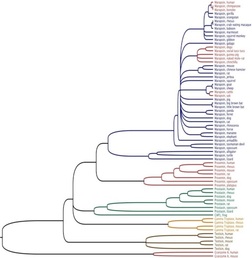
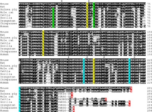
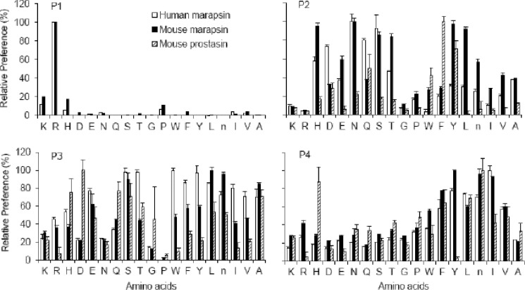
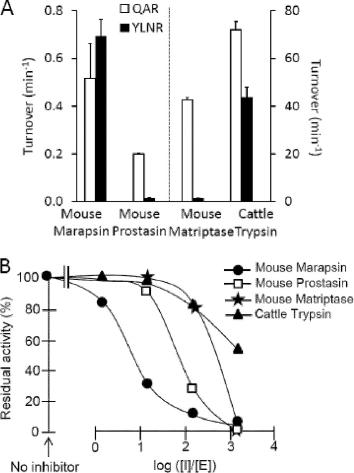
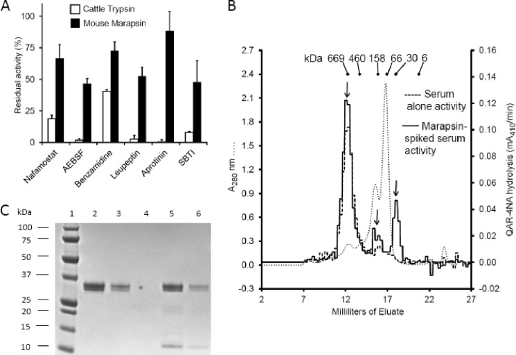
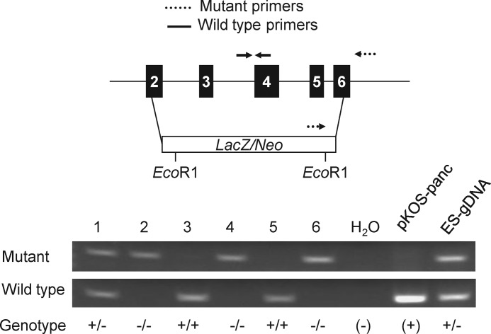
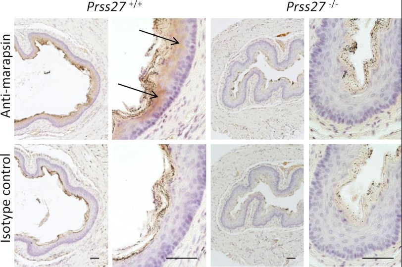
Human and mouse marapsins (Prss27) are serine proteases preferentially expressed by stratified squamous epithelia. However, mouse marapsin contains a transmembrane anchor absent from the human enzyme. To gain insights into physical forms, activities, inhibition, and roles in epithelial differentiation, we traced tail loss in human marapsin to a nonsense mutation in an ancestral ape, compared substrate preferences of mouse and human marapsins with those of the epithelial peptidase prostasin, designed a selective substrate and inhibitor, and generated Prss27-null mice. Phylogenetic analysis predicts that most marapsins are transmembrane proteins. However, nonsense mutations caused membrane anchor loss in three clades: human/bonobo/chimpanzee, guinea pig/degu/tuco-tuco/mole rat, and cattle/yak. Most marapsin-related proteases, including prostasins, are type I transmembrane proteins, but the closest relatives (prosemins) are not. Soluble mouse and human marapsins are tryptic with subsite preferences distinct from those of prostasin, lack general proteinase activity, and unlike prostasins resist antiproteases, including leupeptin, aprotinin, serpins, and α2-macroglobulin, suggesting the presence of non-canonical active sites. Prss27-null mice develop normally in barrier conditions and are fertile without overt epithelial defects, indicating that marapsin does not play critical, non-redundant roles in development, reproduction, or epithelial differentiation. In conclusion, marapsins are conserved, inhibitor-resistant, tryptic peptidases. Although marapsins are type I transmembrane proteins in their typical form, they mutated independently into anchorless forms in several mammalian clades, including one involving humans. Similar pathways appear to have been traversed by prosemins and tryptases, suggesting that mutational tail loss is an important means of evolving new functions of tryptic serine proteases from transmembrane ancestors.


Figures

FIGURE 1.
FIGURE 1.
Cladogram. This rooted tree probing relationships between vertebrate marapsins and related serine proteases was generated by unweighted pair group with arithmetic mean analysis of aligned protein sequences. To allow comparison of proteins with signal peptides, propeptides, and C termini of varying length, the alignment was limited to mature catalytic domains with deletion of C-terminal extensions present in a subset of the proteases. Nodes were assigned if predicted by at least 550 of 1000 iterations of bootstrap resampling. Mouse granzyme A and human granzyme A, which are tryptic serine proteases not closely related to marapsins, prostasins, or tryptases, together serve as an outgroup. Clades of proteases known or predicted to lack a membrane anchor are red. The other proteases are predicted to be type I transmembrane proteins based on the presence of a C-terminal, hydrophobic extension of membrane-spanning length. Note that channel-activating protease (CAP-1) appears to be a frog ortholog of mammalian prostasins. Accession numbers of sequences used in tree construction are given in supplemental Table S1.
FIGURE 2.
FIGURE 2.
Alignment of marapsins. Selected mammalian marapsin sequences beginning with Met1 of the predicted mature catalytic domain after activation are aligned. The nine absolutely conserved Cys residues, one of which (Cys110) is predicted to link with propeptide Cys−9 (not shown), are marked with a “#.” Consensus conserved sites of N-glycosylation sites are green. The absolutely conserved catalytic triad residues (His41, Asp90, and Ser195) and specificity triad residues (Asp189, Gly216, and Gly226) essential for S1 serine protease function and tryptic specificity are yellow and cyan, respectively. Other aligned residues that are identical to those in the mouse marapsin sequence are black. Sequence-terminating stop codons are red, revealing marked length variations.
FIGURE 3.
FIGURE 3.
Comparison of marapsin and prostasin subsite preferences. Mouse and human marapsins and mouse prostasin were profiled using fluorogenic tetrapeptide substrates. For the marapsins, primary specificity was tested in a combinatorial library in which each of the designated 20 amino acids was held constant in turn as residues P2 through P4 were varied. Therefore, each assay condition tested a mixture of 8000 different peptide substrates for a given P1 residue. The amidolytic activity of marapsin liberates 7-amino-4-carbamoylmethylcoumarin. The initial readout in relative fluorescence units was normalized to the result for the most preferred amino acid in each subsite. The other panels show results of similar profiling at positions P2, P3, and P4, respectively, by fixing amino acids at the designated position and varying the residues at the remaining positions. Prostasin was profiled at positions P2, P3, and P4 with P1 fixed at Arg. Abbreviations for amino acids are given in standard one-letter code; n is norleucine (substituting for Met). Error bars represent S.D.
FIGURE 4.
FIGURE 4.
Selectivity of a tetrapeptide substrate and inhibitor synthesized based on mouse marapsin subsite preferences. A shows the relative preference of mouse marapsin, prostasin, and matriptase and bovine trypsin for custom tetrapeptide substrate YLNR-4NA versus nonspecific tripeptide substrate QAR-4NA (both substrates 0.5 mm). YLNR-4NA synthesized based on results of combinatorial peptide substrate profiling of marapsin (as shown in Fig. 3) was highly selective for mouse marapsin over prostasin and matriptase but less so in relation to trypsin in which subsite preferences are less pronounced. QAR-4NA was preferred by all proteases except marapsin. Specific activity for both substrates was much lower for marapsin and prostasin than for matriptase and trypsin. Error bars represent S.E. B shows the effect on QAR-4NA-hydrolyzing activity of preincubating marapsin, prostasin, matriptase, and trypsin with custom tetrapeptide-based inhibitor YLNR-chloromethyl ketone in various inhibitor/enzyme ([I]/[E]) molar ratios. Residual activity after incubation with inhibitor is shown as a percentage of activity without inhibitor.
FIGURE 5.
FIGURE 5.
Inhibitor susceptibility and resistance. A compares the susceptibility of cattle trypsin (1 nm) and mouse marapsin (36 nm) to inhibitors of tryptic serine proteases. Enzymes were preincubated with nafamostat (300 nm), 4-(2-aminoethyl)benzenesulfonyl fluoride (AEBSF; 1 mm), benzamidine (1 mm), leupeptin (0.1 mm), aprotinin (0.1 mm), and soybean trypsin inhibitor (SBTI; 0.3 and 11 μm for trypsin and marapsin, respectively). Results are expressed as the percentage of activity after preincubation with inhibitor relative to activity without inhibitor. Error bars represent S.E. B shows the activity and effective size of marapsin added to mouse serum and loaded on a gel filtration column calibrated with globular proteins, elution positions and molecular masses of which are shown in kDa. The dotted line shows absorbance at 280 nm (A280) of unspiked serum. QAR-4NA-hydrolyzing tryptic activity was measured in eluted fractions of native and marapsin-spiked serum (dashed and solid lines, respectively). Arrows indicate elution positions of potential marapsin activity, the majority of which appeared at ∼30 kDa in the expected position of the monomer. C shows a Coomassie-stained gel after SDS-PAGE of marker proteins (lane 1) and recombinant mouse marapsin under native (lanes 1–3) and reducing (lanes 5 and 6) conditions. Lane 4 contains buffer only. Lanes 2 and 5 contain 3 μg of marapsin; lanes 3 and 6 contain 1 μg. The <10-kDa band appearing in reduced marapsin lanes is likely the disulfide-linked propeptide of activated marapsin.
FIGURE 6.
FIGURE 6.
Generation and genotyping of marapsin-deficient Prss27−/− mice. The top portion of the figure is a schematic representation of exons 2 through 6 of the mouse Prss27 gene showing locations of the LacZ/Neo cassette deleting all exons encoding the pancreasin catalytic domain. Locations of wild type and mutant-specific PCR primers in relation to the genomic sequence are depicted. The bottom portion of the figure is an agarose gel showing results of genotyping of six mice (lanes 1–6, including the three genotypes: Prss27+/+, Prss27+/−, and Prss27−/−) with plasmid containing wild type pancreasin/marapsin gene (pKOS-panc) as a positive/negative control and contains targeted embryonic stem cell genomic DNA (ES-gDNA) as a positive control for heterozygote detection.
FIGURE 7.
FIGURE 7.
Esophageal histology and marapsin immunoreactivity in Prss27+/+ and Prss27−/− mice. The top panels are low and higher power photomicrographs of esophageal tissue sections incubated with anti-mouse marapsin antibody. Arrows indicate immunoreactive stratified squamous epithelial cells lining the esophageal lumen of Prss27+/+ mice. The granular brown staining in the most superficial layer represents lumenal debris and non-nucleated, keratinized cell-derived material. Prss27−/−-derived sections lacked specific staining. The bottom panels show the lack of immunoreactivity in serial sections of Prss27+/+ and Prss27−/− esophageal squamous epithelium incubated with non-immune (isotype control) antibody. Sections were counterstained with hematoxylin. Scale bars, 50 μm.

References

    1. Puente X. S., Sánchez L. M., Overall C. M., López-Otín C. (2003) Human and mouse proteases: a comparative genomic approach. Nat. Rev. Genet. 4, 544–558 - PubMed
    1. Netzel-Arnett S., Hooper J. D., Szabo R., Madison E. L., Quigley J. P., Bugge T. H., Antalis T. M. (2003) Membrane anchored serine proteases: a rapidly expanding group of cell surface proteolytic enzymes with potential roles in cancer. Cancer Metastasis Rev. 22, 237–258 - PubMed
    1. Wong G. W., Stevens R. L. (2005) Identification of a subgroup of glycosylphosphatidylinositol-anchored tryptases. Biochem. Biophys. Res. Commun. 336, 579–584 - PubMed
    1. Wong G. W., Tang Y., Feyfant E., Sali A., Li L., Li Y., Huang C., Friend D. S., Krilis S. A., Stevens R. L. (1999) Identification of a new member of the tryptase family of mouse and human mast cell proteases which possesses a novel COOH-terminal hydrophobic extension. J. Biol. Chem. 274, 30784–30793 - PubMed
    1. Caughey G. H., Raymond W. W., Blount J. L., Hau L. W., Pallaoro M., Wolters P. J., Verghese G. M. (2000) Characterization of human γ-tryptases, novel members of the chromosome 16p mast cell tryptase and prostasin gene families. J. Immunol. 164, 6566–6575 - PubMed

Publication types

MeSH terms