

The thalamus and brainstem act as key hubs in alterations of human brain network connectivity induced by mild propofol sedation
- PMID: 23447611
- PMCID: PMC4162411
- DOI: 10.1523/JNEUROSCI.3480-12.2013
The thalamus and brainstem act as key hubs in alterations of human brain network connectivity induced by mild propofol sedation
Abstract
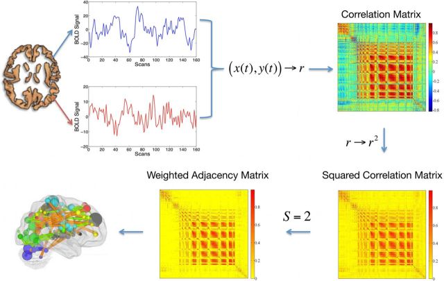
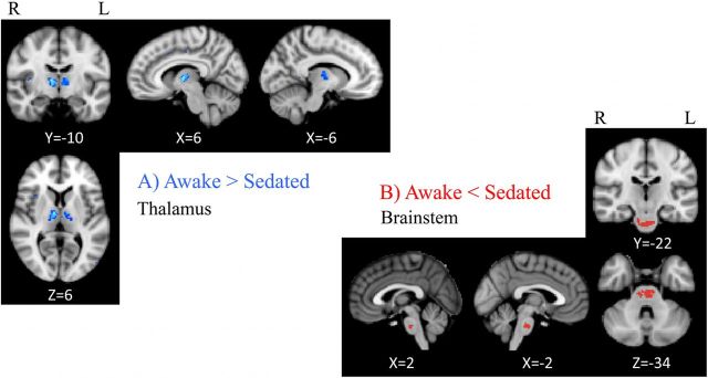
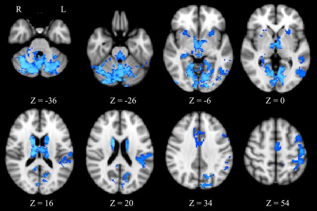
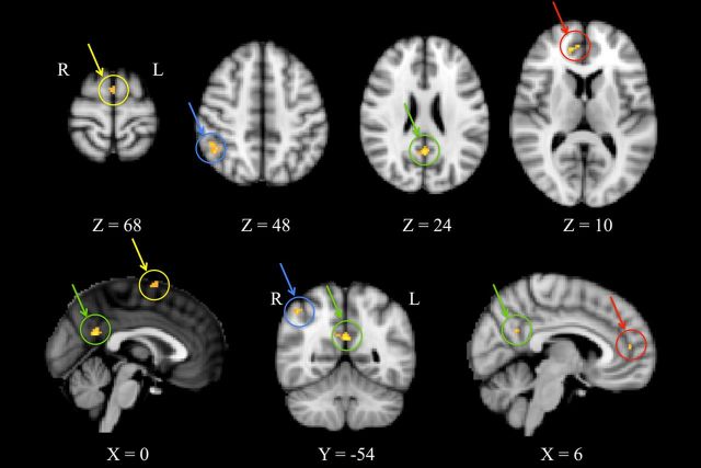
Despite their routine use during surgical procedures, no consensus has yet been reached on the precise mechanisms by which hypnotic anesthetic agents produce their effects. Molecular, animal and human studies have suggested disruption of thalamocortical communication as a key component of anesthetic action at the brain systems level. Here, we used the anesthetic agent, propofol, to modulate consciousness and to evaluate differences in the interactions of remote neural networks during altered consciousness. We investigated the effects of propofol, at a dose that produced mild sedation without loss of consciousness, on spontaneous cerebral activity of 15 healthy volunteers using functional magnetic resonance imaging (fMRI), exploiting oscillations (<0.1 Hz) in blood oxygenation level-dependent signal across functionally connected brain regions. We considered the data as a graph, or complex network of nodes and links, and used eigenvector centrality (EC) to characterize brain network properties. The EC mapping of fMRI data in healthy humans under propofol mild sedation demonstrated a decrease of centrality of the thalamus versus an increase of centrality within the pons of the brainstem, highlighting the important role of these two structures in regulating consciousness. Specifically, the decrease of thalamus centrality results from its disconnection from a widespread set of cortical and subcortical regions, while the increase of brainstem centrality may be a consequence of its increased influence, in the mildly sedated state, over a few highly central cortical regions key to the default mode network such as the posterior and anterior cingulate cortices.
Figures
References
-
- Alkire MT, Haier RJ, Fallon JH. Toward a unified theory of narcosis: brain imaging evidence for a thalamocortical switch as the neurophysiologic basis of anesthetic-induced unconsciousness. Conscious Cogn. 2000;9:370–386. - PubMed
-
- Angel A. The G. L. Brown lecture. Adventures in anesthesia. Exp Physiol. 1991;76:1–38. - PubMed
-
- Antognini JF, Schwartz K. Exaggerated anesthetic requirements in the preferentially anesthetized brain. Anesthesiology. 1993;79:1244–1249. - PubMed
-
- Bassett DS, Bullmore E. Small-world brain networks. Neuroscientist. 2006;12:512–523. - PubMed
Publication types
MeSH terms
Substances
Grants and funding
LinkOut - more resources
Full Text Sources
Other Literature Sources