. 2013 Feb 27;33(9):4032-43.
doi: 10.1523/JNEUROSCI.4702-12.2013.

Neutralization of inhibitory molecule NG2 improves synaptic transmission, retrograde transport, and locomotor function after spinal cord injury in adult rats

Affiliations

Neutralization of inhibitory molecule NG2 improves synaptic transmission, retrograde transport, and locomotor function after spinal cord injury in adult rats

Hayk A Petrosyan et al. J Neurosci. .

Abstract

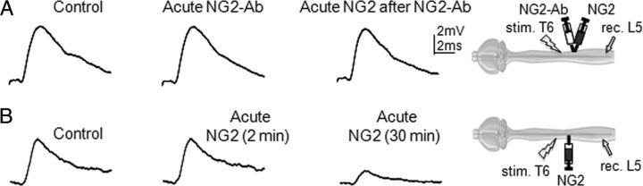
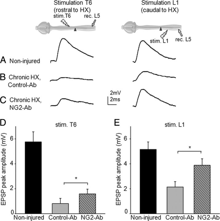
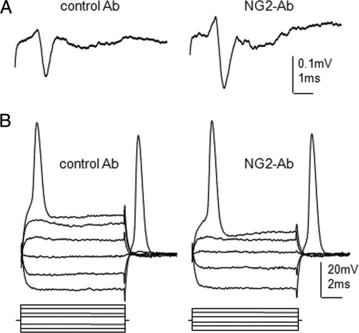
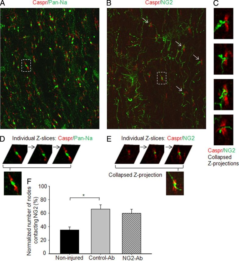
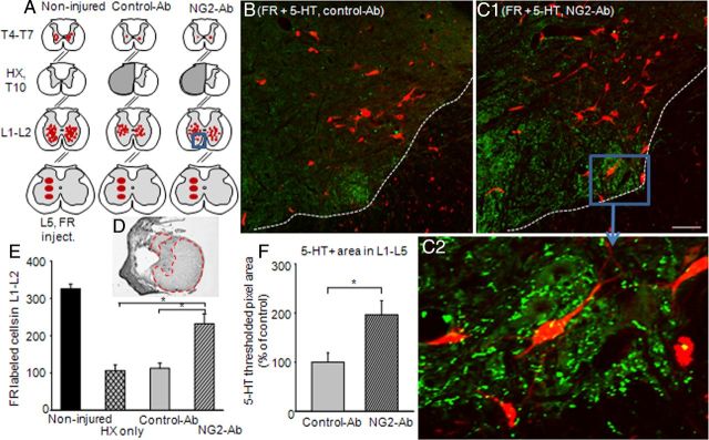
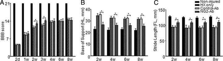
NG2 belongs to the family of chondroitin sulfate proteoglycans that are upregulated after spinal cord injury (SCI) and are major inhibitory factors restricting the growth of fibers after SCI. Neutralization of NG2's inhibitory effect on axon growth by anti-NG2 monoclonal antibodies (NG2-Ab) has been reported. In addition, recent studies show that exogenous NG2 induces a block of axonal conduction. In this study, we demonstrate that acute intraspinal injections of NG2-Ab prevented an acute block of conduction by NG2. Chronic intrathecal infusion of NG2-Ab improved the following deficits induced by chronic midthoracic lateral hemisection (HX) injury: (1) synaptic transmission to lumbar motoneurons, (2) retrograde transport of fluororuby anatomical tracer from L5 to L1, and (3) locomotor function assessed by automated CatWalk gait analysis. We collected data in an attempt to understand the cellular and molecular mechanisms underlying the NG2-Ab-induced improvement of synaptic transmission in HX-injured spinal cord. These data showed the following: (1) that chronic NG2-Ab infusion improved conduction and axonal excitability in chronically HX-injured rats, (2) that antibody treatment increased the density of serotonergic axons with ventral regions of spinal segments L1-L5, (3) and that NG2-positive processes contact nodes of Ranvier within the nodal gap at the location of nodal Na(+) channels, which are known to be critical for propagation of action potentials along axons. Together, these results demonstrate that treatment with NG2-Ab partially improves both synaptic and anatomical plasticity in damaged spinal cord and promotes functional recovery after HX SCI. Neutralizing antibodies against NG2 may be an excellent way to promote axonal conduction after SCI.


Figures

Figure 1.
Figure 1.
Intracellular recordings demonstrating acute effects of NG2 antibody on removing inhibitory effect of NG2 on axonal conduction. Representative traces of EPSPs recorded intracellularly from L5 motoneurons and evoked by stimulation of T6 ventrolateral funiculus in same noninjured adult rat, before and after intraspinal injections of NG2-Ab or NG2. Diagrams show positions of stimulation (stim.) electrode in T6, recording (rec.) electrode in L5 and injection micropipette at T10. A, Superimposed averaged responses showing absence of inhibitory effect of NG2 on conduction in presence of NG2-Ab; control (before injection), 30 min after injection of NG2-Ab, and 30 min after injection of NG2, which was injected 30 min after NG2-Ab injection, respectively. B, Superimposed averaged responses recorded from the opposite side of the cord in same rat, showing depression of EPSPs after injection of NG2 in absence of NG2-Ab; control (before injection), 2 min after injection of NG2, and 30 min after injection of NG2, respectively.
Figure 2.
Figure 2.
Intracellular recordings from L5 motoneurons demonstrating that chronic treatment with NG2-Ab partially restores transmission to L5 motoneurons. A, Superimposed averaged traces demonstrating monosynaptic responses recorded from L5 motoneuron and evoked by electric stimulation of T6 and L1 in same intact spinal cord, respectively. B, Superimposed averaged EPSP responses recorded from chronic HX-injured rats treated with control-Ab. C, Superimposed averaged EPSP responses recorded from NG2-Ab-treated animals showing larger amplitude of synaptic responses compared with control-Ab-treated animal. D, E, Summary of results demonstrating significant improvement of transmission to L5 motoneurons from T6 segment rostral to HX injury and L1 segments caudal to injury in NG2-Ab-treated animals, respectively. Diagrams show positions of the recording electrode in L5 and stimulation electrodes at T6 and L1. Asterisks represent significant difference between corresponding graphs (p < 0.05).
Figure 3.
Figure 3.
Intra-axonal and extra-axonal recordings from lateral white-matter axons to demonstrate effect of NG2-Ab treatment on physiological properties of axons. A, Extracellular recordings of AP volley responses to demonstrate improved conduction in NG2-Ab-treated animal. Representative traces of the volley of APs recorded extracellularly from ventrolateral funiculi at L1 segment and evoked by electric stimulation of VLF at T6 contralateral to HX injury in NG2-Ab-treated and control-Ab-treated chronic HX-injured rats. B, Intra-axonal recordings from L1 VLF axons. Representative traces recorded from single axons in control-Ab-treated and NG2-Ab-treated animals, respectively. Both axons had a resting membrane potential of ∼60 mV. Current steps (displayed below the voltage traces) of a 0.2 nA increment were applied through the recording electrode in both hyperpolarizing (to measure membrane resistance) and depolarizing directions (to trigger an AP). Note the higher rheobase but similar membrane resistance in the axon from control-Ab-treated spinal cord.
Figure 4.
Figure 4.
Confocal images of alternating longitudinal sections representing double immunostaining with Caspr/Pan-Na (A) and Caspr/NG2 (B) to show localization of Na+ channels and NG2-positive processes in relation to nodes of Ranvier in the lateral white matter across to chronic HX injury at depth corresponding to VLF. A, Na+ channel immunoreactivity was found within almost all nodes of Ranvier. B, Many NG2-positive processes make close contacts with nodes of Ranvier. C, High-power views show NG2-positive processes in close relation to nodal gap at nodes indicated by arrows in B. D, E, Individual Z-slices separated by 1 μm to show localization of Na+-channel clusters (D) and NG2-positive processes (E) within nodal gap highlighted in A and B, respectively. F, Summary of results demonstrating significantly higher number of nodes that make close contact with NG2-positive processes within nodal gap in injured versus noninjured animals. Asterisks represent significant difference between corresponding graphs (p < 0.05). Scale bar, 10 μm.
Figure 5.
Figure 5.
Chronic treatment with NG2-Ab improved retrograde transport of FR and density of 5-HT-positive fibers in lumbar segment of chronic HX-injured animals. A, Schematic representation of FR injection site at L5 gray matter and distribution of FR-labeled cells at L1–L2 and T4–T7 spinal segments in noninjured and HX-injured rats treated with NG2-Ab and control-Ab, respectively. B, C, Representative images of lumbar sections from FR experiment stained for 5-HT-positive (5-HT+) fibers of injured animals treated with control-Ab (B) or NG2-Ab (C1), respectively. Dotted lines in B and C represent white matter–gray matter boundary. Scale bar, 100 μm. C2, Higher-power image of boxed area in C1 to demonstrate high density of 5-HT-positive fibers in lumbar ventral horn in the vicinity of neurons. D, Example of cresyl violet-stained spinal cord cross section at the injury epicenter to show extend of injury with highlighted spared white matter. E, Summary of results demonstrating a significantly higher number of labeled cells in L1–L2 segment of NG2-Ab-treated animals compared with control-Ab-treated or HX-only animals. F, Summary of results demonstrating a significantly higher 5-HT+ area in L1–L5 segments in NG2-Ab versus control-Ab animals. Asterisks represent significant difference between corresponding graphs (p < 0.05). Number of animals in each group after exclusion is denoted in Results.
Figure 6.
Figure 6.
Effect of NG2-Ab treatment on locomotor function after chronic HX injury. A, BBB open-field locomotor scores. At 2 d postinjury, animals from both groups showed similar scores, indicating consistency of injury. At 2 and 3 weeks postinjury, NG2-Ab-treated animals showed significantly better performance compared with control-Ab-treated rats. Starting at 4 weeks, however, no significant difference was found between groups in BBB score. B, CatWalk gait analysis showed that treatment with NG2-Ab improved base of support of hindlimbs. C, CatWalk gait analysis showed that NG2-Ab-treated group demonstrated significant improvement in stride length of forelimbs compared with control-Ab-treated and HX-only groups. Asterisks represent significant difference between corresponding bars (p < 0.05). Note that results of CatWalk analysis demonstrate that, beginning from 2 weeks and through 8 weeks postinjury (last time point of experiment), animals from NG2-Ab group showed significant improvement of function compared with control-Ab-treated or HX-only group. Note that control-Ab-treated group did not show any difference versus HX-only group on any test.

Similar articles

Cited by

References

    1. Alilain WJ, Horn KP, Hu H, Dick TE, Silver J. Functional regeneration of respiratory pathways after spinal cord injury. Nature. 2011;475:196–200. - PMC - PubMed
    1. Andrews EM, Richards RJ, Yin FQ, Viapiano MS, Jakeman LB. Alterations in chondroitin sulfate proteoglycan expression occur both at and far from the site of spinal contusion injury. Exp Neurol. 2012;235:174–187. - PMC - PubMed
    1. Arvanian VL, Manuzon H, Davenport M, Bushell G, Mendell LM, Robinson JK. Combined treatment with neurotrophin-3 and LSD facilitates behavioral recovery from double-hemisection spinal injury in neonatal rats. J Neurotrauma. 2006;23:66–74. - PubMed
    1. Arvanian VL, Schnell L, Lou L, Golshani R, Hunanyan A, Ghosh A, Pearse DD, Robinson JK, Schwab ME, Fawcett JW, Mendell LM. Chronic spinal hemisection in rats induces a progressive decline in transmission in uninjured fibers to motoneurons. Exp Neurol. 2009;216:471–480. - PMC - PubMed
    1. Ballermann M, Fouad K. Spontaneous locomotor recovery in spinal cord injured rats is accompanied by anatomical plasticity of reticulospinal fibers. Eur J Neurosci. 2006;23:1988–1996. - PubMed

Publication types

MeSH terms