. 2013 Feb 27;33(9):4118-27.
doi: 10.1523/JNEUROSCI.4187-12.2013.

Increased agonist affinity at the μ-opioid receptor induced by prolonged agonist exposure

Affiliations

Increased agonist affinity at the μ-opioid receptor induced by prolonged agonist exposure

William T Birdsong et al. J Neurosci. .

Abstract

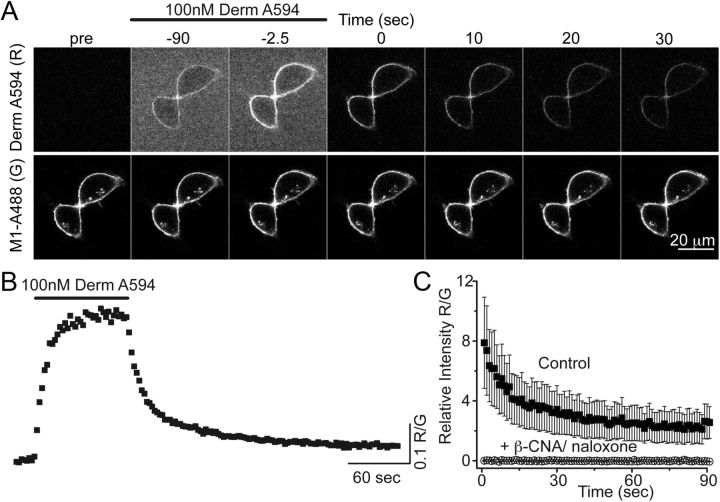
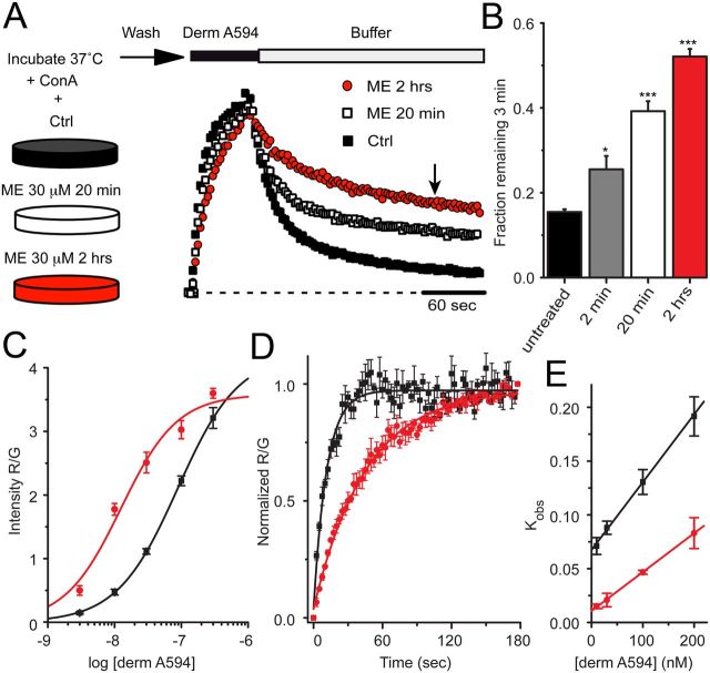
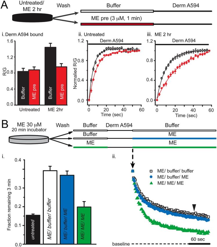
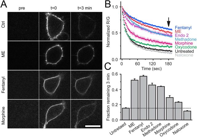
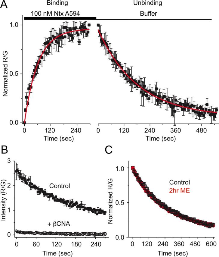
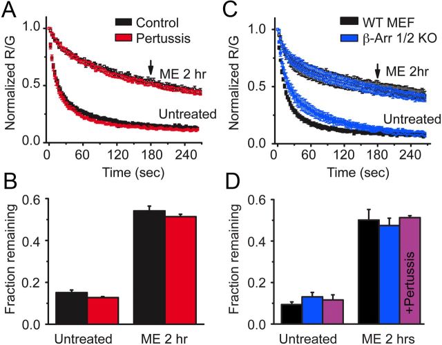
Prolonged exposure to high-efficacy agonists results in desensitization of the μ-opioid receptor (MOR). Desensitized receptors are thought to be unable to couple to G-proteins, preventing downstream signaling; however, the changes to the receptor itself are not well characterized. In the current study, confocal imaging was used to determine whether desensitizing conditions cause a change in agonist-receptor interactions. Using rapid solution exchange, the binding kinetics of fluorescently labeled opioid agonist, dermorphin Alexa594 (derm A594), to MORs was measured in live cells. The affinity of derm A594 binding increased after prolonged treatment of cells with multiple agonists that are known to cause receptor desensitization. In contrast, binding of a fluorescent antagonist, naltrexamine Alexa594, was unaffected by similar agonist pretreatment. The increased affinity of derm A594 for the receptor was long-lived and partially reversed after a 45 min wash. Treatment of the cells with pertussis toxin did not alter the increase in affinity of the derm A594 for MOR. Likewise, the affinity of derm A594 for MORs expressed in mouse embryonic fibroblasts derived from arrestin 1 and 2 knock-out animals increased after treatment of the cells with the desensitization protocol. Thus, opioid receptors were "imprinted" with a memory of prior agonist exposure that was independent of G-protein activation or arrestin binding that altered subsequent agonist-receptor interactions. The increased affinity suggests that acute desensitization results in a long-lasting but reversible conformational change in the receptor.


Figures

Figure 1.
Figure 1.
Imaging binding and unbinding of dermorphin A594. A, FLAG-MOR in HEK 293 cells were labeled with M1 anti-FLAG antibody-conjugated Alexa-488 (M1 A488) to visualize receptors localized on the plasma membrane. Dermorphin Alexa-594 (100 nm, derm A594) was applied for 90 s (wash in at t = −90 washout at t = 0 in A). derm A594 binding and unbinding were assessed by imaging M1 A488 and derm A594 every 2.5 s with images from selected time points shown. Scale bar, 20 μm. B, Ratiometric imaging of derm A594: M1 A488 (R/G) intensity was used to quantify binding and unbinding of agonist to receptor from the experiment shown in A. C, Specificity of binding was assessed by determining the relative fluorescence intensity (R/G) of derm A594 bound immediately after application of derm A594 (5 min, 500 nm) under control conditions or on cells pretreated with β-CNA (1 μm, 5 min) and bathed in naloxone (10 μm). Data are mean ± SEM (ctrl, n = 5; β-CNA/naloxone, n = 6).
Figure 2.
Figure 2.
derm A594 binding to FLAG-MOR in live cells is relatively low affinity. A, Steady-state displacement of [3H]diprenorphine (0.2 nm) with increasing concentrations of derm A594 (0–10 μm) from FLAG-MOR HEK 293 cell membranes was measured in Tris alone (blue) or Tris with Na+, Mg2+, and GTPγS (green) and fit with one site nonlinear regression to define high- and low-affinity binding of derm A594 to FLAG-MOR. The best fit of the data are plotted. Average of 3 experiments performed in duplicate. B, Steady-state binding of derm A594 in live FLAG-MOR HEK 293 cells was relatively low affinity. Cells were incubated in stated concentrations of derm A594, imaged, and relative fluorescence intensity was measured. Data were fit with the Hill equation, and the best fit is plotted (black); n = 5–11. High- and low-affinity binding curves from [3H]diprenorphine competition binding experiments shown in A are overlaid in blue and green, respectively, using the Ki values from competition binding experiments and the slope and R/Gmax values from steady-state whole-cell derm A594 binding to simulate expected results. C, Kinetics of derm A594 binding. derm A594 was applied at 10 (blue), 30, 100 (red), and 200 nm (black), and the apparent association rates were determined by fitting to a single exponential decay function. For display, binding was normalized relative to the amount of derm A594 bound at the end of the 1 min period and averaged results are plotted; n = 4–6. D, Linear fit of the apparent association rate from average data in C plotted as a function of derm A594 concentration. The plotted line has a slope of 612,000 mol−1 s−1 and an intercept of 0.068 s−1, giving a calculated Kd of 110 nm. E, Binding and unbinding kinetics were unaffected by pertussis toxin treatment. Cells were either untreated (black) or treated with pertussis toxin (red) (100 ng/ml, overnight). derm A594 (100 nm) was applied for 90 s at the indicated time and rapidly washed. Data are mean ± SEM (ctrl, n = 6; pertussis toxin, n = 5).
Figure 3.
Figure 3.
MOR retained a memory of previous agonist exposure. A, FLAG-MOR-expressing cells were incubated either under control (■) conditions or in the presence of ME 30 μm for 20 min (□) or 2 h (red circles) as indicated. After washing out ME, cells were imaged while derm A594 (100 nm, 60 s) was applied. Individual examples are shown normalized to the relative fluorescence intensity (R/G) immediately after washout of derm A594. B, Summary data quantifying the fraction of derm A594 that remained bound to the receptor 3 min after washout (indicated by arrow in A) in cells that were untreated or treated with ME 30 μm for 2 min, 20 min, or 2 h, and results are shown (n = 6–11). *p < 0.05, compared with untreated (one-way ANOVA, Tukey post hoc). ***p < 0.001, compared with untreated (one-way ANOVA, Tukey post hoc). C, Steady-state binding was performed as described in Figure 2B after pretreatment of cells with ME for 2 h and was fit using the Hill equation and plotted (n = 7–19 cells for each concentration). Data from untreated cells (black) shown in Figure 2B and data from cells incubated with ME (red) are plotted. D, Average observed association of derm A594 (30 nm) in untreated (black) and ME pretreated (30 μm 2 h, red) cells. E, Plot of average observed association rate of derm A594 after ME treatment (red) is plotted as a function of derm A594 concentration. The linear fit has a slope of 353,000 mol−1 s−1 and an intercept of 0.011 s−1 yielding a Kd of 32 nm; n = 4 each. Data from untreated cells (black) shown in Figure 2D are also plotted.
Figure 4.
Figure 4.
ME unbinds slowly after prolonged ME treatment. A, The amount of derm A594 bound relative to M1 A488 was measured in cells either untreated (Aii) or treated with ME (30 μm, 2 h) (Aiii). After treatment (untreated or ME 2 h), cells were washed with buffer and then subjected to a short pulse of buffer (black) or ME (3 μm, 60 s, red) immediately preceding derm A594 application (200 nm, 60 s) as diagrammed. Ai, Binding of derm A594 was imaged, and the relative amount of derm A594 bound after 60 s (R/G) was measured and plotted the average normalized amount of derm A594 that bound is plotted. Aii, Aiii, Binding of derm A594 (200 nm) is plotted as a function of time in untreated (Aii) or ME (30 μm, 2 h) treated cells (Aiii) and prepulsed with either buffer (black) or ME (3 μm, 60 s, red); n = 5 or 6 each. B, derm A594 (100 nm, 60 s) was applied to cells that had been treated with ME (30 μm, 20 min) and washed with buffer (see diagram). Unbinding of derm A594 was monitored while washing with either buffer (ME/buffer/buffer, □) or ME (3 μm, ME/buffer/ME, blue). ME (3 μm, 1 min) was also pulsed both immediately before and after derm A594 binding to cells that were modulated by ME (30 μm, 20 min) to determine whether residual ME had an effect on derm A594 unbinding kinetics (green, ME/ME/ME). Averaged data in Bi show fraction of derm A594 remaining 3 min after washout (n = 7–11, indicated by arrowhead in Bii), and individual examples of derm A594 unbinding under each condition are shown in Bii.
Figure 5.
Figure 5.
Ligand dependence of receptor modulation. A, Images taken either before (pre), immediately after (t = 0), or 3 min after (t = 3 min) application and rapid washout of derm A594 (100 nm, 90 s). Images show derm A594 bound to the plasma membrane of cells expressing FLAG-MOR that were either untreated (ctrl), or treated with ME, fentanyl, or morphine for 2 h. Scale bar, 20 μm. B, Quantification of unbinding of derm A594 as shown in A after 2 h pretreatment with the indicated opioid ligands. Images were taken every 2.5 s imaging both M1 488 and derm A594. Averaged, normalized ratiometric intensity is plotted. Data are mean ± SEM; n = 6–9). Arrow indicates time = 3 min after washout. C, Fraction of derm A594 remaining at t = 3 min relative to t = 0 from data shown in B. Data are mean ± SEM. All treatments except naloxone resulted in a significant change in the fraction bound after 3 min (p < 0.05, ANOVA, Tukey post hoc test).
Figure 6.
Figure 6.
Unbinding of ntx A594 was not affected by previous agonist exposure. A, Binding kinetics of the antagonist ntx A594 were measured by applying ntx A594 (100 nm, 5 min) and imaging binding and washout relative to M1–488 intensity (R/G). Binding and unbinding were plotted and fit with a single exponential decay formula (τ association = 65.8 ± 5.5 s, τ off = 184 ± 6.1 s, n = 4 each, mean ± SEM). B, Preincubation of cells with the irreversible antagonist β-CNA (1 μm, 5 min) prevented all binding when measured by the fluorescence intensity immediately after washout of ntx A594 (300 nm, 90 s), verifying the specificity of binding (n = 2 ctrl, n = 4 β-CNA mean ± SD). C, ntx A594 (300 nm, 90 s) was applied, and unbinding of ntx A594 was imaged from cells either untreated (black) or pretreated with ME (30 μm, 2 h) (red); n = 5 each, mean ± SEM.
Figure 7.
Figure 7.
Modulation was independent of G-protein signaling and β-arrestins. A, FLAG-MOR-expressing HEK 293 cells were either untreated (black) or treated with pertussis toxin (100 ng/ml overnight) (red), and the unbinding of derm A594 (100 nm, 90 s) was imaged under control conditions or on cells subjected to agonist pretreatment (ME 30 μm, 2 h). Normalized average data are plotted; n = 3–6. B, The fractional amount of derm A594 that remained bound to cells 3 min after washout was quantified relative to the receptor fluorescence intensity. C, MEFs cultured from β-arrestin 1/2 double knock-out mouse embryos (blue) or their WT controls (black) were either untreated or pretreated with ME (30 μm, 2 h), and unbinding of derm A594 (100 nm, 90 s) was imaged; normalized data are shown. D, Quantification of the fraction of derm A594 remaining 3 min after washout from WT (black) and β-arrestin 1/2 d knock-out MEF cells (blue) shown in C; n = 5 each. Additionally, β-arrestin 1/2 d knock-out MEF cells were treated with pertussis toxin (100 ng/ml overnight), and the fraction of derm A594 remaining in untreated and ME treated cells is shown (purple); n = 4 each. All points are mean ± SEM.

References

    1. Ahles A, Rochais F, Frambach T, Bünemann M, Engelhardt S. A polymorphism-specific “memory” mechanism in the β(2)-adrenergic receptor. Sci Signal. 2011;4:ra53. - PubMed
    1. Arttamangkul S, Alvarez-Maubecin V, Thomas G, Williams JT, Grandy DK. Binding and internalization of fluorescent opioid peptide conjugates in living cells. Mol Pharmacol. 2000;58:1570–1580. - PubMed
    1. Arttamangkul S, Lau EK, Lu HW, Williams JT. Desensitization and trafficking of μ-opioid receptors in locus ceruleus neurons: modulation by kinases. Mol Pharmacol. 2012;81:348–355. - PMC - PubMed
    1. Bailey CP, Llorente J, Gabra BH, Smith FL, Dewey WL, Kelly E, Henderson G. Role of protein kinase C and μ-opioid receptor (MOPr) desensitization in tolerance to morphine in rat locus coeruleus neurons. Eur J Neurosci. 2009;29:307–318. - PMC - PubMed
    1. Benovic JL, Kühn H, Weyand I, Codina J, Caron MG, Lefkowitz RJ. Functional desensitization of the isolated β-adrenergic receptor by the β-adrenergic receptor kinase: potential role of an analog of the retinal protein arrestin (48-kDa protein) Proc Natl Acad Sci U S A. 1987;84:8879–8882. - PMC - PubMed

Publication types

MeSH terms

LinkOut - more resources