. 2013 Feb 27;33(9):4165-80.
doi: 10.1523/JNEUROSCI.4185-12.2013.

Tbr2 expression in Cajal-Retzius cells and intermediate neuronal progenitors is required for morphogenesis of the dentate gyrus

Affiliations

Tbr2 expression in Cajal-Retzius cells and intermediate neuronal progenitors is required for morphogenesis of the dentate gyrus

Rebecca D Hodge et al. J Neurosci. .

Abstract

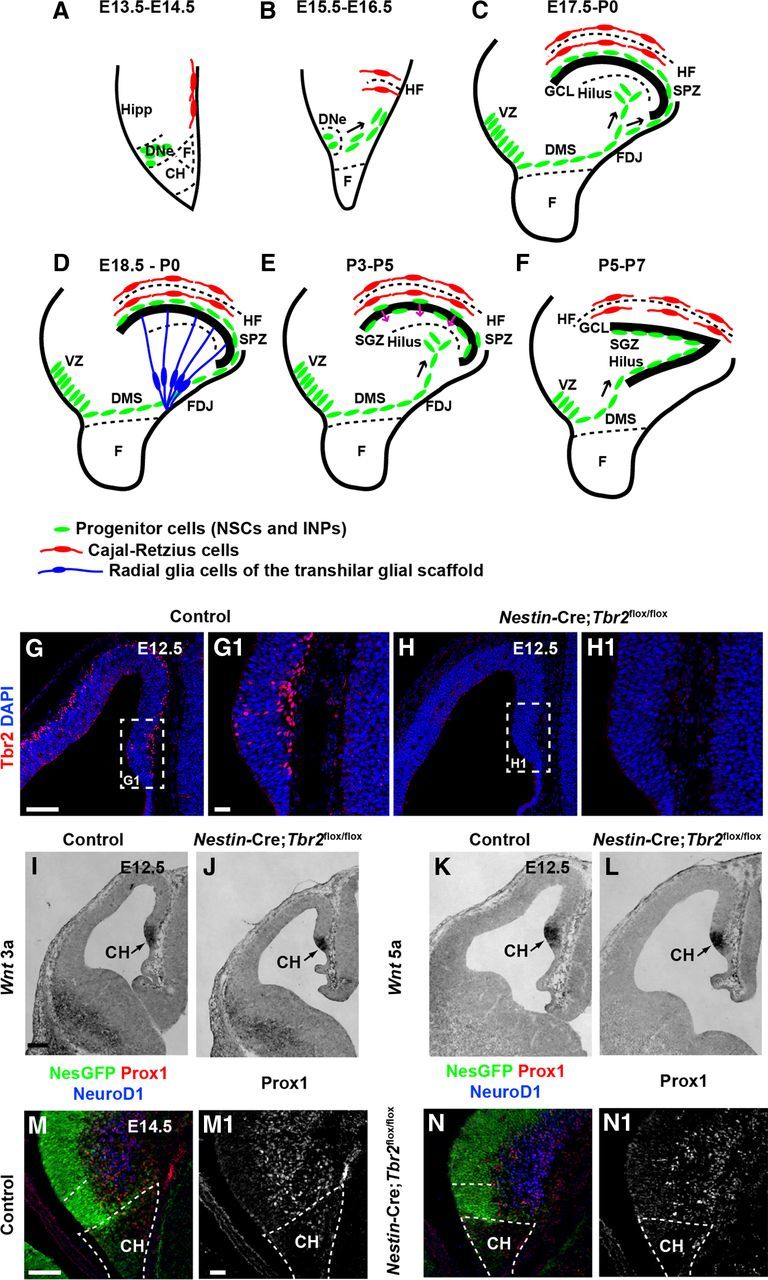
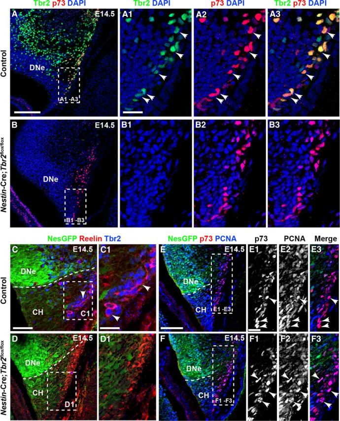
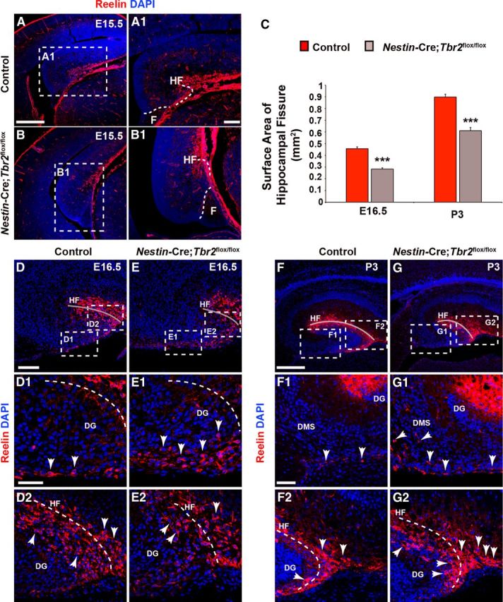
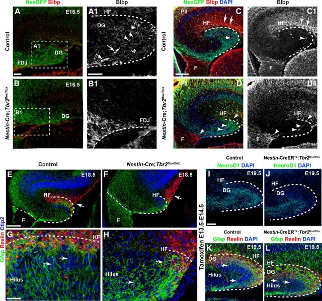
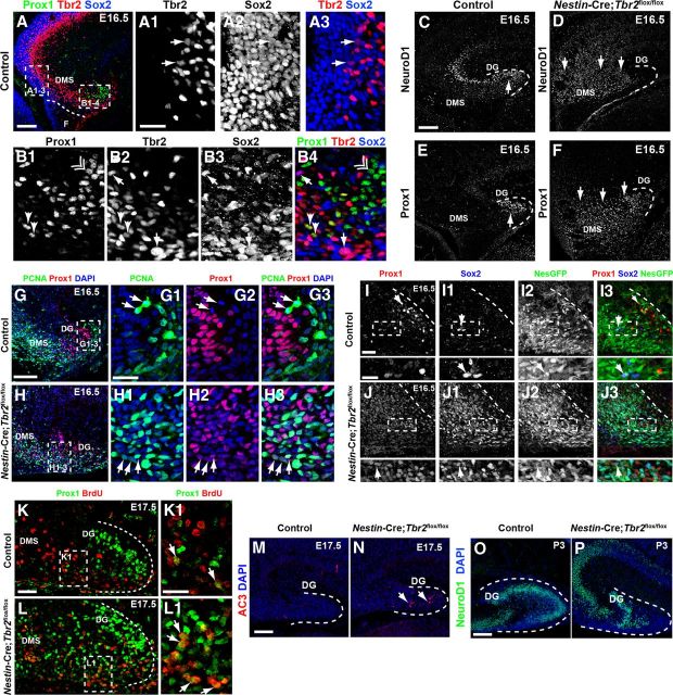
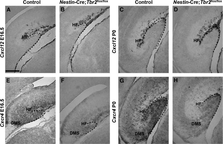
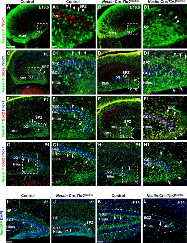
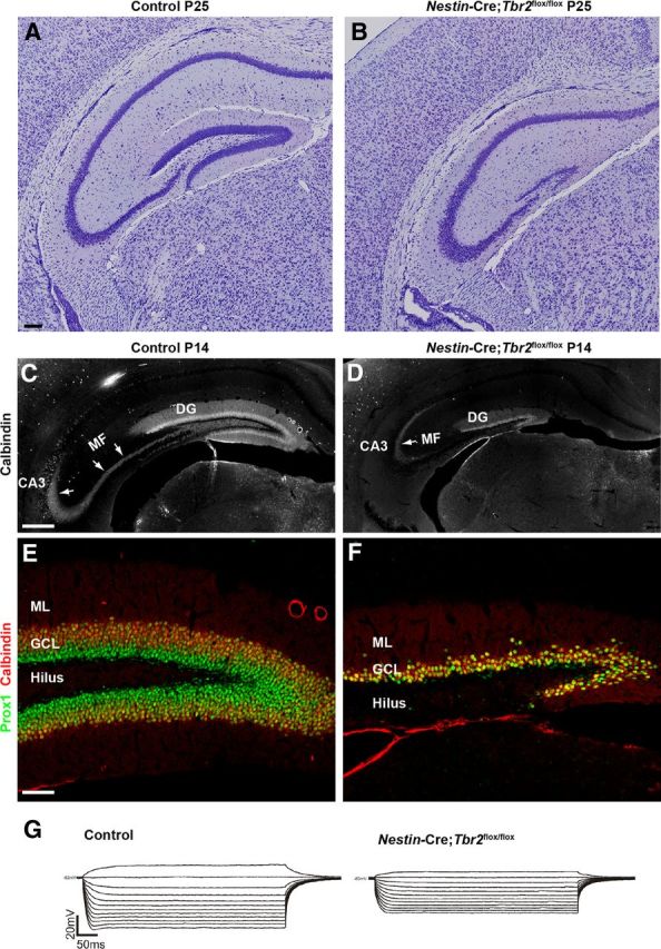
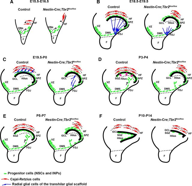
The dentate gyrus (DG) is a unique cortical region whose protracted development spans the embryonic and early postnatal periods. DG development involves large-scale reorganization of progenitor cell populations, ultimately leading to the establishment of the subgranular zone neurogenic niche. In the developing DG, the T-box transcription factor Tbr2 is expressed in both Cajal-Retzius cells derived from the cortical hem that guide migration of progenitors and neurons to the DG, and intermediate neuronal progenitors born in the dentate neuroepithelium that give rise to granule neurons. Here we show that in mice Tbr2 is required for proper migration of Cajal-Retzius cells to the DG; and, in the absence of Tbr2, formation of the hippocampal fissure is abnormal, leading to aberrant development of the transhilar radial glial scaffold and impaired migration of progenitors and neuroblasts to the developing DG. Furthermore, loss of Tbr2 results in decreased expression of Cxcr4 in migrating cells, leading to a premature burst of granule neurogenesis during early embryonic development accompanied by increased cell death in mutant animals. Formation of the transient subpial neurogenic zone was abnormal in Tbr2 conditional knock-outs, and the stem cell population in the DG was depleted before proper establishment of the subgranular zone. These studies indicate that Tbr2 is explicitly required for morphogenesis of the DG and participates in multiple aspects of the intricate developmental process of this structure.


Figures

Figure 1.
Figure 1.
Early development of the DG is essentially normal in Nestin-Cre;Tbr2flox/flox mice. A–F, Schematic diagram illustrating the steps in DG development. Green represents progenitor cells (NSCs and INPs); red, Cajal-Retzius cells; blue, transhilar radial glial scaffold. A, B, Progenitor cells initially located in the DNe migrate to the primordial DG concurrent with invagination of the pial surface and migration of Cajal-Retzius cells to the HF between E13.5 and E16.5. C, Continued migration of progenitors through the DMS contributes to formation of the SPZ during later stages of development (E17.5-P0). D, The transhilar radial scaffold forms at approximately the same time as radial glia become localized to the FDJ. E, Transition of progenitor cells out of the SPZ occurs between P3-P5. F, The SGZ neurogenic niche is established by P5-P7. G, G1, Tbr2 protein (red) is expressed in cells in the cortical hem (CH) at E12.5 in control mice. H, H1, Tbr2 protein is ablated in the cortical hem of mutant mice by E12.5. White dashed boxes in G and H represent areas shown at higher magnification in G1 and H1, respectively. I–L, Expression of Wnt3a and Wnt5a, markers of the cortical hem, are present at approximately normal levels in Nestin-Cre;Tbr2flox/flox mice. M, N, M1, N1, Markers of DG granule neurons (NeuroD1, Prox1) are present in the primordial DG of mutant mice but are slightly reduced compared with controls as early as E14.5. Scale bars: G, 100 μm; G1, 20 μm; I, 100 μm; M, 75 μm; M1, 15 μm.
Figure 2.
Figure 2.
Tbr2 expression is ablated in cortical hem-derived Cajal-Retzius cells in Nestin-Cre;Tbr2flox/flox mice. A–A3, At E14.5, the majority of p73+ Cajal-Retzius cells in the cortical hem (CH) coexpress Tbr2 (arrows). B–B3, p73 expression is maintained in the CH of Nestin-Cre;Tbr2flox/flox mice at E14.5; however, Tbr2 protein is absent from these cells (B2, B3). C–D1, Similarly, Tbr2+ cells coexpress Reelin in the CH of control mice at E14.5, whereas Tbr2 protein is absent from Reelin+ Cajal-Retzius cells in Nestin-Cre;Tbr2flox/flox mice (D1). Regardless, the density of Cajal-Retzius cells in the CH of Nestin-Cre;Tbr2flox/flox mice appears approximately equivalent to controls at E14.5, as evidenced by Reelin (D1) and p73 staining (B3). E–F3, Likewise, proliferation of Cajal-Retzius cells in the CH appears unaffected by ablation of Tbr2, as an approximately equivalent number of p73+ cells coexpress PCNA (arrows) in controls (E1–E3) and Nestin-Cre;Tbr2flox/flox mice (F1–F3). Scale bars: A, 150 μm; A1, 50 μm; C, 100 μm; C1, 30 μm; E, 100 μm; E1, 15 μm. Regions delineated by dashed white boxes are shown in higher magnification in their respective adjacent panels.
Figure 3.
Figure 3.
Rotation of the hippocampus and formation of the HF are abnormal in Nestin-Cre;Tbr2flox/flox mice. A, A1, In control mice, invagination of the pial surface to form the HF occurs concurrent with rotation of the developing hippocampus. B, B1, In Nestin-Cre;Tbr2flox/flox mice, rotation of the hippocampus is impaired, such that the DG appears to be rotated 90° in mutant mice compared with controls (compare A and B). B, B1, As well, invagination of the pial surface is delayed in mutant mice, resulting in a reduction in the size of the HF. C, Consistent with delayed invagination of the pial surface in Nestin-Cre;Tbr2flox/flox mice, the surface area of the HF is persistently and significantly reduced in mutant mice (ANOVA, N = 3, p < 0.001). Graphs represent the mean ± SEM. **p < 0.01. ***p < 0.001. D–D2, By E16.5, the majority of Reelin+ Cajal-Retzius cells are localized to the HF (gray line) in control mice (D2), and a smaller population of these cells can be seen in the DMS. E–E2, In contrast, the DMS is rich in Reelin+ Cajal-Retzius cells in Nestin-Cre;Tbr2flox/flox mice, and there are fewer of these cells located in the HF in mutant mice, suggesting impaired migration and ectopic localization of these cells in mutants. The reduction in HF surface area is also evident in mutant mice (compare gray lines in D, E vs D2, E2). F–F2, By P3, very few Reelin+ Cajal-Retzius cells remain in the DMS in control mice (arrows), and the bulk of these cells are located in the HF (F2), which has increased in surface area with continued growth of the DG compared with controls at E16.5. G–G2, However, in Nestin-Cre;Tbr2flox/flox mice, ectopic Reelin+ cells can been seen in the DMS (G1, arrows) at P3, suggesting impaired migration of these cells to the HF. G2, In mutant mice, there is also an increased concentration of Reelin+ cells at the tip of the DG arrows compared with controls (F, F2), further supporting ectopic localization of Cajal-Retzius cells in Nestin-Cre;Tbr2flox/flox mice. Scale bars: A, 200 μm; A1, 50 μm; D, 175 μm; D1, 50 μm; F, 175 μm; F1, 75 μm. Regions delineated by dashed white boxes are shown in higher magnification in their respective adjacent panels. D–G, Gray lines highlight the HF in each panel.
Figure 4.
Figure 4.
Development of the transhilar radial glial scaffold is perturbed after ablation of Tbr2. A, A1, In E16.5 control mice, Blbp+ radial glia exit the dentate VZ and migrate to the HF to take up residence in the dentate marginal zone. They can be seen exiting the FDJ region and migrating across the hilus (A1, cell bodies are marked by arrows and associated processes by arrowheads). B, B1, Migration of Blbp+ cells is delayed in Nestin-Cre;Tbr2flox/flox mice, and accumulation of Blbp+ cells is apparent in the fimbria and FDJ. Regions delineated by dashed white boxes are shown in higher magnification in their respective adjacent panels (A1, B1). C, C1, By P0, Blbp+ cell migration is essentially complete in control mice, and enrichment of Blbp+ processes is apparent in the HF. D, D1, In Nestin-Cre;Tbr2flox/flox mice (arrows), Blbp+ cells remain concentrated in the fimbria and FDJ and only rarely can be seen migrating to the HF (arrowheads). E–H, Gfap+ processes making up the transhilar glial scaffold are aberrant in Nestin-Cre;Tbr2flox/flox mice. In control mice (E, G), Gfap+ processes exhibit clear radial orientation across the hilus, whereas Gfap+ processes are oriented either randomly or toward the reduced HF where Reelin+ Cajal-Retzius cells are located in Nestin-Cre;Tbr2flox/flox mice (F, H, arrows). These processes do not exhibit the clear radial orientation typically seen in control mice (G, H, arrows). I–L, Tbr2 ablation on E13.5-E14.5 rescues DG morphogenetic defects. Inducible conditional deletion of Tbr2 (Nestin-CreERT2;Tbr2flox/flox) was achieved by treating mice with tamoxifen on E13.5 and E14.5. Control mice were Nestin-CreERT2;Tbr2flox/+. I, J, Consistent with the known role of Tbr2 in regulating granule cell neurogenesis, NeuroD1+ neuroblasts are reduced in the developing DG of Nestin-CreERT2;Tbr2flox/flox mice, resulting in a decrease in the overall size of the DG (J) at E19.5. However, the HF is approximately the same size in controls (K) and Nestin-CreERT2;Tbr2flox/flox mice (L). Reelin+ Cajal-Retzius cells are abundant in the dentate marginal zone adjacent to the HF in both controls (K) and Nestin-CreERT2;Tbr2flox/flox mice (L), and Gfap+ processes are radially oriented across the hilus in both groups (K, L, arrows) consistent with normal development of the transhilar radial scaffold. Scale bars: A, 50 μm; A1, 30 μm; C, 75 μm; E, 100 μm; G, 30 μm; I, 50 μm.
Figure 5.
Figure 5.
Premature granule cell neurogenesis is apparent in Nestin-Cre;Tbr2flox/flox mice. A–A3, In control mice, Sox2 and Tbr2 are coexpressed in a subset of cells (arrows) in the dentate VZ, whereas Prox1 is not expressed in this region but rather is restricted to the DMS and developing DG (A). However, Sox2+/Tbr2+ cells represent a relatively minor fraction of the total Sox2+ population, consistent with the notion that Sox2 is predominantly expressed in NSCs, whereas Tbr2 is largely restricted to INPs (A1–A3). B1–B4, Within the DMS, Tbr2 and Sox2 continue to be coexpressed in a subset of cells, and these cells are Prox1-negative (arrows). However, coexpression of Tbr2 and Prox1 is apparent in a small subset of Tbr2+ cells, but these Tbr2+/Prox1+ cells are Sox2-negative (arrowheads). Prox1 and Sox2 coexpression is very rare in control animals at E16.5, consistent with downregulation of Sox2 during neuronal differentiation. Accordingly, most Prox1+ cells do not coexpress Sox2 or Tbr2 (double arrowhead). C, In control mice, NeuroD1+ neuroblasts are present in the forming suprapyramidal blade of the DG (arrow) and in the hippocampal CA3 field. Only scattered NeuroD1+ cells are present in the DMS in controls. D, In Nestin-Cre;Tbr2flox/flox mice, numerous NeuroD1+ cells are present in the DMS, which appears to be expanded compared with controls, but relatively few are in the DG itself. E, Prox1 protein is also mostly restricted to the forming DG in control mice, whereas Prox1 is expressed extensively in the expanded DMS of Nestin-Cre;Tbr2flox/flox mice, indicative of an early burst of neurogenesis in mutant animals at E16.5 (F). G–G3, In control mice, Prox1+ neuroblasts rarely proliferate as evidenced by the scarcity of Prox1+/PCNA+ cells in these mice. Arrows illustrate proliferating progenitors (PCNA+) that lack Prox1 expression in a control animal at E16.5 However, in Nestin-Cre;Tbr2flox/flox mice at E16.5, the number of proliferating (PCNA+) Prox1+ neuroblasts is increased (H1–H3, arrows) within the expanded DMS and developing DG. Although PCNA+/Prox1+ cells appear to express Prox1 at low levels, these cells still represent a minority of the total population of Prox1+ neuroblasts in mutant mice (H1–H3). I–I3, In control mice at E16.5, Prox1+ neuroblasts very rarely coexpress the NSC markers Sox2 and Nestin-GFP (NesGFP), consistent with downregulation of these NSC markers in neuroblasts. J–J3, Conversely, in Nestin-Cre;Tbr2flox/flox mice, there is extensive, unusual colocalization of Prox1, Sox2, and Nestin-GFP in the DG. Similar to our observations of Prox1+/PCNA+ cells in mutant mice (H1–H3), Prox1 appears to be expressed at low levels in these mutant Prox1+/Sox2+/NesGFP+ cells (J–J3). K–L1, BrdU pulse-chase labeling (BrdU injected on E15.5. with embryo collection on E17.5) shows that the percentage of Prox1+/BrdU+ cells is increased in Nestin-Cre;Tbr2flox/flox mice (L, L1, arrows) compared with controls (K, K1, arrows), confirming premature neurogenesis in mutant mice during embryonic DG development. M, N, The density of AC3 is increased in Nestin-Cre;Tbr2flox/flox mice, indicating that cell death is increased during this period of premature neurogenesis in mutants (N, arrows). O, P, By P3, the suprapyramidal blade of the DG is easily identifiable in control mice and contains numerous NeuroD1+ neuroblasts, whereas these cells are reduced and misplaced in Nestin-Cre;Tbr2flox/flox mice. Scale bars: A, 100 μm; A1, 50 μm; C, 100 μm; G, 100 μm; G1, 35 μm; I, 35 μm; I (inset), 20 μm; K, 100 μm; K1, 35 μm; M, 75 μm; O, 75 μm. Regions delineated by dashed white boxes are shown in higher magnification in their respective adjacent panels.
Figure 6.
Figure 6.
Cxcr4 is downregulated in the DG of Nestin-Cre;Tbr2flox/flox mice. A–D, Cxcl12 expression is apparent in the meninges in the HF in both control and Nestin-Cre;Tbr2flox/flox mice at E16.5 and P0. Expression of Cxcl12 on a per cell basis appears normal in Nestin-Cre;Tbr2flox/flox mice at both ages, although reduction of the HF and decreased numbers of Cxcl12-expressing cells in the HF are readily apparent in mutants. E–H, Cxcr4, the receptor for Cxcl12 that is expressed on migrating progenitors and neuroblasts, is downregulated in the DG of Nestin-Cre;Tbr2flox/flox mice compared with controls at both E16.5 and P0. Scale bar: A, 100 μm.
Figure 7.
Figure 7.
Development of the SGZ neurogenic niche is disrupted in Nestin-Cre;Tbr2flox/flox mice. A, A1, The SPZ is apparent in controls by E16.5 as a layer of Nestin-GFP+ (NesGFP) cells immediately above the Prox1+ neuroblasts populating the forming upper DG blade (A1, arrows). B, B1, In Nestin-Cre;Tbr2flox/flox mice, there is a delay in the formation of the SPZ (B1, arrows). In controls, NesGFP+ NSCs localize to the SPZ above the forming suprapyramidal blade on P0 (C, C1, arrows). In Nestin-Cre;Tbr2flox/flox mice, increased numbers of NesGFP+/Sox2+ cells are apparent in the SPZ on P0 (D, D1, arrows). By P3, NesGFP+ progenitors begin to transition out of the SPZ in control mice (E, E1, arrows). In contrast, many NesGFP+ NSCs remain concentrated in the SPZ in Nestin-Cre;Tbr2flox/flox mice, although some do migrate through the GCL (F, F1). By P4, transition of NSCs out of the SPZ is largely complete in control mice, and the SGZ is apparent adjacent to the hilus (G, G1, arrows). In mutant mice on P4, NesGFP+ NSCs remain concentrated in the SPZ and are reduced in the SGZ (H, H1, arrows). In controls, NesGFP+ NSCs are apparent in the SGZ, and the transitory SPZ is essentially gone by P7 (I). At P7, many NesGFP+ NSCs remain in the SPZ in Nestin-Cre;Tbr2flox/flox mice (J, arrows) and are also present in the hilus. By P14, NesGFP+ progenitor cells are exclusively found in the SGZ in control mice (K). In Nestin-Cre;Tbr2flox/flox mice, the SGZ is present, but is reduced in size (L) with very few NesGFP+ NSCs present. Scale bars: A, 100 μm; A1, 25 μm; I, 75 μm; K, 30 μm. Regions delineated by dashed white boxes are shown in higher magnification in their respective adjacent panels.
Figure 8.
Figure 8.
Tbr2 ablation has severe consequences for DG morphogenesis. A, B, By early postnatal development, the DG of control mice is fully formed, whereas the DG of Nestin-Cre;Tbr2flox/flox mice consists only of a truncated and thinned suprapyramidal blade. B, The infrapyramidal blade of the GCL essentially fails to form in Nestin-Cre;Tbr2flox/flox mice. C, The mossy fiber pathway (MF), the main output of the DG granule neurons to CA3, is easily distinguished by immunostaining for calbindin at P14 in control animals (C, arrows). D, In mutant mice, the MF pathway is greatly reduced (D, arrow) because of the decrease in DG size. E, F, The cells that remain in the DG of Nestin-Cre;Tbr2flox/flox mice appear to be granule neurons as evidenced by their expression of Prox1 and calbindin. However, the number of granule neurons populating the DG is clearly greatly reduced in mutant mice (F) compared with controls (E). G, Representative I-V curves from control and Nestin-Cre;Tbr2flox/flox mice illustrate that input resistance is significantly decreased in granule neurons remaining in the reduced DG of mutant mice compared with controls. Scale bars: A, 100 μm; C, 100 μm; E, 100 μm.
Figure 9.
Figure 9.
Schematic diagram summarizing DG defects in Nestin-Cre;Tbr2flox/flox mice. A, Initial invagination of the pial surface is delayed in Nestin-Cre;Tbr2flox/flox mice, and the migration of Cajal-Retzius cells (red) to the forming HF is delayed in mutants. B, Development of the transhilar radial glial scaffold (blue cells) is abnormal in Nestin-Cre;Tbr2flox/flox mice by E18.5-E19.5, and the number of Cajal-Retzius cells (red) populating the HF is reduced. C, Blbp+ radial glial cells (blue) that contribute to the transhilar radial glial scaffold complete their redistribution to the HF by E19.5-P0 in control mice, but this migration is delayed in Nestin-Cre;Tbr2flox/flox mice. D, During early postnatal development (P3-P4), progenitor cells (green) populating the transient SPZ are redistributed to form the SGZ niche. In Nestin-Cre;Tbr2flox/flox mice, these cells are retained in the SPZ and fail to migrate to the SGZ. E, Formation of the SGZ (green, progenitor cells) is complete by P5-P7 in control mice, and the SPZ is no longer apparent. In mutant mice, retention of progenitors in the SPZ persists. F, By P10-P14, both the suprapyramidal and infrapyramidal blades of the DG are formed in control mice, and progenitor cells (NSCs and INPs, green) are localized to the SGZ. In Nestin-Cre;Tbr2flox/flox mice, the infrapyramidal blade fails to form, the suprapyramidal blade is reduced in size, and progenitor cells are nearly absent from the SGZ (green).

Similar articles

Cited by

References

    1. Arnold SJ, Huang GJ, Cheung AF, Era T, Nishikawa S, Bikoff EK, Molnár Z, Robertson EJ, Groszer M. The T-box transcription factor Eomes/Tbr2 regulates neurogenesis in the cortical subventricular zone. Genes Dev. 2008;22:2479–2484. - PMC - PubMed
    1. Bagri A, Gurney T, He X, Zou YR, Littman DR, Tessier-Lavigne M, Pleasure SJ. The chemokine SDF1 regulates migration of dentate granule cells. Development. 2002;129:4249–4260. - PubMed
    1. Bedogni F, Hodge RD, Elsen GE, Nelson BR, Daza RA, Beyer RP, Bammler TK, Rubenstein JL, Hevner RF. Tbr1 regulates regional and laminar identity of postmitotic neurons in developing neocortex. Proc Natl Acad Sci U S A. 2010;107:13129–13134. - PMC - PubMed
    1. Belvindrah R, Nissant A, Lledo PM. Abnormal neuronal migration changes the fate of developing neurons in the postnatal olfactory bulb. J Neurosci. 2011;31:7551–7562. - PMC - PubMed
    1. Berger O, Li G, Han SM, Paredes M, Pleasure SJ. Expression of SDF-1 and CXCR4 during reorganization of the postnatal dentate gyrus. Dev Neurosci. 2007;29:48–58. - PubMed

Publication types

MeSH terms

LinkOut - more resources