. 2013 May;26(5):347-57.
doi: 10.1093/protein/gzt006. Epub 2013 Feb 26.

Design and properties of efficient tRNA:EF-Tu FRET system for studies of ribosomal translation

Affiliations

Design and properties of efficient tRNA:EF-Tu FRET system for studies of ribosomal translation

Maxim Chudaev et al. Protein Eng Des Sel. 2013 May.

Abstract

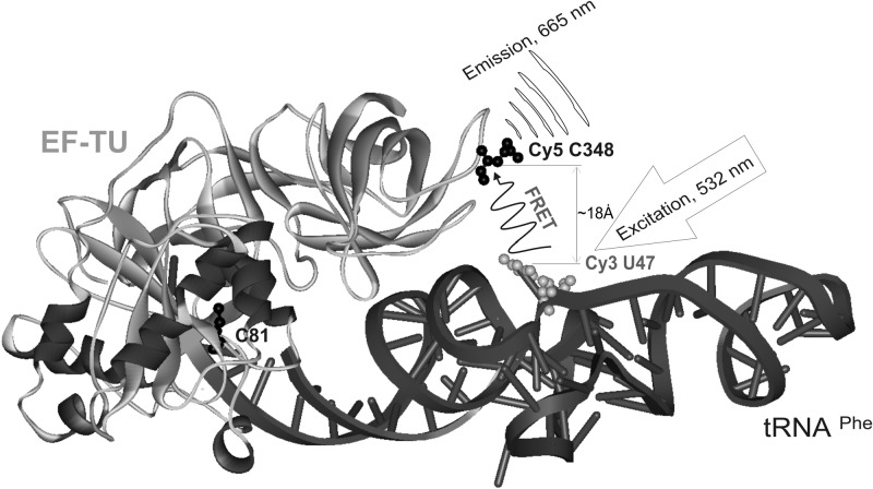
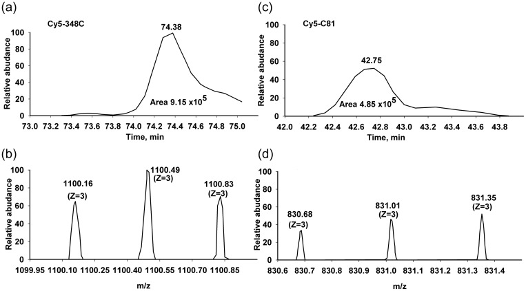
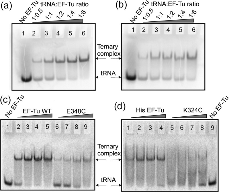
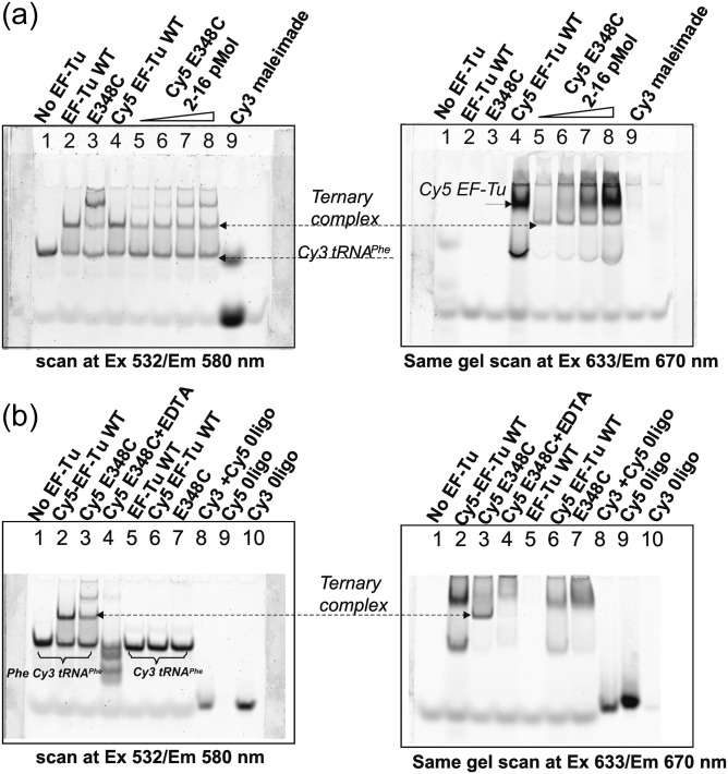
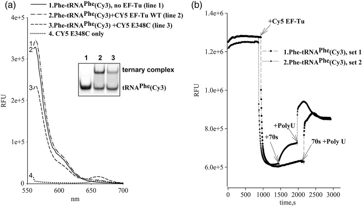
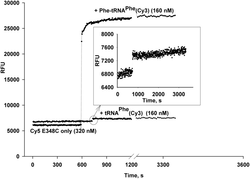
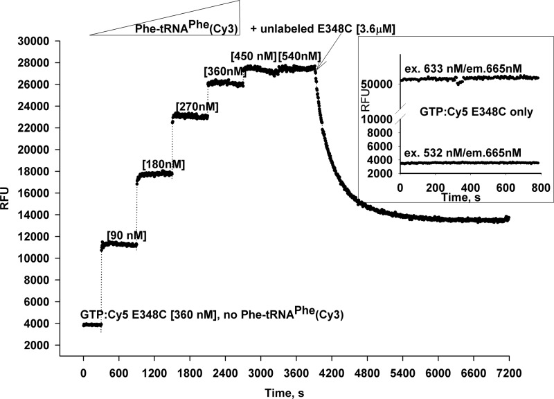
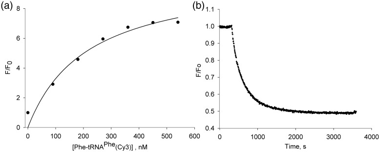
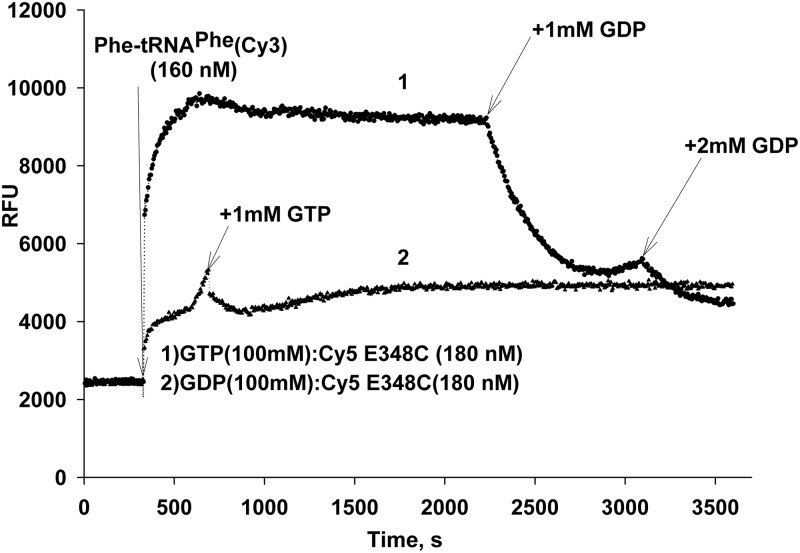
Formation of the ternary complex between GTP-bound form of elongation factor Tu (EF-Tu) and aminoacylated transfer RNA (aa-tRNA) is a key event in protein biosynthesis. Here we show that fluorescently modified Escherichia coli EF-Tu carrying three mutations, C137A, C255V and E348C, and fluorescently modified Phe-tRNA(Phe) form functionally active ternary complex that has properties similar to those of the naturally occurring (unmodified) complex. Similarities include the binding and binding rate constants, behavior in gel retardation assay, as well as activities in tRNA protection and in vitro translation assays. Proper labeling of EF-Tu was demonstrated in MALDI mass spectroscopy experiments. To generate the mutant EF-Tu, a series of genetic constructions were performed. Two native cysteine residues in the wild-type EF-Tu at positions 137 and 255 were replaced by Ala and Val, respectively, and an additional cysteine was introduced either in position 324 or 348. The assembly FRET assay showed a 5- to 7-fold increase of Cy5-labeled EF-Tu E348C mutant fluorescence upon formation of ternary complex with charged tRNA(Phe)(Cy3-labeled) when the complex was excited at 532 nm and monitored at 665 nm. In a control experiment, we did not observe FRET using uncharged tRNA(Phe)(Cy3), nor with wild-type EF-Tu preparation that was allowed to react with Cy5 maleimide, nor in the absence of GTP. The results obtained demonstrate that the EF-Tu:tRNA FRET system described can be used for investigations of ribosomal translation in many types of experiments.


Figures

Fig. 1.
Fig. 1.
Functional assays of mutant EF-Tu proteins. Non-enzymatic hydrolysis of Phe-tRNAPhe in the presence of Escherichia coli wild type or mutant EF-Tu proteins. Each reaction contained 2.5 µM EF-Tu and 0.5 µM [14C] Phe-tRNAPhe.
Fig. 2.
Fig. 2.
Structure of the ternary complex. The sites chosen for labeling of the recombinant Escherichia coli EF-Tu mutants and tRNAPhe with Cy5 and Cy3, respectively, are shown. A cysteine residue was introduced at position 348 in EF-Tu. The distance between the Cy5-labeled cysteine residue and Cy3-labeled U47 on tRNAPhe is indicated. The coordinates for the E. coli EF-Tu:GDPNP:kirromycin:Phe-tRNAPhe ternary complex at 3.3 Å resolution are from the 1ob2 PDB file (Kavaliauskas et al., 2012). WebLab ViewerPro software was used to create the image.
Fig. 3.
Fig. 3.
Purification of tRNA: reverse phase HPLC on the C4 Delta Pack column. The dashed lines represent an optimized buffer B gradient, with the % buffer B indicated by the scale on the right axis. (a) Separation of tRNAPhe and tRNAPhe(Cy3). After the labeling reaction, the pool of tRNAPhe (4.75 A260 units) was resolved using the conditions described in Materials and Methods. (b) Isolation of aminoacylated Cy3-labeled tRNAPhe. The phenol-extracted and ethanol-precipitated aliquot of 5 A260 units of E. coli tRNAPhe(Cy3) after aminoacylation reaction with 14C Phe was applied to the C4 Delta Pack column. The retention time was 33 and 28 min for charged Phe-tRNAPhe(Cy3) and uncharged tRNAPhe(Cy3), respectively. Fractions containing tRNAPhe(Cy3) and [14C ]Phe-tRNAPhe(Cy3) were collected and ethanol-precipitated. A large peak (>1600 RU A260) at the void volume was observed after the phenol extraction. The peak likely originated from oxidized phenol derivatives.
Fig. 4.
Fig. 4.
Poly(Phe) synthesis using Phe-tRNAPhe(Cy3). (a) The reaction mixtures contained 20 mM magnesium acetate, 10 mM potassium phosphate, pH 7.4, 100 mM potassium glutamate, pH 7.7, 95 mM potassium chloride, 10 mM ammonium chloride, 0.5 mM calcium chloride, 1 mM spermidine, 8 mM putrescine, 1 mM DTT, 1 mM ATP, 4.5 mM PEP, 1 mM GTP, 100 µg/ml pyruvate kinase, 2 µM EF-G, 2 μM 70S ribosomes, 60 pmol [14C] Phe-tRNAPhe or [14C] Phe-tRNAPhe(Cy3) and 30 pmol of E. coli EF-Tu. The reaction was carried out at 37°C for 5 min, and then incubated with an equal volume of 0.4 M NaOH at 37°C for 10 min. After hydrolysis of unreacted [14C]Phe-tRNAPhe was completed, the sample was applied onto Whatman filter paper or GF filter, washed in 5% ice cold TCA and in ethanol. The radioactivity in the precipitates was determined by scintillation counting. (b) The reaction was carried out at the same conditions for 1 min in two sets for [14C] Phe-tRNAPhe(Cy3) and [14C] Phe-tRNAPhe. Concentrations of 70S ribosome and EF-Tu were 1 and 30 µM, correspondingly. The concentration of labeled and unlabeled aa-tRNA was in the 0.2–12.8 µM range.
Fig. 5.
Fig. 5.
LCMS-MS analysis of Cy5 maleimide-labeled EF-Tu (E348C mutant). Extracted ion chromatograms representing Cy5 modified Cys 348 (a), with m/z ratio 1100.1668 (b) and Cy5 modified Cys 81 (c), with m/z ratio 830.6873 (d). On base integration of EIC's peaks, the Cy5 dye distribution in EF-Tu is 65% at C348, and 35% at C81.
Fig. 6.
Fig. 6.
Gel retardation assay: monitoring of ternary complex formation by autoradiography. After electrophoresis, the gels were placed in a phosphorimager cassette and scanned after an overnight exposure. [14C] Phe-tRNAPhe(Cy3) (0.4 µM) was incubated in the presence of GTP and increasing concentrations of native EF-Tu (Panel a), His-tagged recombinant wild-type EF-Tu (Panel b), E348C(AV) mutant (Panel c) or K324C(AV) mutant (Panel d) were run on the gel as described in Materials and Methods. Each line contains 4 pmol of 14C Phe tRNAPhe (except 20 pmol in line 1, panels a, b). The amount of EF-Tu was varied between 0.4–2.8 µM.
Fig. 7.
Fig. 7.
Gel retardation assay to monitor ternary complex formation. After native gel electrophoresis, two gels were scanned to monitor Cy3 (left) or Cy5 (right) fluorescence. (a) Each line contains 2 pmol of Phe-tRNAPhe(Cy3). Unlabeled EF-Tu WT, E348C mutant or Cy5 EF-Tu WT were added in 8-fold molar excess (lines 2–4). Cy5 EF-Tu E348C concentration was varied in 1–8 molar excess (lines 5–8). Cy3 maleimide dye itself was loaded as a control for Cy3 fluorescence in line 9. The total Cy3 fluorescence intensity of labeled tRNA in the ternary complex with Cy5-EF-Tu E348C was decreased approx 27% (calculation is based on the band intensity in lines 1 and 2 vs. lines 5 and 6). The quenching of Cy3 fluorescence in the complex is close to 60% (calculation is based on the band intensity in lines 2 and 4 vs. lines 5 and 6). Panel b. Phe-tRNAPhe(Cy3) (0.2 µM) was incubated with four-fold molar excess of Cy5 EF-Tu WT or Cy 5 E348C. No ternary complex was observed in presence of EDTA (line 4) as well as in presence of uncharged tRNAPhe(Cy3) (lines 5, 6 and 7). The total Cy3 fluorescence intensity of the labeled tRNA in the ternary complex with Cy5-EF-Tu E348C was decreased approx 24% (calculation is based on the band intensity in line 1 vs. line 3). Quenching of Cy3 fluorescence in the complex is close to 60% – calculation is based on the intensity of the ternary complex band in line 2 (peak 1) and line 3 (peak 4). Approximately 46% of Phe-tRNAPhe(Cy3) was deacylated after the reaction. The band intensity was calculated using the ImageQuant software.
Fig. 8.
Fig. 8.
Spectral (a) and time-based (b) evidence of FRET upon addition of Phe-tRNAPhe(Cy3) to Cy5 EF-Tu E348C in presence of GTP. (a) The ternary complex was formed in a 24 µl volume using Phe-tRNAPhe(Cy3) [0.2 µM], GTP [100 µM], Cy5-EF-Tu WT or E348C [0.4 µM]. The conditions and incubation buffer are as described in Materials and Methods for the gel retardation assay. A 12-µl volume was loaded on a native 10% polyacrylamide gel (insert), and remaining volume was adjusted to 140 µl. The Cy3 fluorescence was monitored on a Photon Technology International fluorescence spectrofluorometer in the spectrum mode at excitation/emission 549/565 nm. (b) A time-base titration experiment was carried out in a 150 µl black cuvette at RT. Phe-tRNAPhe(Cy3) [60 nM] was in the incubation buffer supplemented with 1 mM ATP and 5 µg/ml of Phe-tRNA synthetase. Cy3 fluorescence (excitation/emission 549/565 nm) was monitored at five sec interval up to 1 h. Cy5 EF-Tu E348C was added from concentrated stock solution to reach final concentration [180 nM]. The 70s ribosome concentration was [240 nM] and poly(U) was 64 µg/ml.
Fig. 9.
Fig. 9.
Effect of aminoacylation of tRNAPhe(Cy3) on FRET during ternary complex formation. Cy5-labeled EF-Tu [320 nM] (GTP form) in a 300-µl volume was placed in 1× incubation buffer. The reaction was supplemented with 100 µM GTP and split into two parts. The FRET upon formation of ternary complex was monitored at 665 nm (suitable for the Cy5 dye) in the time-base mode using the 532 nm excitation for the Cy3 dye. Plot 1 indicates a 4.7-fold increase in Cy5 fluorescence upon addition (10 min) of Phe-tRNAPhe(Cy3). Plot 2: an addition of the same amount of the uncharged tRNAPhe(Cy3) minimally affects the Cy5 fluorescence due to absence of FRET (see inset).
Fig. 10.
Fig. 10.
Titration of GTP:Cy5-E348C by Phe-tRNAPhe(Cy3) and displacement of labeled EF-Tu from the ternary complex by the unlabeled form. Cy5-labeled EF-Tu [360 nM] (GTP form) in a 300 µl volume was placed in 1× incubation buffer supplemented with 100 µM GTP. The Cy5 fluorescence was monitored for 5 min at 25°C using the 532 nm excitation wavelength. After addition of Phe-tRNAPhe(Cy3), the concentration was increased step-wise in the 90–540 nM range, and fluorescence was monitored at 10-min intervals. After the saturation point was reached (1 h), a 10-fold molar excess of unlabeled EF-Tu E348C was added to the cuvette, and the fluorescence was monitored for one additional hour. The inset shows differences in Cy5 fluorescence of Cy5-EF-Tu E348C excited directly at either 532 nm (bottom line) or 633 nm (top line) wavelength.
Fig. 11.
Fig. 11.
EF-Tu fluorescence in ternary complex. (a) The relative fluorescence F/Fo plotted against the concentration of elongation factor Tu in titration of GTP: Cy5-E348C by Phe-tRNAPhe(Cy3). (b) Normalized fluorescence intensity upon displacement of labeled form EF-Tu from the ternary complex in presence of 10 molar excess of unlabeled EF-Tu.
Fig. 12.
Fig. 12.
Displacement of GTP:EF-Tu from ternary complex by GDP. Cy5-labeled EF-Tu [180 nM] (GTP form) in a 300-µl volume was placed in 1× incubation buffer. The reaction volume was split into two parts, and was supplemented with 100 µM GTP (plot 1, top line) or GDP (plot 2, bottom line) and incubated at 25°C with monitoring of Cy5 fluorescence. The GTP:Cy5-labeled EF-Tu (I) shows approximately 4-fold increase in the Cy5 fluorescence upon an addition of Phe-tRNAPhe(Cy3) [160 nM] (addition made after 5 min incubation, arrow). An addition of 10-fold molar excess of GDP led to a 60% decrease in the Cy5 fluorescence. When the Cy5-labeled EF-Tu was incubated with GDP (plot 2), the FRET response also was significantly lower.

Similar articles

Cited by

References

    1. Abrahams J.P., Kraal B., Bosch L. Nucleic Acids Res. 1988;16:10099–10108. - PMC - PubMed
    1. Abrahamson J.K., Laue T.M., Miller D.L., Johnson A.E. Biochemistry. 1985;24:692–700. - PubMed
    1. Arai K., Kawakita M., Nakamura S., Ishikawa K., Kaziro Y. J. Biochem. 1974;76:523–534. - PubMed
    1. Berchtold H., Reshetnikova L., Reiser C.O., Schirmer N.K., Sprinzl M., Hilgenfeld R. Nature. 1993;365:126–132. - PubMed
    1. Bharill S., Chen C., Stevens B., Kaur J., Smilansky Z., Mandecki W., Gryczynski I., Gryczynski Z., Cooperman B.S., Goldman Y.E. ACS Nano. 2011;5:399–407. - PMC - PubMed

Publication types

MeSH terms

LinkOut - more resources